Figure 1.
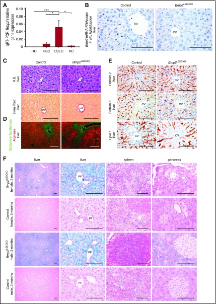
Angiocrine Bmp2 signaling in the liver controls tissue iron content and distribution. (A) Comparative qRT-PCR analysis of Bmp2 mRNA expression in LSECs, hepatocytes (HC), Kupffer cells (KC), and stellate cells (HSC) isolated from livers of wild-type (WT) adult C57Bl/6 mice (n = 5). β-2-Microglobulin was used as housekeeping gene. *P < .05; ***P < .001. (B) Bmp2 mRNA in situ hybridization of liver sections of Bmp2LSECKO mice in comparison with WT controls (n = 3). Scale bar, 100 μm (×60). CV, central vein. (C) Hematoxylin and eosin (H.E.) and Sirius red staining of liver sections of Bmp2LSECKO mice in comparison with WT controls (n = 6). Scale bar, 100 μm (×60). (D) Coimmunofluorescence of GS and arginase in liver (n = 5). Scale bar, 100 μm (×20). (E) Immunohistochemistry of LSEC markers in livers of Bmp2LSECKO and control mice (n = 5). Scale bar, 100 μm (×60). (F) Prussian blue staining demonstrating iron deposition in liver, spleen, and pancreas of Bmp2LSECKO (female, n = 5; male, n = 5). Scale bar, 100 μm (first column, ×10; third column, ×40; second and fourth columns, ×60). PF, portal field.

