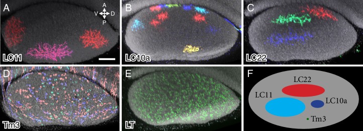
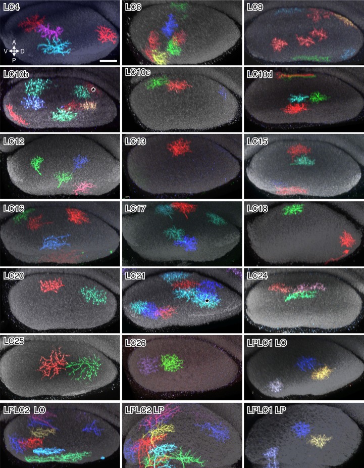
Figure 6. Column spread of LC neurons and other cell types in the lobula in cross-section views.
Cells were labeled using MCFO. Cross-section views of the lobula were generated using Vaa3D. The AP and DV axes of the lobula are indicated. Anti-Brp reference marker is shown in grey. LC neuron arbor sizes and shapes in the lobula are diverse across different cell types but similar within each type. LC cell types shown are LC11 (A), LC10a (B) and LC22 (C). The remaining LC cell types are shown in Figure 6—figure supplement 1. MCFO labeled cells of a columnar medulla neuron type (Tm3) present in each of ~750 visual columns (D) and a lobula tangential cell (LT) that spans the entire lobula (E) are shown for comparison. All LC arbors are multicolumnar with estimated sizes from about 10 (LC10a) to over 60 (LC11) visual columns. LC22 cells were similar in size to LC11 along the long (DV) but not the short (AP) axis of the lobula. (F) Schematic summary of the column spread of different cell types in the lobula. Scale bar represents 20 µm.


