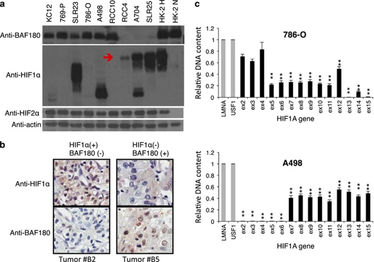
Figure 1.
Mutually exclusive expression of full-length BAF180 and HIF1α proteins in ccRCC cell lines and some primary tumors. (a) Western blot analysis of BAF180, HIF1α and HIF2α protein in ccRCC cell lines. The red arrow indicates the position of the full-length HIF1α protein. A704 cells express full-length as well as truncated HIF1α protein. Normoxic and hypoxic HK2 cells were used as positive controls for BAF180, HIF1α and HIF2α protein detection. (b) Immunohistochemistry staining of HIF1α or BAF180 proteins in the #B2 and #B5 ccRCC tumors in the ccRCC tumor microarray (US Biomax, Derwood, MD, USA, cat. #BC07014a). (c) qPCR analysis of the abundance of individual exons of the HIF1A gene in genomic DNAs isolated from the indicated ccRCC cell lines. Relative DNA content was normalized to LMNA and USF1 genes as these genes were not amplified or deleted in ccRCC cells. In addition, DNA content of HIF1A exons from HK2 cells were used as calibrators as HIF1A gene is maintained at two copies in HK2 cells. Exons with relative DNA content at 1, 0.5 or close to zero indicate normal, loss of one copy or loss of both alleles in ccRCC cell lines.

