Abstract
A supernumerary tooth is one that is supplementary to the normal dentition. It can be found anywhere at the dental arch. A mesiodens is a supernumerary tooth located between the two maxillary central incisors usually palatally or within the alveolar process. Less frequently, the mesiodens is in relation with the nasal floor and the nasopalatine canal walls. This paper presents a very rare case of an impacted inverted mesiodens located inside the nasopalatine canal and found incidentally with a cone-beam computed tomography examination.
Key words: Cone-beam computed tomography, mesiodens, nasopalatine canal
Introduction
By definition, supernumerary teeth are ones formed in addition to the normal dentition.1,2 They can be single or multiple, unilateral or bilateral, erupted or impacted, and located in the mandible and/or the maxilla.3
Mesiodens, which is a supernumerary tooth located between the two central incisors, is the most common type.1,3,4 It can occur as isolated entity or as a part of a syndrome (Gardner’s syndrome, cleidocranial dysostosis, etc.).5,6
Rare in the mandible and in the primary dentition, it is mostly found in the maxilla and in the permanent dentition with a prevalence of 0.15-1.9% in the general population and a higher frequency in males.7 The shape is usually conical and the position can be normal/inclined or inverted.1
Mesiodens can induce many complications, among others, teeth eruption alteration, root resorption and cystic lesions formation.1,2,6
The literature reported that most mesiodens were located palatally; yet very few cases were found in contact with the cortical bone of the nasal floor and the walls of the nasopalatine canal.1
Impacted mesiodens may be detected by conventional imaging techniques used in dental practice such as panoramic, occlusal and periapical radiographs. However, some might not be noticed due to radiographic technical parameters that reflect a lack of clarity in the midline region.6
In order to overcome the limitations of the conventional imaging techniques, cone-beam computed tomography (CBCT) technology has been used providing alternatives detailed and accurate assessments.8
This report describes a case of an exceptional mesiodens located within the nasopalatine canal incidentally found through a CBCT prior to dental treatment.
Case Report
A 50-year-old healthy male presented to our clinic for implant placement.
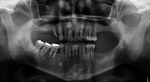
Panoramic radiograph (Figure 1) showed multiple edentulous upper and lower sites with advanced periodontal disease.
Figure 1.

A 50-year-old man panoramic radiograph showing multiple edentulous upper and lower sites with advanced periodontal disease.
A CBCT was requested for better assessment before implant placement decision.
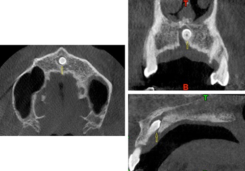
An inverted mesiodens located in the path of the nasopalatine canal was incidentally noticed. No clinical signs were correlated to this pathology. No familial history of supernumerary teeth was reported.
Its exact location and path are shown in the axial, coronal and sagittal views (Figure 2), as well in cross sections (Figure 3).
Figure 2.

A 50-year-old man cone-beam computed tomography image at the level of the premaxilla region, showing an inverted mesiodens located in the path of the nasopalatine canal (yellow arrows) in the axial, coronal and sagittal views.
Figure 3.

A 50-year-old man cone-beam computed tomography image at the level of the premaxilla region, showing an inverted mesiodens located in the path of the nasopalatine canal in cross sections views.
Discussion
According to Mossaz et al.,1 mesiodens represents 48.52% (49/101) of supernumerary teeth. This finding is consistent with Montenegro et al.,9 and Padró et al.,10 who found in their studies that prevalence among the supernumerary teeth was respectively 46.9% and 53.16%.
Seventy five percent (75%) are impacted.11 For Nazif et al.,12 6% are in a labial position, 80% palatally and the remaining 14% located between the roots of the central incisors.
Mossaz et al.,1 observed that 20.5% of the mesiodens are in contact with the cortical bone of the nasal floor and 49% in relation with the nasopalatine canal; the latter falling under 3 categories: i) 38.8% being in external contact with the canal; ii) 8.2% perforated the canal; and iii) 2% located within the canal.
Their morphology is variable. The conical shape is the most frequent followed by the tuberculate and the supplemental (toothlike).1,2,6
Generally, conical mesiodens develop with complete root formation and can erupt,13 unless when they are inverted, with the crown directed superiorly, where they are more likely to remain impacted or erupt occasionally into the nasal cavity.2,14
The tuberculate type presents numerous tubercles/cusps and incomplete or abnormal root.15 Unlike conical mesiodens, they can rarely erupt, inducing delayed eruption of the permanent incisors.6
The third type, which is much rarer, is the supplemental referring to a duplication of a normal tooth with a totally formed root (permanent maxillary lateral incisor, premolar, and molar).13,16
Regarding the position, Mossaz et al.,1 reported a normal/inclined position in 53% of cases against 36.75% in an inverted position.
Mesiodens may cause many complications such as adjacent teeth impaction/ectopic eruption, root resorption, cystic lesions formation, teeth crowding, etc.17,18
In front of such complications, thorough clinical and radiologic assessments are essential. Conventional two-dimensional radiography can be used.
The problem with the panoramic radiography is that structures outside the focal trough may be totally obscured by other structures and thus do not appear. In our case, the mesiodens was asymptomatic, so nothing would make the clinician suspects its presence.
However, the identification and/or the localization relative to neighboring structures and adjacent teeth of some mesiodens would require mainly the use of the occlusal technique. But this remains sometimes difficult due to superimposition of multiple anatomical structures, making a three-dimensional imaging (e.g., CBCT) essential to surmount the weakness of the two-dimensional radiographs.8
In this case report, an asymptomatic impacted inverted mesiodens located inside the nasopalatine canal of a 50-year-old male patient was presented with CBCT images (Figures 2 and 3).
Mesiodens within this canal have been very rarely reported. To our knowledge, no such entity was described in the literature, especially when not associated with pathology such as a nasopalatine duct cyst. The vast majority does not produce any complications and due to their high position and to the radiological unclear maxillary midline region, they can go unnoticed by the classic radiographic techniques generally used by the dental practitioner. Nowadays, with the technological enhancement of the imaging techniques used in dentistry like CBCT, more details and better evaluation are provided and consequently, these uncommon impacted structures could be noticed.
Conclusions
The incidence of a mesiodens located within the nasopalatine canal is very rare. Without clinical symptoms, patients and their dentists may remain unaware of this situation. In these cases, CBCT taken for other different purposes such as implant placement can be of great interest and help to discover this entity.
References
- 1.Mossaz J, Kloukos D, Pandis N, et al. Morphologic characteristics, location, and associated complications of maxillary and mandibular supernumerary teeth as evaluated using cone beam computed tomography. Eur J Orthod 2014;36:708-18. [DOI] [PubMed] [Google Scholar]
- 2.Meighani G, Pakdaman A. Diagnosis and management of supernumerary (mesiodens): a review of the literature. J Dent (Tehran) 2010;7:41-9. [PMC free article] [PubMed] [Google Scholar]
- 3.Parolia A, Kundabala M, Dahal M, et al. Management of supernumerary teeth. J Conserv Dent 2011;14: 221-4. [DOI] [PMC free article] [PubMed] [Google Scholar]
- 4.Alberti G, Mondani PM, Parodi V. Eruption of supernumerary permanent teeth in a sample of urban primary school population in Genoa, Italy. Eur J Paediatr Dent 2006;7:89-92. [PubMed] [Google Scholar]
- 5.Rajab LD, Hamdan MA. Supernumerary teeth: review of the literature and a survey of 152 cases. Int J Paediatr Dent 2002;12:244-54. [DOI] [PubMed] [Google Scholar]
- 6.Russell KA, Folwarczna MA. Mesiodens - diagnosis and management of a common supernumerary tooth. J Can Dent Assoc 2003;69:362-6. [PubMed] [Google Scholar]
- 7.Van Buggenhout G, Bailleul-Forestier I. Mesiodens. Eur J Med Genet 2008;51:178-81. [DOI] [PubMed] [Google Scholar]
- 8.Kapila S, Conley RS, Harrell WE., Jr The current status of cone beam computed tomography imaging in orthodontics. Dentomaxillofac Radiol 2011;40:24-34. [DOI] [PMC free article] [PubMed] [Google Scholar]
- 9.Fernández-Montenegro P, Valmaseda-Castellón E, Berini-Aytés L, Gay-Escoda C. Retrospective study of 145 supernumerary teeth. Med Oral Patol Oral Cir Bucal 2006;11:E339-44. [PubMed] [Google Scholar]
- 10.Ferrés-Padró E, Prats-Armengol J, Ferrés-Amat E. A descriptive study of 113 unerupted supernumerary teeth in 79 pediatric patients in Barcelona. Med Oral Patol Oral Cir Bucal 2009;14:146-52. [PubMed] [Google Scholar]
- 11.Tay F, Pang A, Yuen S. Unerupted maxillary anterior supernumerary teeth: report of 204 cases. ASDC J Dent Child 1984;51:289-94. [PubMed] [Google Scholar]
- 12.Nazif MM, Ruffalo RC, Zullo T. Impacted supernumerary teeth: a survey of 50 cases. J Am Dent Assoc 1983;106:201-4. [DOI] [PubMed] [Google Scholar]
- 13.Garvey MT, Barry HJ, Blake M. Supernumerary teeth - an overview of classification, diagnosis and management. J Can Dent Assoc 1999;65:612-6. [PubMed] [Google Scholar]
- 14.Atasu M, Orguneser A. Inverted impaction of a mesiodens: a case report. J Clin Pediatr Dent 1999;23:143-5. [PubMed] [Google Scholar]
- 15.Umweni AA, Osunbor GE. Non-syndrome multiple supernumerary teeth in Nigerians. Odontostomatol Trop 2002;25:43-8. [PubMed] [Google Scholar]
- 16.Srivatsan P, Babu AN. Mesiodens with an unusual morphology and multiple impacted supernumerary teeth in a non-syndromic patient. Indian J Dent Res 2007;18:130-40. [DOI] [PubMed] [Google Scholar]
- 17.Hyun HK, Lee SJ, Lee SH, et al. Clinical characteristics and complications associated with mesiodentes. J Oral Maxillofac Surg 2009;67:2639-43. [DOI] [PubMed] [Google Scholar]
- 18.Lee DH, Lee JS, Yoon SJ, Kang BC. Three dimensional evaluation of impacted mesiodens using dental cone beam CT. Korean J Oral Maxillofac Radiol 2010;40:109-14. [Google Scholar]


