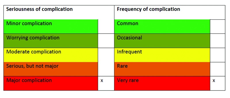
Definition
Any impairment or loss of vision (temporary or permanent) secondary to retinal or retinal branch occlusion occurring as a direct consequence of percutaneous injection for aesthetic treatment (based on methods of 2012 review1)
Introduction
Blindness after facial injection is extremely rare and was first reported by von Bahr more than 50 years ago after scalp injection of a hydrocortisone suspension to treat alopecia.2 The first cases after aesthetic “filling” treatments were reported in the 1980s (four cases) and rose to at least 16 reported cases in the 2000s, presumably related to the increase in the number of treatments being performed.1
Occlusion of the CRA (central retinal artery) due to facial injection is likely due to retrograde displacement of product. For this to happen, the injection pressure must exceed the arterial pressure causing product to move through the vasculature against the flow of blood until it passes the origin of the CRA. When pressure from the plunger is released, blood will flow once again pushing the product into the CRA, cutting off blood supply to the optic nerve.
Incidence
Globally, at least 50 cases of blindness after aesthetic facial injection have ever been reported.1,3,4 In the Lazzeri review,1 15 of 32 cases were after injection of fat. Of the remaining 17 cases, two involved hyaluronic acid and one was from a temple injection (of silicone oil). By far the most common area injected that resulted in blindness is the nose (seven cases).
In 2012, the United Kingdom reported its first case of blindness after aesthetic facial injection (to the temple with poly-L-lactic acid, the first report with this product). In 2013, the first two cases of bilateral blindness were reported (calcium hydroxyapatite to the nose and hyaluronic acid to the glabella, which also led to cerebral infarction).4
Signs and Symptoms
Sudden onset of severe pain (ocular, facial, headache, or any combination) after injection accompanies complete loss of vision (most common) or visual field defects. Other ocular signs may be present, such as deviation of the globes and pupillary defect.
Cerebral infarction can accompany retinal artery occlusion and signs and symptoms of this may also be present, such as aphasia or even hemiparesis.5
Areas of Caution
Intra-arterial injection of particulate material or suspensions must be avoided at all cost. Areas of particular concern are the nose (lateral and dorsal nasal arteries), glabella (supratrochlear and supraorbital arteries), cheek (facial, angular, and infraorbital arteries), and temple (superficial temporal artery), which have significant anastamoses between the internal and external carotid systems. However, no area is “safe” and so every injection should be performed with the knowledge that an important vessel could be nearby.
The equation for the volume of a cylinder (πr2h) tells us that just 0.01mL of product would be enough to fill 5cm of a 0.05cm diameter vessel (assuming that the vessel did not dilate). This combined with our anatomical knowledge explains why injection of very small amounts of product can reach the retinal artery after injection at these areas resulting in blindness.
Minimizing the Risk
Careful aspiration before any facial injection is important, looking at the barrel of the needle for any sign of blood. It is vital that aspiration is done carefully without moving the tip of the needle within the tissue to ensure that area aspirated is indeed the area that is injected. This can be done with any product that allows a bubble to appear in the tip of the syringe on aspiration. When performing retrograde injection, aspirating while inserting the needle will ensure that the entire injection path is aspirated and not just the starting point.
Always inject slowly and use the smallest amount of product necessary. If there is unexpected resistance or pain from the client, immediately stop injection and assess. It is important to note that the absence of a flashback on aspiration does NOT guarantee avoiding intravascular injection.
The use of blunt cannulae decreases (but does not eliminate) the risk of intravascular injection as it is more difficult for them to enter a vessel. These must be used gently as they can still tear vessels, particularly the larger gauge (thinner) cannulae. Aspiration should be performed with cannulae in the same way as with needles for the same reasons.
Good knowledge of vascular anatomy (particularly in the areas of caution listed in the previous section) is important as is remembering that there can be large variations between individuals. Lohn et al5 showed that the branches of the facial artery were symmetrical in only 53 percent of 201 cadaveric dissections.
Treatment of Blindness After Facial Injection
Once the retinal artery has been occluded, there is a window of 90 minutes before blindness is irreversible. It is advisable to transfer the patient to the nearest specialist eye hospital via ambulance as quickly as possible. Give medical staff as much information as possible about the product, area, and volume of injection. Ensure that you know which are your closest hospitals and how to contact them.
Although there is no generally agreed upon treatment regimen,6 there are actions that may help while waiting for an ambulance. Prolonged occular massage attempts to dislodge emboli by rapidly changing intraocular pressure (IOP), thereby changing the pressure and flow in the retinal arteries. Increasing the IOP also causes a reflexive dilation of the retinal arterioles and dropping it suddenly increases the volume of flow significantly.7 Occular massage is performed with the patient looking straight ahead with eyes closed. Gentle pressure is applied over the sclera with a finger, indenting the globe by a few millimeters and then releasing at a frequency of 2 to 3 times a second.8 This should be continued until advised otherwise by staff at the eye hospital. Commonly, firm ocular massage is advised for several seconds and repeated a few times. The alternative advice originates from two case studies where embolized retinal arteries were directly visualized during the massage process. This showed that even when the emboli where dislodged, more would occlude the vessel when massage stopped. Prolonged high frequency massage (up to 3 hours) had a better clearing effect. If hyaluronic acid has been used, administer hyaluronidase to the treatment area according to the Aesthetic Complications Group guideline titled, “The Use of Hyaluronidase in Aesthetic Practice.” Remember, do not let any of the above delay referral to a specialist eye hospital.
In the Lazzeri 20121 review, only two patients reported improvement following remedial treatment so there is not enough data to state that these treatments are effective. The priority is always to get the patient to an eye hospital quickly—ocular massage was ineffective in all four cases in the review, although it is most probable that this was not prolonged high frequency massage.
Treatment aims to lower IOP, increase retinal perfusion, and increase oxygen delivery to the hypoxic tissues.
-
º Lowering IOP
Carbonic anhydrase inhibitors, such as intravenous acetazolamide, reduce the rate of aqueous humor formation. In the Lazzeri review, this was effective in only one of the cases.
Other hyperosmotic diuretics, such as mannitol, can reduce IOP.
-
º Increasing blood flow
Topical and systemic corticosteroids can reduce inflammation and the restriction of blood flow. These were successful in one case with full recovery of sight, but persistent dilated pupil.
-
º Increasing oxygen delivery
-
Hyperbaric oxygen treatment and carbogen (5% carbon dioxide, 95% oxygen) can also provoke retinal dilation.
Hyperbaric oxygen was not successful and only one patient had carbogen therapy in the study, which was also not successful.2
-
Time spent accessing these unproven treatments would be better spent getting the patient to a specialist eye hospital.
References
- 1.Lazzeri D, Agonstini T, Figus M, et al. Blindness following cosmetic injections of the face. Plast Reconstr Surg. 2012;129(4):995–1012. doi: 10.1097/PRS.0b013e3182442363. [DOI] [PubMed] [Google Scholar]
- 2.von Bahr G. Multiple embolisms in the fundus of an eye after an injection in the scalp. Acta Ophthalmol (Copenh.) 963;41:85–91. doi: 10.1111/j.1755-3768.1963.tb02425.x. [DOI] [PubMed] [Google Scholar]
- 3.Woo SJ, Park SW, Park KH, et al. Iatrogenic retinal artery occlusion caused by cosmetic facial filler injections. Am J Ophthalmol. 2012;154(4):653–662. doi: 10.1016/j.ajo.2012.04.019. [DOI] [PubMed] [Google Scholar]
- 4.Carle MV, Roe R, Novack R, Boyer DS. Cosmetic facial fillers and severe vision loss. JAMA Ophthalmol. doi: 10.1001/jamaophthalmol.2014.498. Published online March 06, 2014. [DOI] [PubMed] [Google Scholar]
- 5.Lohn JW, Penn JW, Norton J, Butler PE. The course and variation of the facial artery and vein: implications for facial transplantation and facial surgery. Ann Plast Surg. 2011;67(2):184–188. doi: 10.1097/SAP.0b013e31822484ae. [DOI] [PubMed] [Google Scholar]
- 6.Fraser SG, Adams W. Interventions for acute non-arteritic central retinal artery occlusion. Cochrane Database Syst Rev. 2009;21(1):CD001989. doi: 10.1002/14651858.CD001989.pub2. [DOI] [PMC free article] [PubMed] [Google Scholar]
- 7.Jain N, Juang SC, Sayah AJ. Retinal Artery Occlusion Treatment & Management. http://emedicine.medscape.com/article/799119-treatment#a1126 Updated April 13, 2012.
- 8.Baker DL. Gentle, prolonged ocular massage can restore vision after retinal artery occlusion. Ocular Surgery News U.S. Edition. July 1, 2004.


