Figure 7.

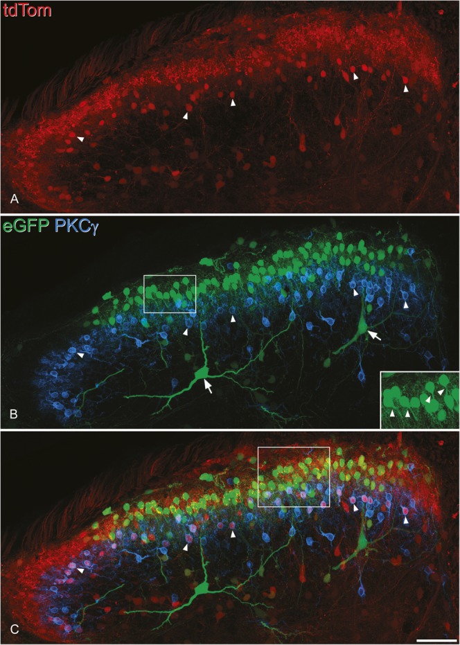
The distribution of tdTomato (tdTom)- and enhanced green fluorescent protein (eGFP)–positive cells in the dorsal horn after intraspinal injection of AAV.flex.eGFP into a Tac1Cre;Ai9 mouse. The section has been scanned to reveal tdTom (red), eGFP (green), and protein kinase C (PKC) γ (blue). (A) The TdTom+ neurons are concentrated in the superficial laminae and scattered through the deep dorsal horn. (B) The distribution of eGFP+ neurons is more restricted, as most of these lie dorsal to the band of neurons immunoreactive for PKCγ, which occupy lamina IIi. Note that none of the eGFP+ cells are PKCγ immunoreactive. In addition, 2 very large eGFP+ neurons (arrows) are located in laminae III-IV, and both have dendrites that extend into the superficial laminae. The inset (corresponding to the box) shows some of the eGFP+ cells at higher magnification, and primary dendrites can be seen leaving the ventral surface of the soma in several cases (arrowheads). (C) In the merged image, it can be seen that there are many tdTom+ neurons that lack eGFP (and therefore appear red) and that these include PKCγ-immunoreactive cells (some of these indicated with arrowheads). The images are projected from 45 optical sections at 1 μm z-spacing. The box in (C) indicates the region shown in Figure 8. Scale bar = 50 μm.
