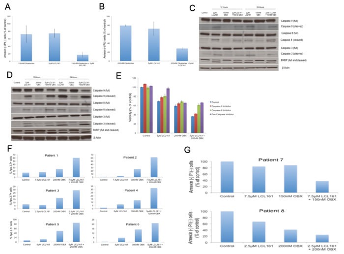
Figure 2.

A. MM1.R was incubated with 2μM of LCL161, 150nM of OBX or the combination for 72hrs. B. MM1.S was incubated with 5μM of LCL161, 200nM of OBX or the combination for 72hrs. In both A. and B. apoptosis induction was measured using Annexin/PI staining and in both cases the drug combination induced more apoptosis than either of the individual drugs. C. MM1.R and D. MM1.S cells were incubated with indicated concentrations of LCL161, OBX or the combination for 12 or 24 hours and caspases 9, 8 and 3 and PARP levels were examined by western blotting. β Actin was used as a loading control. All experiments were performed thrice and the results presented are from a representative experiment. E. We incubated MM1.S cells with indicated doses of LCL161, OBX or the combination for 72hrs alone or in the presence of 10μM of either caspase 9 inhibitor (Ac-LEHD-CMK), caspase 8 inhibitor (Z-IETD-FMK) or pan-caspase inhibitor (Q-VD-OPH) and measured the cytotoxicity induced by MTT assays. F. Primary cells from 6 MM patients were incubated with indicated doses of LCL161, OBX or the combination for 72 hrs. Apo 2.7 staining was done to examine apoptosis induction post drug treatment. G. Primary cells from 2 MM patients were incubated with indicated doses of LCL161, OBX or the combination for 72hrs. Annexin/PI staining was done to examine apoptosis induction post drug treatment.
