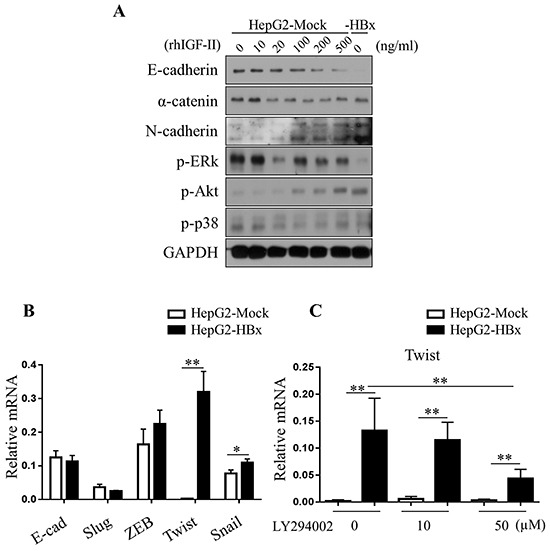
Figure 6. Loss of E-cadherin in HepG2 cells are dependent on Akt pathways.

A. Activation of Akt pathways (p-Akt) were observed in the serum starved HepG2-Mock and HBx cells treated with rhIGF-II (0-500 ng/ml). B. Relative mRNA expression levels of EMT-inducing transcription factors in HepG2-Mock and HBx cells were measured. Mean ± SD; * P< 0.05, ** P< 0.01, n= 6. C. Relative mRNA expression level of twist was measured in HepG2-Mock and HBx cells treated with an Akt inhibitor (LY294002, 0, 25 or 50μM) for 24 hours. Mean ± SD; * * P< 0.01, n= 9.
