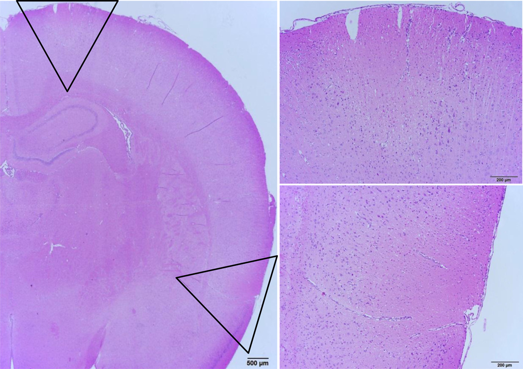
Figure 2.
(Left) Examples of lesions found in rats that spontaneously auto-resuscitated. Discrete, bilateral, wedge-shaped regions of cortical necrosis were observed in the dorsal frontal cortex and the ventral parietal cortex adjacent to the piriform lobe. Necrotic foci were found approximately at the level of the rhinal fissure extending from cortical layers into the middle grey and endopiriform neucleus, but sparing the remaining piriform cortex and amygdala (outlined wedge regions shown in panels to the right). In the cortical regions between the focal lesions, moderate necrosis was observed as both individual neurons and discrete, roughly circular geographic foci. Furthermore, nearly diffuse, severe, and acute bilaterally symmetric neuronal necrosis affects the superficial layers of the frontoparietal (motor agranular) and infralimbic cerebral cortex (not shown, additional description in results). The deeper cortex displays marked neuropil edema, with sparing of the deepest third of the cortical grey. Additionally, mild, acute neuronal necrosis is observed bilaterally and symmetrically in the medial anterior olfactory nucleus, while the piriform cortex was not affected (not shown).

