Abstract
Tmem88a is a transmembrane protein that is thought to be a negative regulator of the Wnt signalling pathway. Several groups have used antisense morpholino oligonucleotides in an effort to characterise the role of tmem88a in zebrafish cardiovascular development, but they have not obtained consistent results. Here, we generate an 8 bp deletion in the coding region of the tmem88a locus using TALENs, and we have gone on to establish a viable homozygous tmem88aΔ8 mutant line. Although tmem88aΔ8 mutants have reduced expression of some key haematopoietic genes, differentiation of erythrocytes and neutrophils is unaffected, contradicting our previous study using antisense morpholino oligonucleotides. We find that expression of the tmem88a paralogue tmem88b is not significantly changed in tmem88aΔ8 mutants and injection of the tmem88a splice-blocking morpholino oligonucleotide into tmem88aΔ8 mutants recapitulates the reduction of erythrocytes observed in morphants using o-Dianisidine. This suggests that there is a partial, but inessential, requirement for tmem88a during haematopoiesis and that morpholino injection exacerbates this phenotype in tmem88a morpholino knockdown embryos.
Introduction
The requirement for Wnt signalling in haemovascular development in vertebrates is complex, with activation of the Wnt pathway capable of both promoting and of inhibiting haematopoiesis. Early in vitro studies showed that overexpression of β-catenin increased proliferation of haematopoietic stem cells (HSCs), whereas use of Wnt inhibitors prevented HSC growth and reduced their ability to reconstitute the haematopoietic system when transplanted into irradiated mice [1]. Subsequently, conditional expression of constitutively active β-catenin, specifically in HSCs, caused a transient expansion of the HSC pool, but it did so at the expense of self-renewal and differentiation, resulting in blood cell depletion and death [2]. Furthermore, HSCs expressing a stable form of β-catenin failed to develop into downstream erythromyeloid lineages and they lost repopulation activity [3].
In vitro analyses of mouse HSCs with different hypomorphic mutations in Apc show that these cells have increased Wnt levels, increased rates of differentiation, and reduced proliferation [4]. Similar results were obtained in Wnt3a-/- mice, where HSCs were significantly fewer in number, with poor self-renewal and repopulation potential [5]. In contrast, the non-canonical Wnt ligand Wnt5a enhances self-renewal and promotes quiescence of HSCs, and this is thought to occur by interfering with the ability of Wnt3a to activate the canonical pathway [6]. Indeed, this might be conserved in mammals because human HSCs transplanted into irradiated mice had a greater reconstitution capacity when treated with Wnt-5a conditioned medium [7].
Together, these data suggest that dynamic regulation of canonical and non-canonical Wnt signalling is necessary to balance both blood cell expansion and differentiation. However, we note that many of these studies make use of exogenous activation of the Wnt pathway and the way in which Wnt signalling is modulated during haematopoiesis in vivo is poorly understood.
Transmembrane protein 88 (TMEM88; ENSG00000167874) is a two-transmembrane protein with a valine-tryptophan-valine (VWV) motif at the C-terminus that binds the PDZ domain of dishevelled (Dvl). In HEK293 cells, RNAi knockdown of TMEM88 increased Wnt activity, while overexpression of TMEM88 attenuated Wnt1-induced activation. A Siamois reporter in Xenopus showed that TMEM88 inhibits Wnt activation by xDvl, also suggesting that TMEM88 negatively regulates Wnt signalling [8].
The zebrafish orthologue, tmem88a (ENSDARG00000056920), is expressed in the heart fields, vasculature, and blood islands. This suggests that it might regulate Wnt signalling in these tissues and thereby influence their development in vivo [9,10]. Three studies have explored this question. Our own work has shown that tmem88a is enriched in fli1a:gfp+ve endothelial cells between 26–28 hpf, and morpholino oligonucleotide (MO) knockdown of tmem88a inhibited primitive blood development at 48 hpf [9].
Novikov and Evans showed that tmem88a is expressed downstream of gata5/6 in the heart field but is expressed independently of gata5/6 in the posterior blood island. MO knockdown of tmem88a reduced expression of nkx2.5 in the heart field at the 8-somite stage (8ss), reduced the expression of cardiomyocyte markers at the 23-somite stage, and reduced the number of cardiomyocytes in Tg(myl7:DsRed2-nuc) embryos at 48 hpf. Heat-shock inducible expression of the Wnt antagonist dikkopf 1 (dkk1) was sufficient to rescue expression of nkx2.5 and to restore the number of myl7:DsRed2+ve cardiomyocytes. In addition, overexpression of full-length tmem88a reduced nkx2.5 expression in cardiac progenitors and this expression was rescued by heat-shock inducible wnt8 expression. This study also observed a significant reduction in gata1 and spi1 expression in embryos at the 8-somite stage, further suggesting a requirement for tmem88a in primitive haematopoiesis [10].
Most recently, Musso and colleagues showed that the hearts of Tmem88a-deficient zebrafish embryos had increased ventricular conduction velocity at 48 hpf, although the decrease in the number of ventricular nuclei reported by Novikov and Evans was not observed. In addition, Musso and colleagues report the up-regulation of erythrocyte marker hbbe3 expression in tmem88a knockdown embryos, in contrast to the down-regulation of hbbe1 observed in our own experiments. Finally, the development of cyclopia in embryos lacking Wnt11 was exacerbated when tmem88a was also knocked down [11]. These data suggest a role for Tmem88a in regulating the non-canonical Wnt cascade, either directly or indirectly by inhibiting canonical Wnt activation [12].
The different results obtained by different groups might be explained in part by off-target effects of anti-sense morpholino oligonucleotides. Indeed, several studies have described variation between the phenotypes of MO knockdown and genetic mutants [13–20]. To define definitively the role of tmem88a in zebrafish haematopoiesis, we therefore made a genetic mutant using transcription activator-like effector nucleases (TALENs). We show that the mutants are morphologically normal and viable, but have reduced expression of some genes associated with early cardiovascular development. We also describe possible off-target effects of morpholino injection that appear to exacerbate the phenotype in MO knockdown embryos, but not in mutants.
Materials and methods
Protein alignments
Human, Xenopus, and zebrafish TMEM88 protein sequences were aligned using EMBL-EBI Clustal Omega software. Phylogenetic tree data was organised into an unrooted phylogenetic tree cladogram using the TreeDraw software from the PHYLIP package [21,22].
Ethics statement
All zebrafish work was carried out with approval from the Francis Crick Institute Biological Research Facility Strategic Oversight Committee and the Animal Welfare and Ethical Review Body, and in accordance with the Animals (Scientific Procedures) Act 1986, the Animal Welfare Act (2006) and the Welfare of Animals in Transport Order. Care was taken to minimize the numbers of animals used in these experiments in accordance with the ARRIVE guidelines (http://www.nc3rs.org.uk/page.asp?id=1357).
Zebrafish stocks and maintenance
Zebrafish (Danio rerio) adults were maintained and bred under standard conditions [23] and embryos were staged as described previously [24]. The wild-type (WT) Lon AB line was provided by the Francis Crick Institute, Mill Hill Laboratory aquatics facility (London, UK). The published transgenic line Tg(fli1a:egfp)y1 [25] was a gift from Dr Tim Chico (MRC Centre for Developmental and Biomedical Genetics, University of Sheffield). The Tg(fli1a:egfp)tmem88aΔ8 (“tmem88aΔ8”) strain was created as part of this work.
TALEN synthesis and mutagenesis
Two TALENs were designed to target exon 2 of tmem88a, either side of a spacer region containing a ScaI endonuclease site. TALEN constructs were assembled from the Golden Gate TALEN 2.0 plasmid kit, a gift from Daniel Voytas and Adam Bogdanove (Addgene, Kit #1000000024), as previously described and according to the manufacturer’s protocol [26,27]. Complete TALEN plasmid DNA was linearised with SacI (New England BioLabs) in a 50 μl reaction and purified using QIAquick purification columns (Qiagen) according to the manufacturer’s instructions. mRNA was synthesised from linear plasmid using the T3 mMESSAGE mMACHINE kit (Ambion, Life Technologies) according to the manufacturer’s instructions. It was purified by LiCl precipitation. 1 nl containing 60 ng/nl of each TALEN mRNA was injected into single-cell Tg(fli1a:egfp)y1 embryos.
Genotyping
To extract genomic DNA, single 24 hpf embryos or adult tail fin clips were incubated in 10–50 μL extraction buffer containing 50 mM Tris-HCl (pH 8.5), 1 mM EDTA, 0.5% Tween-20 and 80 mg/ml proteinase K (Ambion, Life Technologies) for 3 hours at 55°C. Proteinase K was inactivated by heating to 95°C for 10 minutes. 1 μl of genomic DNA mix was used in a standard 50 μl PCR reaction with Phusion High-Fidelity DNA polymerase (New England BioLabs) according to the manufacturer’s instructions and using primers spanning the TALEN target site (Fw: 5′-CACACAGCCCAATGCATGAC-3′; Rv: 5′-TCTTTTTCCTTGGCATTGGGTA-3′). PCR products were purified using QIAquick purification columns (Qiagen) and digested overnight at 37°C in a 10 μl reaction containing ScaI-HF endonuclease (New England BioLabs) according to the manufacturer’s instructions. Samples were then run on a 2% agarose gel. Mutant products were undigested at 705 bp, whereas digested wild-type products were detected as fragments of 240 and 465 bp.
Genotypes were also sequence verified using 1μl of genomic DNA in a standard 50 μl PCR reaction with Phusion High-Fidelity DNA polymerase (New England BioLabs) according to the manufacturer’s instructions, and using primers spanning the TALEN target site: primers include the SP6 promoter sequence, which is shown underlined; Fw: 5′–ATTTAGGTGACACTATAGAAGNGAAAACTGGCCCTTCACATAC-3′; Rv: 5′– AATGGCAGAGGAAGCCAAAAAC-3′. PCR products were purified using QIAquick purification columns (Qiagen) according the manufacturer’s instructions, sequenced using SP6 promoter, and aligned to the wild type genomic sequence.
Reverse transcription, quantitative RT-PCR and melting curve analyses
Between 30 and 50 embryos were collected for extraction and purification of RNA using TRIzol and RNeasy Minikit Columns (Qiagen) according to the manufacturer’s protocol. cDNA was reverse transcribed from 0.5–3 μg total RNA using M-MLV RT RNase (-H) Point Mutant (Promega) according to the manufacturer’s instructions and diluted 1:10 for qRT-PCR. Quantitative RT-PCR was performed in duplicate 10 μl reactions using Lightcycler Mastermix (Roche) on a Lightcycler LC480 (Roche) according to the manufacturer’s instructions. Primer sequences were as previously published or designed using NCBI primer-BLAST and are shown in Table 1 [9]. Two primer sets detecting all tmem88a transcript (F1R1) and only wild type tmem88a transcript (F2R2) were used. Expression levels were compared to a standard curve, normalised to total RNA, and expressed as a percentage of the control groups (defined as 100%). Melt curves using cDNA synthesised from RNA extracted from Tg(fli1a:egfp) and tmem88Δ8 mutant embryos in biological triplicate using Light Cycler 480 software version 1.5.0.
Table 1. Sequences of primers used for qRT-PCR.
| Forward primer (5′-3′) | Reverse primer (5′-3′) | |
|---|---|---|
| fli1a | AGCGCTACGCCTACAAGTTC | AGCTCCAGTATGGGGTTGTG |
| gata1a | CTCGTTGGGTGTCCCCCGGT | CGACGAGGCTCGGCTCTGGA |
| hbbe1 | AACTGTGCTCAAGGGTCTGG | TACGTGGAGCTTCTCGGAGT |
| lyz | GCACGGCCTACTGGGAAAGCA | CCCAGGGGTCCCGTCATCACA |
| myoD | GGGCCCAACGTGTCAGACGA | GTTGAGGGCAGCTGGTCGGG |
| nkx2.5 | CTGTGCCAGTTTTGGTTCGG | CGCAGGGTAGGTGTTGTAGG |
| runx1 | AGTGGACGGACCCCGAGAGC | ACCGCATGGCACTTCGCCTC |
| scl | CAACGATGGTTCGCAGCCCA | ACCGCCGACCATGTCGTCCT |
| spi1b | ATGCGGCCAGTGTGCATCGC | CACCGATGTCCGGGGCAAGT |
| tmem88a F1R1 | CCTGCCATCGCTCGTCATGGT | AGACGGCACGGCTGTATGGGA |
| tmem88a F2R2 | TGCCAGTCTGAGGAATCTGCC | GTTTGCTGGAGTACTGGAGGAGA |
| tmem88b | CCTCCACCTTACTCCCCAGA | ATGGGATAAAGCACAGCCCC |
fli1a, friend leukaemia integration 1a; gata1a, GATA-binding factor 1a; hbbe1, haemoglobin beta-embryonic 1; lyz, lysozyme; runx1, runt-related transcription factor 1; scl, stem cell leukaemia; spi1, spleen focus forming virus (sffv) and proviral integration oncogene 1b (also known as pu.1).
For digestion of tmem88a cDNA, 2 μl of cDNA synthesised from wild type and tmem88a mutant RNA was used in a standard 20 μl PCR reaction with Phusion High-Fidelity DNA polymerase (New England BioLabs) according to the manufacturer’s instructions. The primers used spanned the deletion and contained a 5′ SP6 promoter sequence, which is shown underlined (Fw: 5′–ATTTAGGTGACACTATAGAAGNGAAAACTGGCCCTTCACATAC-3′; Rv: 5′– AATGGCAGAGGAAGCCAAAAAC-3). 10 μl of each PCR product was digested overnight at 37°C with ScaI endonuclease (Thermo Fisher Scientific) according to the manufacturer’s instructions. Samples were then run on a 2% agarose gel. Mutant products were undigested at 423 bp, whereas digested wild type products were detected as fragments of 244 and 179 bp.
DIG-labelled probe synthesis and whole mount in situ hybridisation
Anti-sense fli1a probes were synthesised as previously described [9]. Antisense DIG-labelled probes for gata1a were synthesised using the SP6 promoter and cDNA template amplified using the following primers: the SP6 promoter sequence is underlined and the T7 promoter sequence is shown in italics; Fw: 5′-TAATACGACTCACTATAGGGAGACACTCTCACACCTCCACGTC-3′; Rv: 5′-ATTTAGGTGACACTATAGAAGNGATTGAGGGAAACAAAAAGTGTGT-3′. DIG-labelled anti-sense myoD probes were synthesised from the T7 promoter and template plasmid DNA (IRBOp991B1179D, Source Bioscience) linearised with EcoRV according to the manufacturer’s instructions. In situ hybridisation was performed as previously described [28,29].
Whole embryo staining for blood cells
To study erythrocyte development, o-Dianisidine staining was performed on 48 hpf embryos as previously described [30]. Stained embryos were then fixed in 4% paraformaldehyde in PBS (Thermo Scientific) for 20 minutes at room temperature, extensively rinsed in PBST, and mounted for imaging.
To investigate neutrophil development, embryos were fixed for 1 hour at room temperature in 4% glutaraldehyde (Sigma-Aldrich) in borate buffer. Embryos were then stained with Sudan Black B (Sigma-Aldrich): 0.18% (w/v) Sudan Black, in 69% ethanol; for 20 minutes at room temperature with agitation. Embryos were then extensively rinsed in three times in 70% EtOH with agitation, and rehydrated in PBST.
Mounting, imaging and image analysis
For imaging, stained embryos were mounted in 50% glycerol on glass slides with coverslips. 8-somite stage (8ss) embryos were flat mounted as previously described [29]. Whole embryos were imaged using a Leica M165 FC microscope and Leica Application suite 3.4.1 software. o-Dianisidine staining was quantified by dividing the stained area of the yolk by the total yolk area using Adobe Photoshop and Image J software, and presented as a percentage of the yolk. All quantification of blood cells was performed blind.
Morpholino oligonucleotide (MO) knockdown
One-cell Tg(fli1a:egfp)y1 or tmem88aΔ8 embryos were injected with 5 ng of both tmem88a e2i2 splice-blocking MO (SBMO; 5′-GCATTCTCACTCCACACATACCGTT-3′) and p53 MO (5′-GCGCCATTGCTTTGCAAGAATTG-3′) as previously described [9].
Results
TALEN mutagenesis of tmem88a
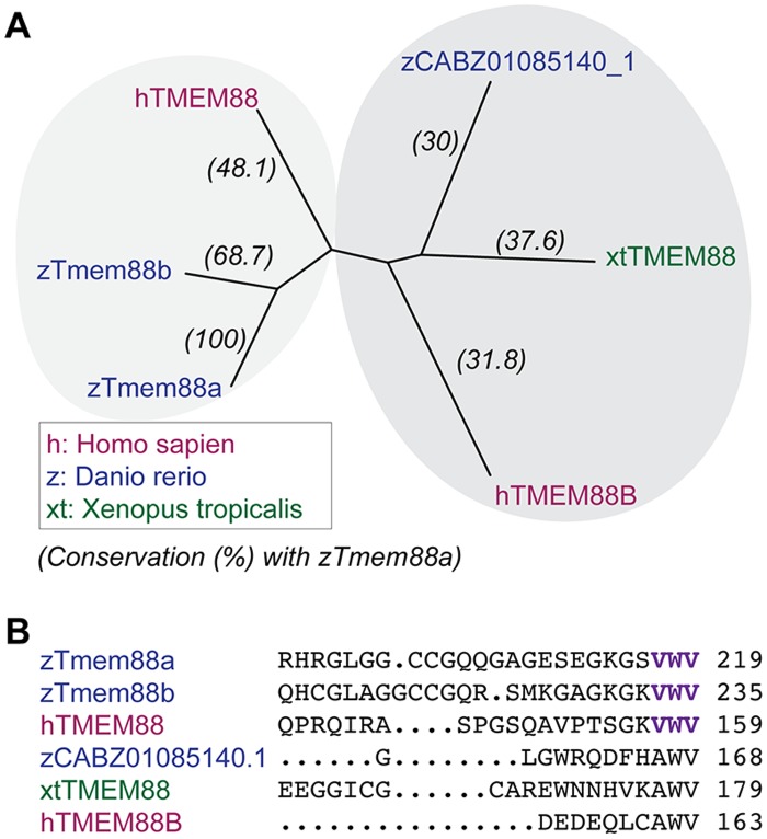
Human (Homo sapiens), Xenopus tropicalis, and zebrafish (Danio rerio) TMEM88 protein sequences were aligned using Clustal Omega and grouped in an unrooted phyologenetic tree based on sequence conservation. The results reveal two main clusters, the first of which includes human TMEM88 and the zebrafish orthologues Tmem88a and Tmem88b (ENSDARG00000069388) (Fig 1A). The zebrafish Tmem88a and Tmem88b proteins share the valine-tryptophan-valine (VWV) motif, which was previously identified as being involved in Dishevelled binding [8]. Members of the second cluster are less closely related and include human TMEM88B (ENSG00000205116), Xenopus TMEM88 (NW_004668240.1), and the zebrafish orthologue CABZ01085140.1 (ENSDARG00000098070), which all lack the conserved VWV motif (Fig 1B).
Fig 1. Species conservation of TMEM88 proteins.
(A) Unrooted phylogenetic tree of aligned TMEM88 protein orthologues from human (Homo sapiens, magenta), Xenopus tropicalis (green), and zebrafish (Danio rerio, blue) show clustering into two branches of sequence conservation. (B) Zebrafish orthologues—Tmem88a and Tmem88b –share the conserved valine-tryptophan-valine (VWV) motif in the Dishevelled (Dvl) binding domain of human TMEM88.
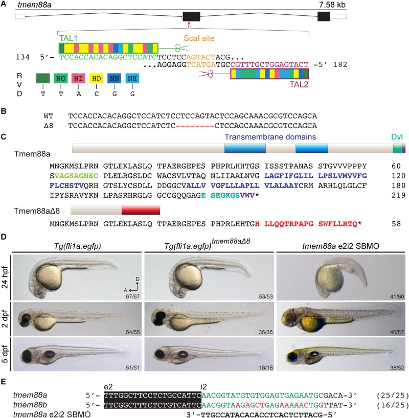
TALENs were designed to target the coding region in exon2 of the tmem88a locus. This target included a ScaI restriction site that would be destroyed by mutations in the spacer region. ScaI digestion was used for genotyping TALEN-injected embryos and subsequent generations of fish (Fig 2A). TALEN constructs were injected into Tg(fli1a:egfp) embryos. F0 adults were screened for tmem88a mutations by PCR amplification of genomic DNA followed by ScaI digestion. An F0 founder female was crossed with a wild type Lon AB male to produce an F1 generation. The F1 generation was in-crossed and F2 adults were screened for mutations by sequencing the target region. F2 mutants were in-crossed and F3 adults with homozygous deletions were selected by sequencing to produce a homozygous mutant line, with an 8 bp deletion at position 120 of the coding sequence, expected to cause a frame-shift and nonsense-mediated decay of the transcript (Fig 2B). The translated sequence of tmem88aΔ8 was predicted to include an early stop codon and truncation of the protein, preventing translation of the transmembrane and Dishevelled-binding domains (Fig 2C).
Fig 2. Mutation of the zebrafish tmem88a gene.
(A) Schematic structure of the zebrafish tmem88a gene shows three exons and two introns. TALEN pairs TAL1 (green) and TAL2 (magenta) were designed and assembled to target the coding region of exon2. There is a ScaI endonuclease restriction site within the spacer region (orange). (B) A tmem88a mutant line with an 8 bp deletion (Δ8) was generated. (C) Predicted translated sequences of wild-type Tmem88a and Tmem88aΔ8 show frame-shift after residue 48 (red), causing truncation of the protein and loss of the trans-membrane domains (blue) and the Dishevelled-binding domain (teal). (D) Brightfield images of control Tg(fli1a:egfp) and tmem88aΔ8 embryos at 24 hpf (n = 67, n = 53 respectively), 2 dpf (n = 54, n = 35), and 5 dpf (n = 51, n = 18) as labelled. (E) Sequence alignment shows that the tmem88a e2i2 SBMO is specific to tmem88a and illustrates a 9 bp mismatch with tmem88b mRNA.
To our surprise, tmem88aΔ8 mutant embryos showed no morphological abnormalities during early development (0–5 dpf), and did not exhibit any developmental defects when compared with Tg(fli1a:egfp) controls of the same genetic background and no developmental delay was observed compared to tmem88a SBMO knockdown embryos (Fig 2D). Indeed, the line proved to be viable and tmem88aΔ8 mutants produced fertile adults. Sequence alignments show morpholino specificity for tmem88a transcripts and not double knockdown of tmem88a and tmem88b because of a 9 bp mismatch (Fig 2E) [31].
Wild type tmem88a transcripts are absent in tmem88aΔ8 mutant embryos
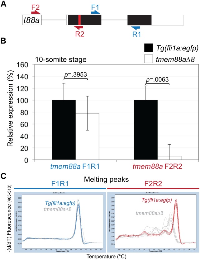
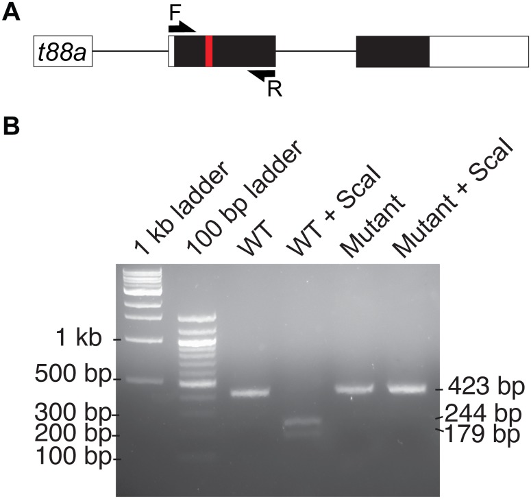
To validate knock out of the tmem88a gene qRT-PCR was performed using RNA recovered from 10-somite stage embryos. Two primer sets were used as illustrated, F1R1 detected all spliced tmem88a transcript, while F2R2 only detects wild type tmem88a transcript because the reverse primer binds across the mutated region (Fig 3A). Results do not show a significant reduction (p = 0.3953, Unpaired Student’s t test) in tmem88a transcript in tmem88aΔ8 mutants compared to controls, suggesting that not all transcripts were degraded by nonsense-mediated decay (Fig 3B). However, a significant decrease (p = 0.0063, Unpaired Student’s t test) in wild type tmem88a transcript was found in tmem88aΔ8 embryos compared to controls (Fig 3B). Analyses of the melting curves generated by the two primers sets showed a shift in F2R2 primers when binding mutant cDNA, suggesting no wild type transcripts were detected (Fig 3C). In addition, cDNA generated from tmem88a mutants is not digested by ScaI endonuclease compared with wild type controls, providing further evidence that wild type transcripts are not present in tmem88a mutant embryos (Fig 4).
Fig 3. The expression of tmem88a in tmem88Δ8 mutant embryos.
(A) Schematic representation of the tmem88a (t88a) mRNA transcript. Non-coding and coding exons are shown as white and black blocks respectively. Introns as depicted as solid lines. The deleted site in tmem88aΔ8 mutants is shown by a red bar. F1R1 primers detected tmem88a transcript and F2R2 primers detected wild type transcript only. (B) qRT-PCR showing the expression of tmem88a in tmem88Δ8 mutants compared to Tg(fli1a:egfp) controls at the 10-somite stage using both primer sets. P-values determined by Unpaired Student’s t test. Error bars show the standard deviation for three biological replicates. (C) Melting curves using two primer sets designed for tmem88a detection as shown in (A). A shift in melting in temperature is observed in primers designed to bind the mutated region in tmem88aΔ8 mutants compared to controls (F2R2, green).
Fig 4. tmem88a cDNA from tmem88a mutants is not digested by ScaI.
(A) Schematic representation of the tmem88a (t88a) mRNA transcript. Non-coding and coding exons are shown as white and black blocks respectively. Introns as depicted as solid lines. The deleted site in tmem88aΔ8 mutants is shown by a red bar. F and R primers used for amplification of cDNA are shown as arrows. (B) 2% agarose gel showing ScaI digestion of wild type PCR product into fragments of 244 and 179 bp in size. Tmem88a mutant cDNA is not digested by ScaI.
Reduced expression of some key cardiac and haematopoietic factors is observed in tmem88a mutant embryos
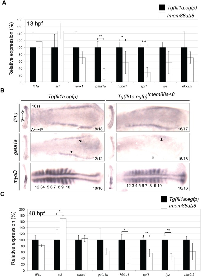
Tmem88a knockdown has previously been shown to reduce expression of haematopoietic genes [9,10]. We therefore investigated the expression of known cardiovascular markers in tmem88aΔ8 embryos. Tg(fli1a:egfp) and tmem88Δ8 embryos were collected at 13 hpf, and RNA extracted for qRT-PCR. Although there was no significant difference between the expression of fli1a, scl, and runx1 at this stage, there was a significant (p<0.05, Unpaired Student’s t test) reduction in gata1a, hbbe1, and spi1 expression (Fig 5A). To confirm this observation we performed in situ hybridisation on 10-somite stage embryos. There was no gross difference in fli1a expression between the two lines (controls: 18/18; tmem88aΔ8 mutants: 16/17), but gata1a expression was greatly reduced in tmem88aΔ8 embryos (15/18) compared to controls (12/12). Expression of MyoD, studied as a control for developmental delay, was slightly weaker in mutant embryos, with expression varying a little between somites (Fig 5B).
Fig 5. Cardiovascular associated genes are downregulated in tmem88aΔ8 embryos.
(A) qRT-PCR showing the expression of key cardiovascular genes in tmem88Δ8 mutants compared to Tg(fli1a:egfp) controls at 13 hpf. (B) In situ hybridisation for fli1a, gata1a, and myoD in control and tmem88aΔ8 mutants as labelled. Gata1a expression is denoted with black error heads, reduced or absent expression is shown with grey or white arrowheads respectively. Somites are marked by asterisks. (C) qRT-PCR showing the expression of key cardiovascular genes in tmem88Δ8 mutants compared to Tg(fli1a:egfp) controls at 48 hpf. All P-values determined by Unpaired Student’s t test. Error bars denote the standard deviation of three biological replicates. P-values determined by Unpaired Students t test. * = p<0.05; ** = p<0.01; *** = p<0.001; unlabelled bars show no significant difference (p>0.05). fli1a, friend leukaemia integration 1a; gata1a, GATA-binding factor 1a; hbbe1, haemoglobin beta-embryonic 1; hpf, hours post fertilisation; lyz, lysozyme; runx1, runt-related transcription factor 1; scl, stem cell leukaemia; spi1, spleen focus forming virus (sffv) and proviral integration oncogene 1b (also known as pu.1).
tmem88a is dispensable for primitive haematopoiesis
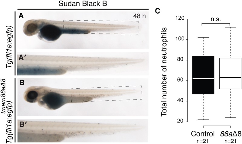
Tmem88aΔ8 mutants showed a reduction of myeloid markers spi1b at 13 and 48 hpf, and lyz at 48 hpf. However, tmem88aΔ8 mutants were otherwise unaffected and survived to adulthood, suggesting that this reduction was not sufficient to prevent blood development. To investigate whether the myeloid cell lineage was affected, 48 hpf Tg(fli1a:egfp) control embryos (n = 21) and tmem88aΔ8 mutants (n = 21) were stained for neutrophils with Sudan Black B solution (Fig 6A and 6B). Neutrophils were counted and results showed no significant difference (p = 0.6769, Unpaired Student’s t test) between the number of cells in control embryos and Tmem88a-deficient embryos (Fig 6C). This suggests that tmem88a is not required for primitive myelopoiesis.
Fig 6. Myelopoiesis was not affected in tmem88aΔ8 zebrafish mutants.
(A-B) Sudan Black B staining of neutrophils at 48 hpf in Tg(fli1a:egfp) controls (A, n = 21), with the highlighted tail region magnified (A′) and tmem88aΔ8 mutants (B, n = 21), magnified in (B′). (C) The number of neutrophils was counted for each condition represented in a Tukey box and whisker plot. Neutrophil numbers were not significantly changed between the two groups (p = 0.6769, Unpaired Student’s t test). A, anterior; h, hours post fertilisation; D, dorsal; n.s., not significant.
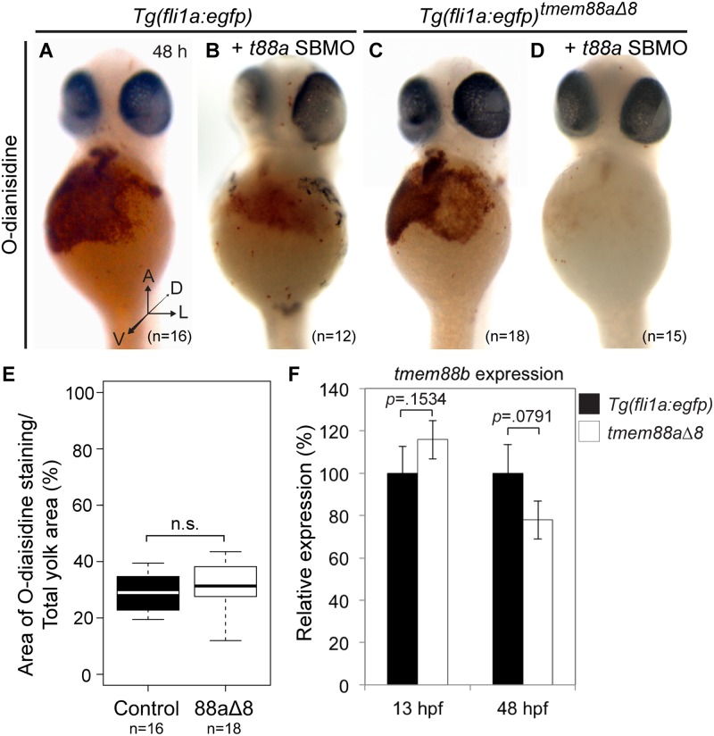
We also observed a reduction in expression of erythrocyte markers gata1a and hbbe1 in tmem88aΔ8 mutants at 13 and 48 hpf. To ask whether primitive erythropoiesis was affected, we performed o-Dianisidine staining on Tg(fli1a:egfp) control (n = 16) and tmem88aΔ8 embryos (n = 18) at 48 hpf (Fig 7A and 7C). Staining was quantified by measuring the area of stained cells as a percentage of the yolk (Fig 7E). There was no significant difference between Tg(fli1a:egfp) controls and Tmem88a-deficient embryos (p = 0.2710, Unpaired Student’s t test).
Fig 7. Erythropoiesis was not affected in tmem88aΔ8 zebrafish mutants.
(A-D) o-Dianisidine staining of erythrocytes at 48 hpf in uninjected Tg(fli1a:egfp) controls (A, n = 16) or controls injected with tmem88a SBMO (B, n = 12), and uninjected tmem88aΔ8 mutants (C, n = 18) or mutants injected with tmem88a SBMO (D, n = 15). (E) The area of o-Dianisine staining was quantified and presented as a percentage of the total yolk area, shown in a Tukey box and whisker plot. No significant difference was found between control embryos and tmem88aΔ8 mutants (p = 0.2710, Unpaired Student’s t test). (F) tmem88b expression is not affected in tmem88aΔ8 mutants. qRT-PCR showing the expression of tmem88b in tmem88Δ8 mutants compared to Tg(fli1a:egfp) controls at 13 and 48 hpf. P-value determined by Unpaired Student’s t test. Error bars denote the standard deviation of three biological replicates. A, anterior; h, hours post fertilisation; D, dorsal; L, lateral; V, ventral; n.s., not significant.
These results contrast with our previous observations that both myelocyte peroxidase and o-Dianisidine staining was reduced when tmem88a was knocked-down using MOs. It is possible that the differences between the mutant and morphant phenotypes reflect genuine biological differences relating to the effects of genetic knockout versus knockdown by interfering oligonucleotides. One recent study has suggested that compensatory transcriptional changes may occur when a gene is inactivated by genetic mutation, but not as a result of knockdown in morphants [14]. One way in which compensation could occur is through the upregulation of a paralogue. We therefore examined the expression of the tmem88a paralogue, tmem88b, in the tmem88a mutant line and did not see a significant change in tmem88b expression (Fig 7F).
Next, we next asked whether the loss of blood cells seen in morphant embryos was due to an off-target effect of MO injection. We injected the tmem88a splice-blocking MO (SBMO) and a p53 MO into Tg(fli1a:egfp) controls (n = 12) and tmem88aΔ8 embryos (n = 15), on the basis that tmem88aΔ8 embryos lack wild-type tmem88a protein, and thus any defects seen cannot be due to loss of protein. Tmem88a SBMO injection caused a clear reduction in o-Dianisidine staining of erythrocytes in both control and in tmem88aΔ8 mutants (Fig 7B and 7D). This suggested that off-target effects from morpholino injection affected o-Dianisidine staining of erythrocytes.
Discussion
The role of tmem88a in zebrafish development has been investigated by three groups using antisense morpholino oligonucleotides. All identify cardiovascular phenotypes, but otherwise the results have been inconsistent [9–11,32]. To resolve this issue we generated an 8 bp deletion and frame-shift mutation in the endogenous tmem88a locus using TALEN technology [26]. In this way we generated a viable tmem88a loss-of-function zebrafish mutant line to study the role of tmem88a in primitive haematopoiesis.
tmem88a is dispensable for zebrafish haematopoiesis
In situ hybridisation and qRT-PCR revealed that the expression of key haematopoietic factors gata1a, hbbe1, and spi1 are reduced in 8-somite stage and 48 hpf tmem88aΔ8 mutants. This is consistent with tmem88a morpholino knockdown data, which also showed a reduction of haematopoietic markers at these time points [9,10]. Bearing in mind the probable role of tmem88a as a regulator of Wnt signalling, these observations suggest that temporal and spatial Wnt inhibition is necessary to promote haematopoiesis [8]. Although we observed that tmem88aΔ8 mutants had reduced expression of haematopoietic markers, equivalent to the reduction seen by tmem88a morpholino knockdown [9], this was not sufficient to prevent the development of mature primitive erythrocytes and neutrophils as determined by histological staining. This suggests that although haematopoietic genes are down-regulated in the absence of Tmem88a, primitive erythrocytes and neutrophils still formed. Thus, there is a partial, but not essential, requirement for tmem88a in primitive blood cell development.
Differences between mutation and knockdown
Our mutant data conflict with our previously-described morpholino knockdown phenotype, in which, according to o-Dianisidine staining and myelocyte peroxidase assays, the numbers of primitive erythrocytes and neutrophils were reduced [9]. No significant morphological heart defects, like those described by Novikov and Evans, were observed in tmem88aΔ8 mutants [10]. We did not observe a significant decrease in nkx2.5 expression in tmem88aΔ8 mutants using qRT-PCR, although we note that our study of cardiac factors was limited.
There have been several examples of differences between MO knockdown and genetic mutant phenotypes, and these differences have been suggested to represent nonspecific off-target effects of morpholinos [13,16–20,33–35]. In some cases mutant alleles may not represent true loss-of-function phenotypes because use of alternate (non-AUG) translation start codons, cryptic splice sites, or translational readthrough prevent complete loss of the target protein [36–40]. rpholino to it;s they are more toxic effects of morpholinos are induced by the binding of the morpholino to it;s they are more Alternatively, one or several proteins with similar functions might be up-regulated to compensate in genetic mutants, but not in morphants [14]. Although tmem88b –an obvious candidate for a compensatory gene in tmem88aΔ8 mutants—was not significantly up-regulated compared with controls, there may be up-regulation of other genes that we have not investigated. Injecting a morpholino specific to tmem88a into tmem88aΔ8 mutants reduced o-Dianisidine staining, suggesting that erythrocyte development was affected by morpholino injection independently of Tmem88a-deficiency. It is known that morpholinos can induce nonspecific toxic effects such as pericardial oedema, reduced cardiomyocyte proliferation, and p53-dependent cell death [31,41–44]. Furthermore, many stressors, including morpholinos, are known to cause heart failure and reduced circulatory flow, which is required for proper vascular development, although the mechanisms by which stressors cause these effects remain unknown [45,46]. Finally, it is possible that in this case the reduced expression of haematopoietic factors in Tmem88a-deficient embryos might increase susceptibly to such stressors, increasing the likelihood of nonspecific vascular abnormalities occurring as a result of morpholino injection.
Conclusion
We observed that although tmem88a mutants had a reduction of some cardiovascular markers equivalent to those seen in tmem88a morphants, tmem88a was dispensable for primitive haematopoiesis because erythrocytes and neutrophils were identified, respectively, by o-Dianisidine and Sudan Black B staining. Injecting a tmem88a MO into mutants reduced o-Dianisidine staining to the level observed in tmem88a morphants. This suggests that MO use might have off-target effects on later erythrocyte development and the that published tmem88a MO knockdown phenotypes might include such nonspecific toxic effects.
Acknowledgments
We thank the Francis Crick Aquatics facility at Mill Hill for maintenance of zebrafish lines, and John Cannon for reagents. We are also grateful to Nick Morrell and other members of our Leducq network for discussion, support and fun.
Data Availability
All relevant data are within the paper.
Funding Statement
This work was supported by the Francis Crick Institute (www.crick.ac.uk), which receives its core funding from Cancer Research UK (FC001-157), the UK Medical Research Council (FC001-157), and the Wellcome Trust (FC001-157). We are also grateful to the Fondation Leducq (www.fondationleducq.org) for a Transatlantic Network of Excellence Award. The funders had no role in study design, data collection and analysis, decision to publish, or preparation of the manuscript.
References
- 1.Reya T, Duncan AW, Ailles L, Domen J, Scherer DC, Willert K, et al. A role for Wnt signalling in self-renewal of haematopoietic stem cells. Nature. 2003;423: 409–414. 10.1038/nature01593 [DOI] [PubMed] [Google Scholar]
- 2.Scheller M, Huelsken J, Rosenbauer F, Taketo MM, Birchmeier W, Tenen DG, et al. Hematopoietic stem cell and multilineage defects generated by constitutive beta-catenin activation. Nat Immunol. 2006;7: 1037–1047. 10.1038/ni1387 [DOI] [PubMed] [Google Scholar]
- 3.Kirstetter P, Anderson K, Porse BT, Jacobsen SEW, Nerlov C. Activation of the canonical Wnt pathway leads to loss of hematopoietic stem cell repopulation and multilineage differentiation block. Nat Immunol. 2006;7: 1048–1056. 10.1038/ni1381 [DOI] [PubMed] [Google Scholar]
- 4.Famili F, Brugman MH, Taskesen E, Naber BEA, Fodde R, Staal FJT. High Levels of Canonical Wnt Signaling Lead to Loss of Stemness and Increased Differentiation in Hematopoietic Stem Cells. Stem Cell Reports. The Authors; 2016;6: 652–659. 10.1016/j.stemcr.2016.04.009 [DOI] [PMC free article] [PubMed] [Google Scholar]
- 5.Luis TC, Weerkamp F, Naber BAE, Baert MRM, De Haas EFE, Nikolic T, et al. Wnt3a deficiency irreversibly impairs hematopoietic stem cell self-renewal and leads to defects in progenitor cell differentiation. Blood. 2009;113: 546–554. 10.1182/blood-2008-06-163774 [DOI] [PubMed] [Google Scholar]
- 6.Nemeth MJ, Topol L, Anderson SM, Yang Y, Bodine DM. Wnt5a inhibits canonical Wnt signaling in hematopoietic stem cells and enhances repopulation. Proc Natl Acad Sci U S A. 2007;104: 15436–41. 10.1073/pnas.0704747104 [DOI] [PMC free article] [PubMed] [Google Scholar]
- 7.Murdoch B, Chadwick K, Martin M, Shojaei F, Shah K V, Gallacher L, et al. Wnt-5A augments repopulating capacity and primitive hematopoietic development of human blood stem cells in vivo. Proc Natl Acad Sci U S A. 2003;100: 3422–7. 10.1073/pnas.0130233100 [DOI] [PMC free article] [PubMed] [Google Scholar]
- 8.Lee HJ, Finkelstein D, Li X, Wu D, Shi DL, Zheng JJ. Identification of transmembrane protein 88 (TMEM88) as a dishevelled-binding protein. J Biol Chem. 2010/11/04. 2010;285: 41549–41556. 10.1074/jbc.M110.193383 [DOI] [PMC free article] [PubMed] [Google Scholar]
- 9.Cannon JE, Place ES, Eve AMJ, Bradshaw CR, Sesay A, Morrell NW, et al. Global analysis of the haematopoietic and endothelial transcriptome during zebrafish development. Mech Dev. 2013;130: 122–131. 10.1016/j.mod.2012.10.002 [DOI] [PMC free article] [PubMed] [Google Scholar]
- 10.Novikov N, Evans T. Tmem88a mediates GATA-dependent specification of cardiomyocyte progenitors by restricting WNT signaling. Development. 2013; 140. [DOI] [PMC free article] [PubMed] [Google Scholar]
- 11.Musso G, Tasan M, Mosimann C, Beaver JE, Plovie E, Carr L a, et al. Novel cardiovascular gene functions revealed via systematic phenotype prediction in zebrafish. Development. 2014;141: 224–35. 10.1242/dev.099796 [DOI] [PMC free article] [PubMed] [Google Scholar]
- 12.Bisson JA, Mills B, Helt JCP, Zwaka TP, Cohen ED. Wnt5a and Wnt11 inhibit the canonical Wnt pathway and promote cardiac progenitor development via the Caspase-dependent degradation of AKT. Dev Biol. Elsevier; 2015;398: 80–96. [DOI] [PubMed] [Google Scholar]
- 13.Kok FO, Shin M, Ni CW, Gupta A, Grosse AS, van Impel A, et al. Reverse Genetic Screening Reveals Poor Correlation between Morpholino-Induced and Mutant Phenotypes in Zebrafish. Dev Cell. Elsevier Inc.; 2015;32: 97–108. [DOI] [PMC free article] [PubMed] [Google Scholar]
- 14.Rossi A, Kontarakis Z, Gerri C, Nolte H, Hölper S, Krüger M, et al. Genetic compensation induced by deleterious mutations but not gene knockdowns. Nature. 2015;0: 1–4. [DOI] [PubMed] [Google Scholar]
- 15.Swift MR, Pham VN, Castranova D, Bell K, Poole RJ, Weinstein BM. SoxF factors and Notch regulate nr2f2 gene expression during venous differentiation in zebrafish. Dev Biol. Elsevier; 2014;390: 116–125. [DOI] [PMC free article] [PubMed] [Google Scholar]
- 16.van Impel A, Zhao Z, Hermkens DMA, Roukens MG, Fischer JC, Peterson-maduro J, et al. Divergence of zebrafish and mouse lymphatic cell fate specification pathways. Development. 2014;141: 1228–1238. 10.1242/dev.105031 [DOI] [PMC free article] [PubMed] [Google Scholar]
- 17.Wakayama Y, Fukuhara S, Ando K, Matsuda M, Mochizuki N. Cdc42 mediates Bmp—Induced sprouting angiogenesis through Fmnl3-driven assembly of endothelial filopodia in zebrafish. Dev Cell. Elsevier Inc.; 2015;32: 109–122. [DOI] [PubMed] [Google Scholar]
- 18.Phng LK, Gebala V, Bentley K, Philippides A, Wacker A, Mathivet T, et al. Formin-mediated actin polymerization at endothelial junctions is required for vessel lumen formation and stabilization. Dev Cell. 2015;32: 123–132. 10.1016/j.devcel.2014.11.017 [DOI] [PubMed] [Google Scholar]
- 19.Faucherre A, Kissa K, Nargeot J, Mangoni ME, Jopling C. Piezo1 plays a role in erythrocyte volume homeostasis. Haematologica. 2014;99: 70–75. [DOI] [PMC free article] [PubMed] [Google Scholar]
- 20.Shmukler BE, Huston NC, Thon JN, Ni C-W, Kourkoulis G, Lawson ND, et al. Homozygous knockout of the piezo1 gene in the zebrafish is not associated with anemia. Haematologica. 2015;100: 294–299. [DOI] [PMC free article] [PubMed] [Google Scholar]
- 21.Dereeper A, Guignon V, Blanc G, Audic S, Buffet S, Chevenet F, et al. Phylogeny.fr: robust phylogenetic analysis for the non-specialist. Nucleic Acids Res. 2008;36: 465–469. [DOI] [PMC free article] [PubMed] [Google Scholar]
- 22.Felsenstein J. PHYLIP-Phylogeny Inference Package (Version 3.2). Cladistics. 1989;5: 163–166. [Google Scholar]
- 23.Nusslein-Volhard C, Dahm R. Zebrafish: a practical approach Nusslein-Volhard2002: New York: Oxford University Press; 2002. [Google Scholar]
- 24.Kimmel CB, Ballard WW, Kimmel SR, Ullman B, Schilling TF. Stages of Embryonic development of the zebrafish. Dev Dyn. 1995;203: 253–310. 10.1002/aja.1002030302 [DOI] [PubMed] [Google Scholar]
- 25.Lawson ND, Weinstein BM. In vivo imaging of embryonic vascular development using transgenic zebrafish. Dev Biol. 2002/08/09. 2002;248: 307–318. Available: http://www.ncbi.nlm.nih.gov/pubmed/12167406 [DOI] [PubMed] [Google Scholar]
- 26.Cermak T, Doyle EL, Christian M, Wang L, Zhang Y, Schmidt C, et al. Efficient design and assembly of custom TALEN and other TAL effector-based constructs for DNA targeting. Nucleic Acids Res. 2011;39: e82 10.1093/nar/gkr218 [DOI] [PMC free article] [PubMed] [Google Scholar]
- 27.Huang P, Xiao A, Zhou M, Zhu Z, Lin S, Zhang B. Heritable gene targeting in zebrafish using customized TALENs. Nat Biotechnol. 2011;29: 699–700. 10.1038/nbt.1939 [DOI] [PubMed] [Google Scholar]
- 28.Thisse C, Thisse B. High-resolution in situ hybridization to whole-mount zebrafish embryos. Nat Protoc. Nature Publishing Group; 2008;3: 59–69. Available: 10.1038/nprot.2007.514 [DOI] [PubMed] [Google Scholar]
- 29.Cheng CN, Li Y, Marra AN, Verdun V, Wingert R a. Flat mount preparation for observation and analysis of zebrafish embryo specimens stained by whole mount in situ hybridization. J Vis Exp. 2014; e51604. [DOI] [PMC free article] [PubMed] [Google Scholar]
- 30.Detrich HW 3rd, Kieran MW, Chan FY, Barone LM, Yee K, Rundstadler JA, et al. Intraembryonic hematopoietic cell migration during vertebrate development. Proc Natl Acad Sci U S A. 1995/11/07. 1995;92: 10713–10717. Available: http://www.ncbi.nlm.nih.gov/pubmed/7479870 [DOI] [PMC free article] [PubMed] [Google Scholar]
- 31.Eisen JS, Smith JC. Controlling morpholino experiments: don’t stop making antisense. Development. 2008/04/12. 2008;135: 1735–1743. 10.1242/dev.001115 [DOI] [PubMed] [Google Scholar]
- 32.Palpant NJ, Pabon L, Rabinowitz JS, Hadland BK, Stoick-Cooper CL, Paige SL, et al. Transmembrane protein 88: a Wnt regulatory protein that specifies cardiomyocyte development. Development. 2013;3808: 3799–3808. [DOI] [PMC free article] [PubMed] [Google Scholar]
- 33.Novodvorsky P, Watson O, Gray C, Wilkinson RN, Reeve S, Smythe C, et al. Klf2ash317 mutant zebrafish do not recapitulate morpholino-induced vascular and haematopoietic phenotypes. PLoS One. 2015;10. [DOI] [PMC free article] [PubMed] [Google Scholar]
- 34.Nicoli S, Standley C, Walker P, Hurlstone A, Fogarty KE, Lawson ND. MicroRNA-mediated integration of haemodynamics and Vegf signalling during angiogenesis. Nature. 2010/04/07. 2010;464: 1196–1200. 10.1038/nature08889 [DOI] [PMC free article] [PubMed] [Google Scholar]
- 35.Swift MR, Weinstein BM. Arterial-venous specification during development. Circ Res. 2009;104: 576–88. 10.1161/CIRCRESAHA.108.188805 [DOI] [PubMed] [Google Scholar]
- 36.Stainier DYR, Kontarakis Z, Rossi A. Making sense of anti-sense data. Dev Cell. Elsevier Inc.; 2015;32: 7–8. [DOI] [PubMed] [Google Scholar]
- 37.Ivanov IP, Firth AE, Michel AM, Atkins JF, Baranov P V. Identification of evolutionarily conserved non-AUG-initiated N-terminal extensions in human coding sequences. Nucleic Acids Res. 2011;39: 4220–4234. 10.1093/nar/gkr007 [DOI] [PMC free article] [PubMed] [Google Scholar]
- 38.Dabrowski M, Bukowy-Bieryllo Z, Zietkiewicz E. Translational readthrough potential of natural termination codons in eucaryotes—The impact of RNA sequence. RNA Biol. 2015;12: 950–958. 10.1080/15476286.2015.1068497 [DOI] [PMC free article] [PubMed] [Google Scholar]
- 39.Kozak M. Pushing the limits of the scanning mechanism for initiation of translation. Gene. 2002;299: 1–34. [DOI] [PMC free article] [PubMed] [Google Scholar]
- 40.Tikole S, Sankararamakrishnan R. A survey of mRNA sequences with a non-AUG start codon in RefSeq database. J Biomol Struct Dyn. 2006;24: 33–42. 10.1080/07391102.2006.10507096 [DOI] [PubMed] [Google Scholar]
- 41.Bedell VM, Westcot SE, Ekker SC. Lessons from morpholino-based screening in zebrafish. Brief Funct Genomics. 2011;10: 181–188. 10.1093/bfgp/elr021 [DOI] [PMC free article] [PubMed] [Google Scholar]
- 42.Robu ME, Larson JD, Nasevicius A, Beiraghi S, Brenner C, Farber S a, et al. P53 Activation By Knockdown Technologies. PLoS Genet. 2007;3: e78 10.1371/journal.pgen.0030078 [DOI] [PMC free article] [PubMed] [Google Scholar]
- 43.Ekker SC, Larson JD. Morphant technology in model developmental systems. Genesis. 2001;30: 89–93. [DOI] [PubMed] [Google Scholar]
- 44.Gerety SS, Wilkinson DG. Morpholino artifacts provide pitfalls and reveal a novel role for pro-apoptotic genes in hindbrain boundary development. Dev Biol. Elsevier; 2011;350: 279–289. [DOI] [PMC free article] [PubMed] [Google Scholar]
- 45.Chen J. Impaired cardiovascular function caused by different stressors elicits a common pathological and transcriptional response in zebrafish embryos. Zebrafish. 2013;10: 389–400. 10.1089/zeb.2013.0875 [DOI] [PMC free article] [PubMed] [Google Scholar]
- 46.North TE, Goessling W, Peeters M, Li P, Ceol C, Lord AM, et al. Hematopoietic stem cell development is dependent on blood flow. Cell. Elsevier Ltd; 2009;137: 736–48. [DOI] [PMC free article] [PubMed] [Google Scholar]
Associated Data
This section collects any data citations, data availability statements, or supplementary materials included in this article.
Data Availability Statement
All relevant data are within the paper.


