

Abstract
Background:
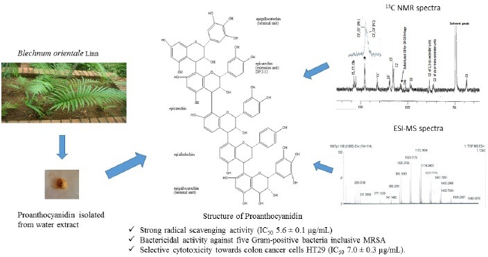
Blechnum orientale Linn. (Blechnaceae), a fern, is traditionally used in the treatment of various ailments, such as skin diseases, stomach pain, urinary bladder complaints, and also as a female contraceptive. Previously, we reported a strong radical scavenging activity, antibacterial activity and cytotoxicity against HT29 colon cancer cells by aqueous extract of B. orientale.
Objective:
In this study, we attempted to isolate and identify the active compound from the aqueous extract of B. orientale.
Materials and Methods:
Aqueous extract of B. orientale was subjected to repeated MCI gel chromatography, Sephadex-LH-20, Chromatorex C18 and semi-preparative high performance liquid chromatography and was characterized using nuclear magnetic resonance and electrospray ionization mass-spectrometry spectroscopic methods. Antioxidant activity was determined using 2, 2-diphenyl-1-picrylhydrazyl radical scavenging assay. Antibacterial assays were conducted using disc diffusion whereas the minimum inhibitory concentration (MIC) and minimum bactericidal concentration were determined using the broth microdilution assay. Cytotoxicity was assessed using thiazolylblue tetrazoliumbromide.
Results:
A polymeric proanthocyanidin consisting of 2-12 epicatechin extension units and epigallocathecin terminal units linked at C4-C8 was elucidated. Bioactivity studies showed strong radical scavenging activity (IC50 = 5.6 ± 0.1 µg/mL), antibacterial activity (MIC = 31.3-62.5 µg/mL) against five gram-positive bacteria and selective cytotoxicity against HT29 colon cancer cells (IC50 = 7.0 ± 0.3 µg/mL).
Conclusion:
According to our results, the proanthocyanidin of B. orientale demonstrated its potential as a natural source of antioxidant with antibacterial and anti-cancer properties.
SUMMARY
A bioactive proanthocyanidin was isolated from the aqueous extract of medicinal fern Blechnum orientale Linn and the structure was elucidated using NMR and ESI-MS spectral studies.
The proanthocyanidin compound possessed strong radical scavenging activity (IC50 5.6 ± 0.1 µg/mL)
The proanthocyaniding compound showed bactericidal activity against five gram-positive bacteria inclusive of MRSA (minimum inhibitory concentration, MIC and minimum bactericidal concentration, MBC 31.3-62.5 µg/mL).
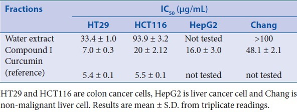
The proanthocyanidin compound is strongly cytotoxic towards cancer cells HT29 (IC50 7.0 ± 0.3 µg/mL), HepG2 (IC50 16 µg/mL) and HCT116 (IC50 20 µg/mL) while weakly cytotoxic towards the non-malignant Chang cells (IC50 48 µg/mL).
Abbreviation used: CC: Column chromatography, DP: degree of polymerization, DPPH: 2,2-diphenyl-1-picrylhydrazyl, ESI-MS: electronsprayionisation mass-spectrometry, MBC: Minimum bactericidal concentration, MIC: Minimum inhibitory concentration, MTT: Thiazolyl Blue Tetrazolium Bromide, MRSA: methicillin-resistant Staphylococcus aureus, NMR: nuclear magnetic resonance, TLC: thin layer chromatography, PD: prodelphinidin
Keywords: Antibacterial, antioxidant, blechnum orientale, colon cancer, cytotoxicity proanthocyanidin
INTRODUCTION
Blechnum orientale Linn. (Blechnaceae), a fern, is commonly distributed in Malaysia and in other Asian countries, Australia and Pacific islands. It is commonly known as the centipede fern (Malays term it as ‘pakuikan’ and the Chinese term it ‘Kuan Chung’). Its leaf is traditionally used as a poultice to treat boils, blisters or abscesses and sores. It is also used as a diaphoretic, for stomach pain, for urinary bladder complaints and as a female contraceptive.[1,2] Its leaf is also eaten as a vegetable by the indigenous people of Malaysia.[3]
Proanthocyanidins (condensed tannins) represent the second most abundant natural phenolics after lignins.[4] Structurally, they are mixtures of polymers consisting of two or more flavan-3-ols, such as afzelechin, epiafzelechin, catechin, epicatechin, gallocatechin, and epigallocatechin, which occasionally incorporate gallic acid. The monomer units are distinguished by the number of hydroxyl (OH) groups on the B ring [Figure 1].[5] The proanthocyanidins consisting exclusively of catechin/epicatechin are designated as procyanidins (PC's). Proanthocyanidins containing afzelechin/epiafzelechin or gallocatechin/epigallocatechin as subunits are named propelargonidins or prodelphinidin, respectively.[5]
Figure 1.
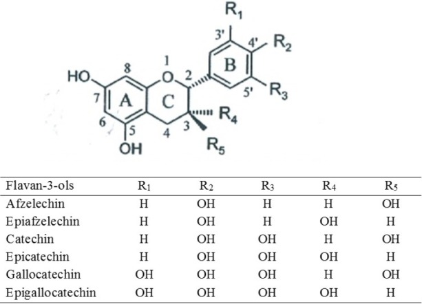
Structures of flavan-3-ol units in proanthocyanidins.[5]
As part of an on-going research on natural antioxidants and antibacterial agents from medicinal ferns, we found that the methanolic extract of the leaves of B. orientale possessed the strongest potential in the aforementioned bioactive properties.[6] Subsequently, we fractionated the methanolic extract with solvents of increasing polarity to facilitate the isolation of bioactive compounds. Previously, we reported a strong antioxidant activity (DPPH radical scavenging activity, IC50 = 13.0 µg/mL), antibacterial activity (minimum inhibitory concentration (MIC), 31.3 - 62.5 µg/mL against five gram-positive bacteria) and cytotoxicity against HT29 colon cancer cells (IC50 = 33.4 µg/mL) using the aqueous extract of B. orientale.[7] Maridass and Gantikumar also reported antibacterial properties of B. orientale.[2] The phytochemical analyses of the aqueous extract of B. orientale has demonstrated the presence of flavonoids, tannins and glycosides.[7] In continuation of our previous study, we attempted to isolate and identify the bioactive compounds of B. orientale. In this study, we report the isolation of proanthocyanidin, which is attributed to the observed bioactivity in the aqueous extract of B. orientale. To the best of our knowledge, this is the first report on the isolation and the potential therapeutic properties of proanthocyanidin from B. orientale.
MATERIALS AND METHODS
Plant material
Leaves of B. orientale Linn were collected from Putrajaya Botanical Garden, Kuala Lumpur in April, August and November 2009. The samples were identified and confirmed by Dr. Richard Chung Cheng Kong of Forest Research Institute Malaysia. A voucher specimen (LAA007) was deposited at the Herbarium of Monash University Sunway Campus, Malaysia.
Sample preparation, extraction and isolation
Fresh leaves of B. orientale were washed from residual soil, lyophilized and pulverized in a blender to give a mass of dried leaf powder. A total of 1.7 kg of the pooled dried leaf powder was then extracted with methanol (1:10, w/v) by agitation using an orbital shaker at room temperature until the extracts obtained were light in colour. The extracts were pooled and filtered through Whatman No. 1 filter paper. Subsequently, the solvent was evaporated under reduced pressure with rotary evaporator (Eyela, Japan) at 40°C. The methanolic extract was freeze-dried and the dark green mass obtained (17% yield on dry weight basis) was suspended in distilled water (1:10, w/v) and partitioned successively at room temperature with petroleum ether (b.p. 40-60°C), chloroform, ethyl acetate and n-butanol. In the final partitioning with n-butanol, the lower aqueous layer was removed and concentrated under reduced pressure. It was then lyophilized to obtain a dry brown powder. The powder was labelled as the aqueous extract (6.5% yield on dry weight basis) and stored at -70°C until further use.
The concentrated aqueous extract was subjected to repeated column chromatography (CC) using MCI gel CHP20P (Supelco), Sephadex-LH20 (25-100µm, Sigma-Aldrich) and Chromatorex C18 (40-75 µm, Fuji Silysia Chemical), each eluting with water-methanol solvent mixture (starting from water and in 10% increment of methanol for one bed volume for each ratio till 100% methanol). Fractions of 10 mL each per tube were collected. Each fraction was then analyzed for its component similarity by spotting onto thin layer chromatographic (TLC) plates as described in the next section. The fractions that gave the same spots with the same retention factor (Rf) and colour were pooled together. Methanol was evaporated from each pooled fraction using rotary evaporator under reduced pressure at 40°C, and moisture was removed by freeze-drying. Each freeze-dried fraction was subjected to bioactivity tests. Fraction 9 from final CC using Chromatorex C18 exhibited the most bioactivity (DPPH assay, IC50= 5.66 µg/mL; cytotoxicity assay, IC50= 7.0 µg/mL; and strong antibacterial activity, MIC = 31.3-62.5 µg/ml). HPLC analysis was performed on C18 column (4.6 × 150 mm, 5 µm, Agilent Eclipse XDB), with methanol acidified with 0.1% trifluoroacetic acid as the mobile phase. Elution was performed by applying a gradient of 5% methanol to 100% methanol for 15 min with a flow rate of 1 mL/min and UV detection at 280 nm. There were two peaks observed at retention times 1.9 and 5.5 min. Fractions corresponding to the first peak at 1.9 min were subsequently collected using semi-preparative HPLC using Chromolith Semi Prep RP18e 100-10 mm (Merck) column and eluted under the same analytical conditions as mentioned earlier and labelled as Compound I.
General experimental procedures
TLC was performed on a precoated Merck Kieselgel 60 F254 plate (0.25 µm). The chromatogram was visualized by observing under ultraviolet lamp at 254 nm and 365 nm or spraying with 10% sulphuric acid (H2SO4) followed by heating on a hot plate. Liquid chromatography-mass spectrometry (LC-MS) was performed using a Thermo Finnigan model LCQDeca liquid chromatography coupled mass spectrometer fitted with an electrospray ionisation (ESI) source. Positive and negative ion mass spectra of the column eluate were recorded in the range of m/z 50-2000. 1H and 13C nuclear magnetic resonance (NMR) analyses were conducted using JEOL NMR Spectrometer 400 MHz in deuterated methanol.
DPPH radical scavenging activity
DPPH assay was performed as per the procedure described previously.[7] An ascorbic acid was used as the reference standard for the determination of antioxidant activity. Briefly, various dilutions of the extract in methanol (100 µL of 2-1000 µg/mL, in triplicate) were added to 100 µL of DPPH (200 µM) in a 96-well plate. The mixture was left in the dark for 30 min, before reading the absorbance at 517 nm. The control reaction mixture consisted of methanol instead of the sample. The percentage radical scavenging activity was calculated as follows: scavenging (%) = (Acontrol-Asample)/Acontrol x 100. A graph was plotted with %scavenging versus concentration of extract (µg/mL). The value of IC50, concentration of the extract to scavenge 50% of the DPPH radical, was obtained from the graph.
Antibacterial assays
Antibacterial assays were conducted using the disc diffusion method whereas the MIC and minimum bactericidal concentration (MBC) were determined using the broth microdilution assay. These methods have been described previously.[7] The following five gram-positive bacteria were tested: Bacillus cereus ATCC14579, Micrococcus luteus ATCC4698, methicillin-susceptible Staphylococcus aureus MSSA ATCC25923, methicillin-resistant Staphylococcus aureus MRSA ATCC33591 and Stapylococcusepidermidis. These bacteria were tested as they were found to be susceptible towards the aqueous extract in an earlier study.[7] All the bacteria were grown on nutrient agar (Oxoid).
Cytotoxic activity
Cytotoxicity was measured using the thiazolyl blue tetrazolium bromide (MTT) assay as described previously.[7] The human colon adenocarcinoma HT29, human colorectal carcinoma HCT116 and liver Chang cells were obtained from Monoclonal Lab, Faculty of Medicine, University of Malaya. Human hepatocellular carcinoma HepG2 was purchased from ATCC. The cells were cultured in a humidified atmosphere at 37°C in 5% CO2. Dulbecco's modified Eagle's medium (DMEM) supplemented with 10% foetal calf serum, 1% (v/v) penicillin/streptomycin and 4 mM L-glutamine was used as the culture medium of HepG2 and liver Chang cells. RPMI-1640 supplemented with 10% foetal calf serum, 2 mM L-glutamine and 1% (v/v) penicillin/streptomycin was used for cell culturing of HT29 and HCT116 cells. Briefly, 96-well plates were seeded with 180 µl of medium containing an appropriate number of cells (6000 for HT29, 2000 for HCT116, 4000 for Chang liver and 4000 for HepG2). The cells were allowed to adhere for 24 h. Samples (20 mg/ml) were dissolved in 20% DMSO and diluted to 1 mg/ml with medium. Aliquots of 20 µL of the extracts at various concentrations (10-100 µg/ml) were added to each well. For the control, 20 µL of 1% DMSO in the medium was used instead of extract. The final concentration of DMSO was less than 1%. Curcumin (Sigma) was used as positive control. After incubation for 72 h, 50 µl of 2 mg/ml MTT (Sigma) was added to each well. The plate was incubated for another 3 h. The medium of each well was pipetted out and 150 µl of DMSO was then added. Absorbance was read at 554 nm using a Bio-TEK Microplate Scanning Spectrophotometer. The cytotoxic effect was expressed as IC50, the concentration of extract that reduces cell viability to 50% of the control.
Phytochemical screening
Phytochemical tests for saponins, tannins, terpenoids, flavonoids, and alkaloids were performed as previously described.[7] Dragendorff reagent was used to screen alkaloids, foam test for saponins, Mg-HCl and Zn-HCl for flavonoids, Salkowski test for terpenoids, and ferric chloride and gelatin for tannins.
Statistical analysis
All the data are expressed as mean ± standard deviation (SD). Statistical analyses were evaluated by one-way analysis of variance (ANOVA) followed by Tukey honest significant difference (HSD) test. A p<0.05 was considered statistically significant.
RESULTS AND DISCUSSION
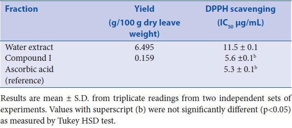
Table 1 shows yield and the antioxidant activity of Compound I in comparison with the aqueous extract and ascorbic acid as the reference compound. Compound I exhibited strong DPPH radical scavenging activity (IC50= 5.6 µg/mL), which is comparable to ascorbic acid (IC50 = 5.3 µg/mL). It should be noted that there were many fractions eluted that were also tested for their bioactivity. However, their results are not shown here as their bioactivity was weaker when compared to that of Compound I and ascorbic acid.
Table 1.
Yield and the antioxidant DPPH scavenging activity of water extract and Compound I
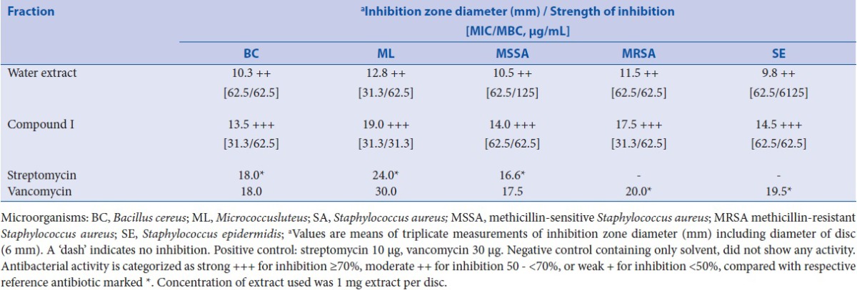
The antibacterial activity of the fractions was measured using the disc diffusion assay to test for the susceptibility of the bacteria and thereafter microbroth dilution assay to determine the MIC and MBC. According to the results, Compound I exhibited strong antibacterial activity against all five bacteria tested [Table 2]. A >70% inhibition (denoted as +++ in Table 2) was observed when compared to the reference antibiotics (streptomycin or vancomycin). These results also complemented with the MIC (31.3-62.5 µg/mL) and MBC (31.3-62.5 µg/mL). Currently, bioactives with strong bactericidal activity against MRSA is of particular interest to the researchers because of the demand for antibiotics, which can target for methicillin-resistant strains of bacteria.
Table 2.
Antibacterial activity of water extract and Compound I
Cytotoxicity analysis demonstrated that Compound I possessed strong cytotoxic potential against HT29 (IC50= 7.0 µg/mL), however it was less active against HCT116 and HepG2 (IC50= 20 and 16µg/mL, respectively) [Table 3]. Furthermore, the contrasting nature of cytotoxicity demonstrated by Compound I against malignant (HT29, HCT116 and HepG2) and non-malignant cells (liver Chang cells) is particularly interesting. Non-malignant Chang cells have been used in cytotoxicity studies to test the effects of drugs/agents on normal cells.[8] In this study, we observe weaker cytotoxic potential of Compound I against the non-malignant Chang cells (IC50= 48 µg/mL). This indicates that Compound I demonstrated selective cytotoxicity towards the cancer cells and less cytotoxicity towards the non-malignant liver Chang cells. This finding has an important implication in the potential use of such compounds as chemotherapeutic agents considering current anticancer drugs, which are found to be cytotoxic to the normal cells thereby causing serious adverse effects.[8] In this study, curcumin was chosen as a positive control (reference) for both HT29 and HCT116 cancer cells as it is widely used in clinical trials for chemoprevention of colon cancer.[9,10]
Table 3.
Cytotoxic activities of water extract and Compound I against cancer cell-lines
Phytochemical analyses
Phytochemical analyses on Compound I revealed presence of proanthocyanidin (condensed tannin) and absence of the other classes such as flavonoid, terpenoid, alkaloid, and saponin. Proanthocyanidins are polymers composed of flavan-3-ol units linked primarily through C4-C8 or C4-C6 linkage (both are called B-type). The flavan-3-ol units can also be doubly linked by an additional ether bond at C2-O7 (A-type). The size of the proanthocyanidin molecule is described by the degree of polymerization (DP).[5]
Structural analyses of proanthocyanidin
The proanthocyanidin was characterized using NMR technique. The spectra of 1H NMR and 13C NMR are shown in Figures 2a and 2b respectively. The primary working region of the 1H NMR spectrum to determine the monomeric unit lies in the region of 5.8-7.5, which is dominated by signals from aromatic protons of the phenolic constituents.[11] The signals of (-)- epicatechin were indicated on the spectrum and corresponded to chemical shifts of 7.07 (singlet), 6.94 (singlet) and 6.18 ppm (singlet) as observed previously.[11] It should be noted that as (-)- epicatechin and (+)- catechin are diastereomers, they may have similar spectrum. However, the presence of the signal at 7.07 ppm implied the occurrence of (-)- epicatechin while the other signals were common to both stereoisomers.[11] Hence, the monomers were deduced to be (-)- epicatechin and/or (+)- catechin.
Figure 2a.
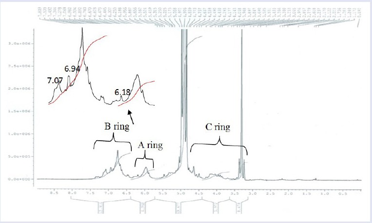
Spectra of 1H NMR of Compound I in deuterated methanol.
Figure 2b.
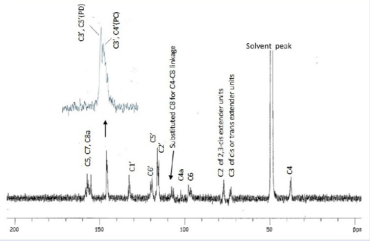
Spectra of 13C NMR of Compound I in deuterated methanol.
The signals were assigned for 13C NMR following the descriptions of previous reports.[12,13,14] The distinct signals (at the right side of the enlarged insert in Figure 2b) centred at 145.3 ppm were assignable to C3’ and C4’ in the B-ring of the PC units (catechin/epicatechin). Further evidence of the PC units was shown by the presence of strong peaks in the region of 115.1-120.0 ppm centered at 115.1 ppm (C2’), 116.2 ppm (C5’) and 119.4 ppm (C6’), which were indicative of the catechol B-ring. The resonance lines centred at 145.8 ppm, which was assignable to C3’ and C5’ in the B-ring of the prodelphinidin (PD) represented a typical indicator for the presence of PD units (gallocatechin/epigallocatechin). The ratio of PC and PD could be determined by using the ratio of the signal intensities at 145 and 146 ppm.[13] However, because of the overlapping of peak signals, this quantitation was not possible.
The other signals in 13C NMR spectrum appeared at 154.5-157.2 ppm (corresponded to C5, C7 and C8a of the aromatic A-ring), 132.6 ppm (C1’), 97.8 ppm (C6), and 102.3 ppm (C4a) and 107.7 ppm (C8). The typical inter-flavonoid linkage of C4-C8 was represented by the resonance at 106.6-107.7 ppm, which was assigned to substituted C8.[15] The observation of an unsubstituted C6 at 97.8 ppm showed the absence of C4-C6 inter-flavonoid linkage[16] and with the band at 107.7 ppm for substituted C8, it was evident that the subunits were linked at C4-C8.
The region between 70 and 90 ppm was sensitive to the stereochemistry of the C-ring. The determination of the ratio of the 2,3-cis to 2,3-trans stereochemistries could therefore be determined through distinct differences in their respective C2 chemical shifts.[14] The signal centred at 77.0 ppm, which was assigned to C2, was indicative of 2,3-cis chain extender units whereas signal centred at 72.6 ppm corresponded to C3 chain extender units.[15] The absence of signal at 84 ppm (assignable to the trans form at C2) obviously ruled out the possibility of a trans isomerism, and thus provided evidence of only epicatechin subunits present in the proanthocyanidin of B. orientale. These results showed that the polymeric proanthocyanidin consisted of a combination of PC and PD with epicatechin and epigallocatechin as the monomer units. The C3 in terminal units generally have chemical shifts around 67 ppm.[13] Theoretically, its intensity relative to the signal of the C3 in extension monomer units at 73 ppm could be used to obtain the number-average molecular weight.[14] However, the absence of signal at 67 ppm, possibly due to the low signal-to-noise ratio of the 13C spectrum, did not allow the quantization of the number-average molecular weight of the polymer. The absence of signal at above 170 ppm indicated the absence of gallic acid linked at C3.[16] Thus, it ruled out the possibility of epicatechingallate as subunits in the polymer.
ESI-mass spectrometry (ESI-MS) was used to further characterize Compound I. The spectra of the positive and negative ion modes were recorded in the range of 50-2000 Da, as shown in Figure 3. The method of the assignment of the molecular ions corresponding to specific m/z values followed those described in earlier works.[14,17,18] From the spectra of the positive ion mode, an ion peak at m/z 289.0736 implied fragmented extension monomer unit of epicatechin or catechin [C15H12O6+H]+ (288+1 Da). As indicated in the 13C NMR, epicatechin (and not catechin) subunits were present in the polymer, and this confirmed the occurrence of PC. Epicatechin (C15H14O6) has molecular mass of 290 Da and with the loss of two protons from inter-flavan linkagecarbons, an intermediate (extension) unit in the polymer has a mass of 288 Da. A series of abundant ions separated by 288 Da were observed at m/z 577; 865; 1,153; 1,441 and 1729, each of which was consistent with [M-H]- of the dimeric, trimeric, tetrameric, pentameric and hexameric (DP 2, 3, 4, 5 and 6) PCs, respectively [Table 4].
Figure 3.

ESI-MS spectra of Compound I recorded in the positive mode (upper) and negative mode (lower) from 50-2000 Da.
Table 4.
ESI-MS data of peak signals of Compound I in negative and positive modes
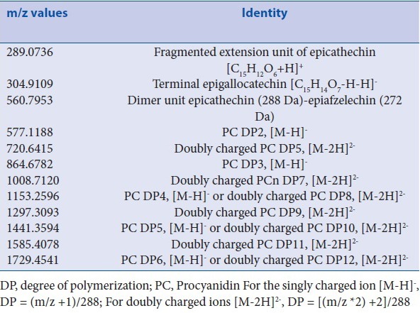
A series of peak signals observed at m/z 720; 1,008; 1,297 and 1,585 were attributed to doubly charged ions [M-2H]2- of odd polymerisation degree, corresponding to pentameric, heptameric, nonameric and undecameric (DP5, 7, 9 and 11) PCs [Table 4]. Such multiple charged species could occur and became more intense in an ESI-MS spectroscopy due to the longer chain length and higher molecular weight of the polymers, which allowed a better charge separation, thus minimizing the electrostatic repulsive forces.[14] It should be noted that signals at m/z 1,153; 1,441 and 1,729 could also correspond to the doubly charged DP8, 10 and 12 PC oligomers, respectively. Higher molecular weight proanthocyanidins were difficult to detect with a good precision as their singly charged ions showed very weak intensity. Hence, the largest detectable polymeric PC with epicatechin monomer subunits was up to DP12 Table 4.
Apart from the peak signals described above, following are the informative peaks shown in [Table 4]:
(a) peak signal at m/z 560.7953 which was 16 Da less than the peak signal of PC dimer (m/z 577.1188), suggested the existence of a subunit with one less hydroxyl group (-OH) than the epicatechin. The flavan-3-ol subunit with one less-OH group than the epicatechin (H atom at the C3 of the B ring), was epiafzelechin. However, only the dimeric form showed 16 Da less whereas this pattern was not observed with the trimer, tetramer or other oligomeric forms. This indicated the presence of a proanthocyanidin dimer unit consisting of epiafzelechin as an extension unit. The dimer form was postulated to be epicatechin→epiafzelechin based on m/z 560.7953.
(b) a deprotonated peak signal at m/z 304.9109 Da showed the occurrence of a terminal epigallocatechin (Mr 306.3) with the removal of one proton for an interflavan linkage [C15H14O7-H-H]-. Hence, it was deduced that a part of the proanthocyanidin was:
→epicatechin (288 Da) →epiafzelechin (272 Da) → epigallocatechin (305 Da)
With this data, it can be deduced that the polymeric proanthocyanidin in B. orientale was composed of predominantly epicatechin subunits as the extension unit (DP = 2-12) with a minor unit of epiafzelechin bound to the PCs and with terminating epigallocatechin subunits [Figure 4]. While PCs with epicatechin subunits are the most widely distributed proanthocyanidins in plants, its co-existence with epiafzelechin and epigallocatechins is common. These heterogeneous proanthocyanidins have been reported in various studies.[5,19]
Figure 4.
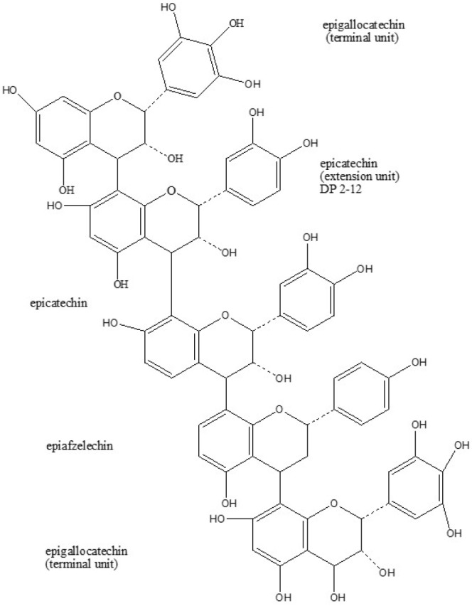
Structure of proanthocyanidin in B. orientale. DP, degree of polymerisation
Various reports on various types of proanthocyanidins have shown free radical scavenging activity,[20,21] cytotoxic activity[22,23] and antimicrobial activity.[24] Many of these studies suggested that the biological activities of proanthocyanidins correlated to their ability to form complexes with macromolecules, which in turn was attributed to the number of OH groups at specific positions and to the degree of polymerization of the molecule.[14,24] In general, more pronounced activities were seen with proanthocyanidins possessing predominantly epicatechin-containing subunits and with galloylation at the C3 OH group, due to the increase of free hydroxyl groups.
The structures containing three B-ring OH groups, a hydroxyl at C3, a saturated C2, C3 bond and the absence of any carbonyl at C4 contributed to maximal trapping efficiency and therefore a good superoxide scavenging activity.[25] The antioxidant ability was also found to be proportional to the degree of polymerization.[26] All these features were observed with the proposed structure of the proanthocyanidins of B. orientale, which showed a polymeric structure with up to 12 epicatechin subunits and the presence of epigallocatechin possessing ortho-trihydroxyl groups in the B-ring.
CONCLUSION
In summary, in this study, we demonstrate the isolation of proanthocyanidin from B. orientale with potent antioxidant and cytototoxic potential towards HT29 cancer cells as well as bactericidal activities for the first time. This polymeric proanthocyanidin possessed strong antioxidant activity (equivalent to that of ascorbic acid), bactericidal against selected gram-positive bacteria such as MRSA and selective cytotoxicity towards colon cancer cell HT29.
Financial support and sponsorship
Nil.
Conflicts of interest
There are no conflicts of interest.
Acknowledgement
This study was supported by research grants from Monash University Sunway Campus and Malaysian Ministry of Higher Education (MOHE) through ERGS/1/2012/SKK03/TAYLOR/02/3.
REFERENCES
- 1.Ahmad F, Holdsworth DK. Medicinal Plants of Sabah, East Malaysia-Part I. Pharm Biol. 2003;41:340–46. [Google Scholar]
- 2.Maridass M, Ghantikumar S. Antibacterial activity of leaves of Blechnum Orientale L. Pharmacologyonline Newslett. 2008;3:58–60. [Google Scholar]
- 3.Piggott AG. Ferns of Malaysia in colour. Kuala Lumpur: Tropical Press; 1996. p. 400. [Google Scholar]
- 4.Behrens A, Nagamitsu M, Knicker H, Kogel-Knaber I. MALDI-TOF mass spectrometry and PSD fragmentation as means for the analysis of condensed tannins in plant leaves and needles. Phytochemistry. 2003;62:1159–70. doi: 10.1016/s0031-9422(02)00660-x. [DOI] [PubMed] [Google Scholar]
- 5.Gu LW, Kelm MA, Hammerstone JF, Beecher G, Holden J, Haytowitz D, et al. Screening of foods containing proanthocyanidins and their structural characterization using LC-MS/MS and thiolytic degradation. J Agric Food Chem. 2003;51:7513–521. doi: 10.1021/jf034815d. [DOI] [PubMed] [Google Scholar]
- 6.Lai HY, Lim YY, Tan SP. Antioxidative, tyrosinase inhibiting and antibacterial activities of leaf extracts from medicinal ferns. Biosci Biotechnol Biochem. 2009;73:1362–6. doi: 10.1271/bbb.90018. [DOI] [PubMed] [Google Scholar]
- 7.Lai HY, Lim YY, Kim KH. Blechnumorientale Linn-a fern with potential as antioxidant, anticancer and antibacterial agent. BMC Complement Altern Med. 2010;10:15. doi: 10.1186/1472-6882-10-15. [DOI] [PMC free article] [PubMed] [Google Scholar]
- 8.Zakaria Y, Rahmat A, Pihie AHL, Abdullah NR, Houghton PJ. Eurycomanone induce apoptosis in HepG2 cells via up-regulation of p53. Cancer Cell Int. 2009;9:16. doi: 10.1186/1475-2867-9-16. [DOI] [PMC free article] [PubMed] [Google Scholar]
- 9.Cen L, Hutzen B, Ball S, DeAngelis S, Chen CL, Fuchs JR, et al. New structural analogues of curcumin exhibit potent growth suppressive activity in human colorectal carcinoma cells. BMC Cancer. 2009;9:99. doi: 10.1186/1471-2407-9-99. [DOI] [PMC free article] [PubMed] [Google Scholar]
- 10.Johnson JJ, Mukhtar H. Curcumin for chemoprevention of colon cancer. Cancer Lett. 2007;255:170–81. doi: 10.1016/j.canlet.2007.03.005. [DOI] [PubMed] [Google Scholar]
- 11.Berregi I, Santos JI, del Campo G, Miranda JI. Quantitative determination of (-)-epicatechin in cider apple juices by 1H NMR. Talanta. 2003;61:139–45. doi: 10.1016/S0039-9140(03)00236-4. [DOI] [PubMed] [Google Scholar]
- 12.Czochanska Z, Foo LY, Newman RH, Porter LJ. Polymeric proanthocyanidins, Stereochemistry, structural units, and molecular weight. J C.S. Perkin Trans 1. 1980:2278–286. [Google Scholar]
- 13.Behrens A, Nagamitsu M, Knicker H, Kogel-Knaber I. MALDI-TOF mass spectrometry and PSD fragmentation as means for the analysis of condensed tannins in plant leaves and needles. Phytochemistry. 2003;62:1159–70. doi: 10.1016/s0031-9422(02)00660-x. [DOI] [PubMed] [Google Scholar]
- 14.Es-Safi NE, Guyot S, Ducrot PH. NMR, ESI/MS and MALDI-TOF/MS analysis of pear juice polymeric proanthocyanidins with potent free radical scavenging activity. J Agri Food Chem. 2006;54:6969–77. doi: 10.1021/jf061090f. [DOI] [PubMed] [Google Scholar]
- 15.Rao LJM, Yada H, Ono H, Ohnishi-Kameyama M, Yoshida M. Occurrence of antioxidant and radical scavenging proanthocyanidins from the Indian minor spice nagkesar (Mammealongifolia planch and trianasyn) Bioorg Med Chem. 2004;12:31–36. [PubMed] [Google Scholar]
- 16.Navarrete P, Pizzi A, Pasch H, Rode K, Delmotte L. MALDI-TOF and 13C-NMR characterization of maritime pine industrial tannin extract. Ind Crop Prod. 2010;32:105–10. [Google Scholar]
- 17.Guyot S, Doco T, Souquet JM, Moutounet M, Drilleau JF. Characterization of highly polymerized procyanidins in cider apple (Malussylvestris var. Kermerrien) skin and pulp. Phytochemistry. 1997;44:351–57. [Google Scholar]
- 18.Roux LE, Doco T, Sarni-Manchado P, Lozano Y, Cheynier V. A-type proanthocyanidins from pericarp of Litchi chinensis. Phytochemistry. 1998;48:1251–58. [Google Scholar]
- 19.Souquet JM, Cheynier V, Broussaud F, Moutounet M. Polymeric proanthrocyanidins from grape skins. Phytochemistry. 1996;43:509–12. [Google Scholar]
- 20.Bagchi D, Garg A, Krohn RL, Bagchi M, Tran MX, Stohs SJ. Oxygen free radical scavenging abilities of vitamins C and E, and a grape seed proanthocyanidin extract in vitro. Res Commun Mol Pathol Pharmacol. 1997;95:179–89. [PubMed] [Google Scholar]
- 21.Masuda K, Hori T, Tanabe K, Kano Y, Hirayama A, Nagase S. Proanthocyanidin promotes free radical-scavenging activity in muscle tissues and plasma. Appl Physiol Nutr Metab. 2007;32:1097–104. doi: 10.1139/H07-073. [DOI] [PubMed] [Google Scholar]
- 22.Carnésecchi S, Schneider Y, Lazarus AS, Coehlo D, Gossé F, Raul F. Flavanols and procyanidins of cocoa and chocolate inhibit growth and polyamine biosynthesis of human colonic cancer cells. Cancer Lett. 2002;175:147–55. doi: 10.1016/s0304-3835(01)00731-5. [DOI] [PubMed] [Google Scholar]
- 23.Miura T, Chiba M, Kasai K, Nozaka H, Nakamura T, Shoji T, et al. Apple procyanidins induce tumor cell apoptosis through mitochondrial pathway activation of caspase-3. Carcinogenesis. 2008;29:585–93. doi: 10.1093/carcin/bgm198. [DOI] [PubMed] [Google Scholar]
- 24.Scalbert A. Antimicrobial properties of tannins. Phytochemistry. 1991;30:3875–883. [Google Scholar]
- 25.Bruyne TD, Pieters L, Witvrouw M, Clercq ED, Berghe DV, Vlietinck AJ. Biological evaluation of proanthocyanidin dimmers and related polyphenols. J Nat Prod. 1999;62:954–8. doi: 10.1021/np980481o. [DOI] [PubMed] [Google Scholar]
- 26.Hagerman AE, Riedl KM, Jones GA, Sovik KN, Ritchard NT, Hartzfeld PW. High molecular weight plant polyphenolics (tannins) as biological antioxidants. J Agric Food Chem. 1998;46:1887–92. doi: 10.1021/jf970975b. [DOI] [PubMed] [Google Scholar]