Abstract
Dentin sialoprotein (DSP) is essential for dentinogenesis and processed into fragments in the odontoblast-like cells and the tooth compartments. Matrix metalloproteinase 9 (MMP9) is expressed in teeth from early embryonic to adult stage. Although MMP9 has been reported to be involved in some physiological and pathological conditions through processing substrates, its role in tooth development and whether DSP is a substrate of MMP9 remain unknown. In this study, the function of MMP9 in the tooth development was examined by observation of Mmp9 knockout (Mmp9−/−) mouse phenotype, and whether DSP is a substrate of MMP9 was explored by in vitro and in vivo experiments. The results showed that Mmp9−/− teeth displayed a phenotype similar to dentinogenesis imperfecta, including decreased dentin mineral density, abnormal dentin architecture, widened predentin and irregular predentin-dentin boundary. The distribution of MMP9 and DSP overlapped in the odontoblasts, the predentin, and the mineralized dentin, and MMP9 was able to specifically bind to DSP. MMP9 highly efficiently cleaved DSP into distinct fragments in vitro, and the deletion of Mmp9 caused improper processing of DSP in natural teeth. Therefore, our findings demonstrate that MMP9 is important for tooth development and DSP is a novel target of MMP9 during dentinogenesis.
Dentin is a major mineralized component of teeth. Odontoblasts, differentiated from neural-crest derived mesenchymal cells1, synthesize and secrete un-mineralized extracellular matrix (ECM) termed predentin containing rich non-collagenous proteins (NCPs). Later, when apatite crystals are deposited, the predentin is transformed to the mineralized dentin. Under normal conditions, a rather uniform layer of predentin is maintained between the mineralized dentin and the odontoblast layer, indicating that the rates of secretion and maturation of the predentin is identical and controlled by accurate mechanisms. NCPs have been proposed to be critical for this process2.
Dentin sialoprotein (DSP) and dentin phosphoprotein (DPP) are the two most abundant NCPs in dentin, and encoded by a single gene, dentin sialophosphoprotein (Dspp)3,4. Dspp knockout (Dspp−/−) mouse displays a phenotype similar to the manifestations of human dentinogenesis imperfecta III (DGI-III)5. Transgenic expression of mouse Dsp in the Dspp−/− background leads to a partial rescue of the Dspp−/− tooth phenotype with restored predentin width, an absence of irregular unmineralized areas in dentin and less frequent pulp exposure6. Heterogeneous mutations in DSP coding domain in humans have been reported to cause DGI-II and dentin dysplasia II7. These studies demonstrate that DSP, the NH2-terminus protein from DSPP, is essential to dentinogenesis.
Only trace amount of full-length DSPP protein was detected in extracts from the pulp/odontoblasts and dentin. Key proteolytic cleavage site of mouse DSPP into DSP and DPP has been determined at the NH2-terminal peptide bond of Asp452. The cleavage of DSPP into DSP and DPP is as an activation step of DSPP function, for failure to make this cleavage results in dentin and periodontal developmental defects8,9,10. Interestingly, DSP has been recently found to be further processed into small fragments in odontoblast-like cells and dental organs11,12,13. Our previous study has reported that cleaved DSP fragments are localized in different compartments of teeth. DSP NH2-terminal fragment(s) are mainly distributed in the non-mineralized predentin and odontoblasts, but weakly in the mineralized dentin, whereas DSP COOH-terminal fragment(s) are mainly restricted to the mineralized dentin rather than the predentin and odontoblasts. The distinct distribution pattern of DSP NH2-terminal and COOH-terminal fragments in the odontoblasts, predentin and dentin suggest that they may play distinct functions during dentinogenesis11. Besides, DSP and DSP-derived peptides are able to activate intracellular signaling transductions through regulating gene expression and protein phosphorylation, and induce dental primary/stem cell differentiation14,15,16. The above evidence suggested that, similar to DSPP, the proteolytic processing of DSP might also be an activation step of DSP biological function. However, the protease(s) catalyzing DSP proteolysis remained unknown.
Matrix metalloproteinases (MMPs) are a family of zinc- and calcium-dependent proteolytic enzymes. The involvement of MMPs in a wide variety of physiological development and pathological diseases has been reported through remodeling and degrading their extracellular, membrane-bound or intracellular substrate repertoire17. Experiments to chemically inhibit MMPs indicated that MMPs (possibly MMP2, 3, 9 and/or 20) played a critical role in the onset of dentin mineralization18. MMP9 belongs to gelatin-binding MMP and is known to be involved in bone development. Ablation of Mmp9 in mice causes delayed skeletal growth plate ossification and defective bone fracture repair, and mutations of Mmp9 in humans result in the human disease metaphyseal anadysplasia19,20,21. Bone and dentin are both mineralized connective tissues and share many common characteristics including composition and mechanisms of formation2. Therefore, we hypothesized that MMP9 may play critical roles during dentin formation through remodeling its substrate(s) in teeth. MMP9 has an overlapping distribution profile with DSP in the odontoblasts, predentin and dentin matrix11,22, suggesting that MMP9 might involve in dentin formation and process DSP. In this study, we showed the function of MMP9 in dentin formation by observation of Mmp9−/− tooth phenotype and revealed the possible underlying mechanism by experiments of in vitro and in vivo cleavage of DSP by MMP9.
Results
Mmp9−/− mouse teeth show severe cusp wear and increased reactionary dentin (RD) formation
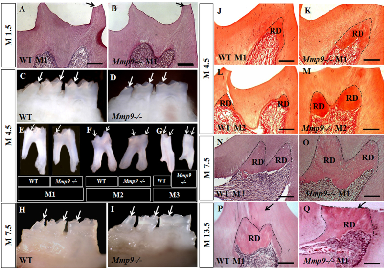
To address the role of MMP9 for tooth development, we first examined the morphology of Mmp9−/− teeth from 1.5 months old (M1.5) to M13.5 by stereomicroscopy and histology. At M1.5, only a very slight wear of molar cusp was seen in the control and Mmp9−/− molars, and no RD was seen in either group (Fig. 1A,B). At M4.5 and M7.5, Mmp9−/− mice showed more severe wear of molar cusps (Fig. 1C–I). In response to severe wear, the amount of RD in Mmp9−/− molars was apparently increased compared to the control (Fig. 1J–O). At M13.5, the cusp wear progressed to the depth of RD in Mmp9−/− mice, but thick dentin tissue remained on the top of RD in the control mice (Fig. 1P,Q).
Figure 1. More severe molar cusp wear and increased reactionary dentin (RD) formation in the Mmp9−/− mice.
(A,B) Slight wear of one cusp (arrows) and no RD formation was seen at M1.5 in both Mmp9−/− and wild-type molars. (C–G) At M4.5 and (H,I) M7.5, the heights of the cusps (arrows) in the Mmp9−/− mice were reduced compared with the controls. (J–Q) More amount of RD (dashed lines) was seen in the Mmp9−/− molars at M4.5, M7.5 and M13.5. At M13.5, dentin on top of RD (arrows) was worn off in the Mmp9−/− mice but not in the wild-type mice. WT, wild-type; M1, the first mandibular molar; M2, the second mandibular molar, M3, the third mandibular molar; RD, reactionary dentin. Scale bars: 100 μm. (A,B,J–Q) HE staining images; (C–I) Stereomicroscopy images.
Abnormal mineralization and structures of Mmp9−/− teeth
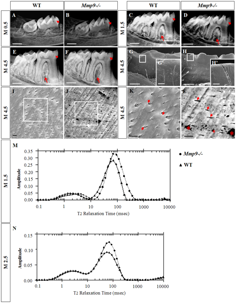
To further understand why severe cusp wear occurred in Mmp9−/− molars, possible changes in the mineralization and structures of Mmp9−/− teeth were examined by radiography and SEM. Decreased mineral density of the dentin and the enamel in the Mmp9−/− molars was seen from M0.5 to M4.5 by radiography (Fig. 2A–F). SEM analysis showed that the molar enamel surfaces in the M4.5 Mmp9−/− mice had abnormal fissures (Fig. 2G,H,G’,H’). Meanwhile, the dentinal tubules and inter-tubular dentin were uniformly distributed in the wild-type mice, but sparsely scattered at an obviously impaired density in Mmp9−/− mice. Numerous abnormal “holes” were also observed in the Mmp9−/− dentin (Fig. 2I–L). Then, NMR proton spin-spin (T2) relaxation time measurement was applied to analyze the porosity and pore size of molars at M1.5 and M2.523. Inversion NMR T2 relaxation time spectra showed that Mmp9−/− molars had larger porosity and pore size compared to the wild-type control (Fig. 2M,N). These results indicated that teeth in Mmp9−/− mice had not only reduced mineral content but also abnormal architecture.
Figure 2. Decreased mineral density of teeth, loss of the alveolar bone in the furcation region, and abnormal tooth structures in the Mmp9−/− mice.
(A–F) Representative radiographs show an overall reduction of the mineralization of the molars (arrowheads) and loss of the alveolar bone in the furcation region (arrows) of the Mmp9−/− mice. (G,G’,H,H’) SEM analysis showed the presence of abnormal fissure on the surface of the Mmp9−/− molars. (I–L) The dentin tubules (arrowheads) and the intertubular dentins were well-distributed in the wild-type mice, but in the Mmp9−/− mice the distribution of the dentine tubules (arrowheads) and the intertubular dentins were not uniform with decreased number of dentin tubules and numerous “holes” (*) in the intertubular dentins. K and L are higher magnifications of the rectangles in (I and J), respectively. (M,N) Inversion T2 relaxation time spectra for the first mandibular molars of the wild-type and the Mmp9−/− mice at M1.5 and M2.5. The lines with “dots” are for the Mmp9−/− molars and the lines with “triangles” are for the wild-type molars. Longer T2 relaxation time and larger area under the curve was found in the Mmp9−/− molars. The longer T2 relaxation time is corresponding to larger pores, and the larger area is corresponding to larger porosity (larger amount of holes). WT, wild-type. Scale bars: (A–J) 500 μm, (K,L) 250 μm, (K’,L’) 20 μm, (M-P) 5 μm.
In addition, examination of the mandibles by radiography also identified alveolar bone defects in Mmp9−/− mice (Fig. 2C–F). The mineral density in the furcation region of mandibular molars decreased in Mmp9−/− mice, which is most notable when comparing the 4.5-month (Fig. 2E,F) samples. The significant alveolar bone loss was confirmed by hematoxylin and eosin (HE)-stained tissue sections (Supplementary Fig. 1).
Delayed differentiation of odontoblasts, widened predentin, and irregular mineralization front in Mmp9−/− mice
To further observe the function of MMP9 during the odontoblast differentiation and secretion at earlier stages, Mmp9−/− teeth at D1 and D19 were analyzed using histological method. In wild-type mice at D1, the odontoblasts were well polarized and elongated at the cusp tip region, where the deposition of the predentin matrix was clearly discernible (Fig. 3A). In contrast, the polarization and elongation of odontoblasts were delayed, and the thickness of predentin at the cusp area was reduced in Mmp9−/− molars at D1 (Fig. 3B). At D19, increased predentin width and irregular predentin-dentin boundary was seen in Mmp9−/− molars versus the control (Fig. 3C,D).
Figure 3. Delayed differentiation, widened predentin and irregular mineralization front in the Mmp9−/− molars.
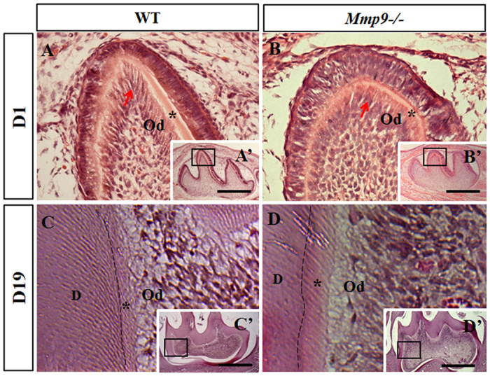
(A,A’,B,B’) At D1, the predentin matrix was deposited by well-differentiated odontoblasts (arrow) at the cusp tip of wild-type first mandibular molar. The polarization and elongation of odontoblasts (arrow) were delayed, and the thickness of predentin at the cusp area was reduced in the Mmp9−/− first mandibular molar cusp. (C,C’,D,D’) Compared to the control tooth, irregular mineralization front (dashed line) and enlarged predentin layer were observed in the Mmp9−/− molar at D19. (A–D) are higher magnifications of the rectangles in A’–D’, respectively. *predentin; D, mineralized dentin; Od, odontoblasts. Scale bars: (A–F) 50 μm, (G,H) 25 μm; (A’–H’) 500 μm.
Overlapping expression of MMP9 and DSP in the odontoblast-like cells and the developing teeth
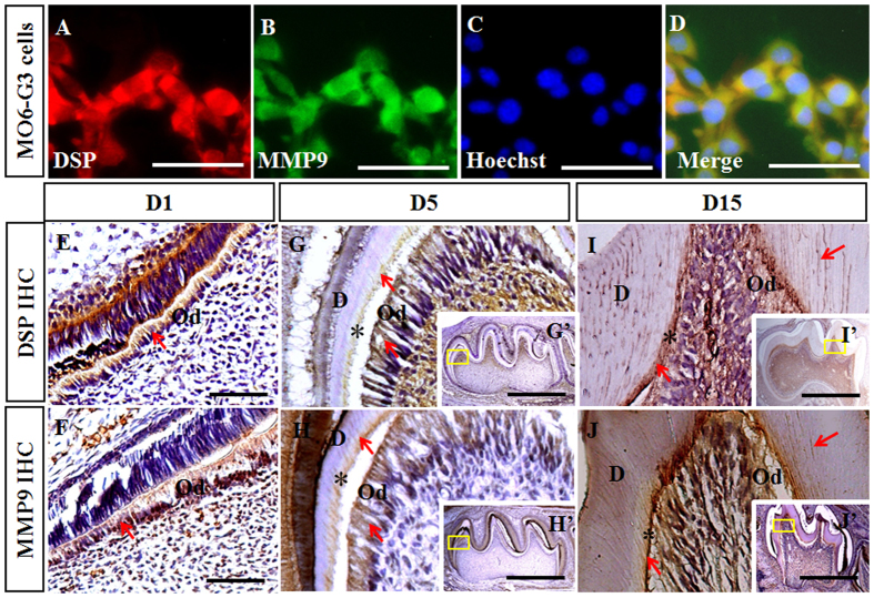
MMP9 and MMP2 belong to gelatinase subgroup of MMP family and share many common substrates24. DSP is critical for dentinogenesis and a substrate of MMP2 in the porcine molars7,12. Therefore, we hypothesized that DSP might act as a substrate of MMP9, which may explain the dentin phenotype of Mmp9−/− mice. To assess whether MMP9 is able to cleave DSP, we first confirmed co-distribution of DSP and MMP9 proteins in the mouse odontoblast-like (MO6-G3) cells and tooth tissues. Double immunofluorescence experiments showed that expression of MMP9 and DSP overlapped in the cytoplasm of MO6-G3 cells (Fig. 4A–D). Immunohistochemistry showed that in molars at D1, DSP and MMP9 were both highly expressed in the odontoblasts (Fig. 4E,F). At D5 and D15, signals of DSP and MMP9 were intense in the odontoblasts and the predentin, and mild in the mineralized dentin (Fig. 4G–J,G’–J’).
Figure 4. Overlapping distribution of DSP and MMP9 in the odontoblast-like cells and the developing teeth.
(A,B) Immunofluorescent signals of DSP and MMP9 were observed in the cytoplasm of MO6-G3 cells. (C) Nuclei of the cells were counterstained with Hoechst. (D) Images of A-C were merged. (E,F) DSP and MMP9 were highly expressed in the odontoblasts at D1. (G-J, G’–J’) Immunoreactions of DSP and MMP9 were strong in the odontoblasts and the predentin, but weak in the mineralized dentin at D5 and D15. (G–J) are higher magnifications of the boxes in G’–J’, respectively. *predentin; Od, odontoblasts; D, the mineralized dentin. Scale bars: (F,G,J,K) 100 μm, (H,I,L,M) 50 μm, (H’,I’,L’,M’) 750 μm.
Binding between DSP and MMP9
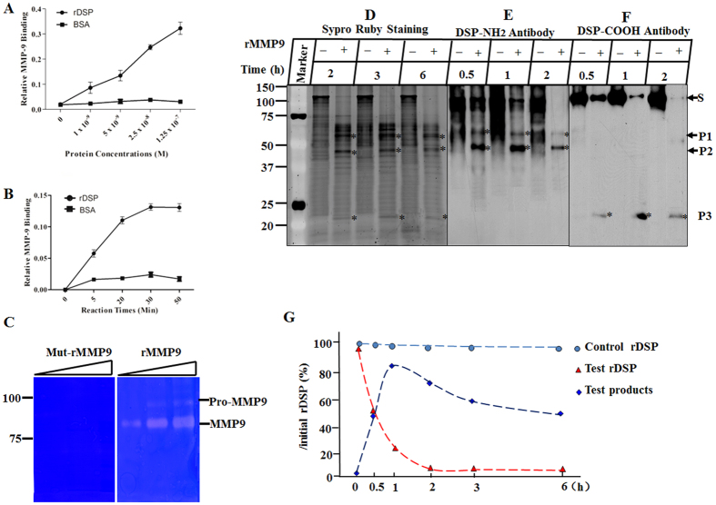
We next determined whether MMP9 was able to bind to DSP. First, rDSP fusion protein was generated, purified, and confirmed by Coomassie Blue staining and Western blot assays using anti-DSP and anti-GST antibodies as described previously16. Then the rDSP protein was labeled with biotin and serial dilutions of the biotinylated rDSP were incubated with either mut-rMMP9 or BSA. The results showed that rDSP bound specifically to MMP9 in the dose- and time-dependent manner whereas there was no binding effect on control BSA (Fig. 5A,B).
Figure 5. Recombinant DSP protein bound to MMP9 and was processed by MMP9 in vitro.
(A,B) DSP bound to MMP9 in the dose- and time-dependent manner. (C) Gelatin zymography was performed to characterize the cleavage activity of rMMP9 and mut-rMMP9 with ascending concentrations of enzyme from the left the right lanes. Two clear bands at 92 kDa and 86 kDa were seen in the gel containing rMMP9, but not in the gel containing mut-rMMP9. (D) rDSP was incubated alone or with activated rMMP9 for 2, 3 and 6 h. The reaction products were run on SDS-PAGE gels and stained with Sypro Ruby dye. (E,F) Western blot assays were performed using anti-DSP-NH2 and -COOH antibodies, respectively. S, P1, P2, and P3 represent the substrate and cleaved products of rDSP. (G) rDSP and cleaved products were quantified when digested for 0, 0.5, 1, 2, 3 and 6 h with initial rDSP as 100%. Approximately 50% of the rDSP substrate was digested after 30 min of incubation, and almost all of the substrate was cleaved after 2 h. The quantity of the products reached the maximum at 1 h, and gradually reduced afterwards.
DSP processed by MMP9 in vitro
To verify that rMMP9 was successfully activated and had the ability to cleave its substrates, gelatin zymography was first performed with rMMP9 and mut-rMMP9. Our data showed that rMMP9 efficiently digested gelatin with two clear bands at the molecular weight of 92 kDa and 86 kDa, whereas mut-rMMP9 failed to do so (Fig. 5C). Next, to determine whether the activated rMMP9 was able to process DSP protein, the rDSP was incubated alone or with rMMP9 for different time periods. The results showed that rDSP was cleaved by rMMP9 into three major fragments, whereas rDSP alone remained intact and stable (Fig. 5D–F). Western blot analyses demonstrated that the intact rDSP and the two cleaved fragments at high molecular weight (HMW) were recognized by anti-DSP-NH2 antibody (Fig. 5E), and the intact rDSP and the smallest cleaved rDSP product were detected by anti-DSP-COOH antibody (Fig. 5F). The cleaved products were quantified when digested for 0, 0.5, 1, 2, 3 and 6 h with initial rDSP substrate as 100%. The results showed that approximately 50% of the substrate was cleaved after 30 min of incubation, and the cleavage reaction was almost complete after 2 h. The products reached their maximum amount at 1 h, and were further processed by rMMP9 when incubated longer resulting in reduced remnant quantity (Fig. 5G).
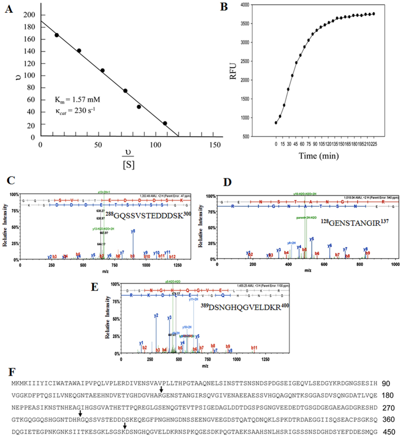
To assess the catalytic efficiency of MMP9, steady state cleavage velocities were measured with a constant amount of rMMP9 and varying amounts of DSP substrate. As expected, the enzymatic reaction displayed reaction kinetics within a Michaelis-Menten analysis. An Eadie-Hofstee plot of these results showed Michaelis-Menten parameters were Km = 1.57 mM and kcat = 230 (s−1), yielding a relative catalytic efficiency (kcat/Km) of 146, 500 M−1 s−1 (Fig. 6A). Similar to rDSP, MMP9 also had high efficiency in catalyzing a known fluorescent MMP9 substrate as a positive control (Fig. 6B)25. These results demonstrated that DSP was a novel substrate of MMP9.
Figure 6. Enzyme kinetics and proteolytic sites of rDSP cleavage by MMP9.
(A) An Eadie-Hofstee plot for MMP9 hydrolysis of rDSP. rDSP substrate ranging from 0.25 μM to 10 μM was incubated with the activated rMMP9 at a constant concentration (13.6 nM) in reaction buffer for 30 min at 37 °C. The reaction products were run on a SDS-PAGE gel and stained with Sypro Ruby dye. The density of the cleaved products was quantitated using imageJ software. (B) Activated rMMP9 was reacted with fluorescence-labeled MMP9 substrates (DNP-Pro-Cha-Gly-Cys(Me)-His- Ala-Lys(N-Me-Abz)-NH2). Gelatinase assays were carried out in 96-well microplates in assay buffer. Reactions were performed at 23 °C and the changes in fluorescence intensity were expressed in relative fluorescent units (RFU) as measured with λexcitation = 365 nm and λemission = 450 nm in a SpectraMAX Gemini XS fluorescent plate reader. Data points represent an average of three separate experiments repeated in duplicate. (C–F) Mass spectrometry determined the sites of DSP cleavage by MMP9, including Arg127-Gly128 (P2), Arg287-Gly288 (P1), and Lys388-Asp389 (P3) peptide bonds.
To further identify the specific cleavage sites of DSP by MMP9, the three cleaved rDSP products were analyzed by mass spectrometry. The sites of DSP cleavage by MMP9 were determined as indicated in Fig. 6C–F. These results indicated that MMP9 selectively cleaved DSP protein.
DSP processed by MMP9 in vivo
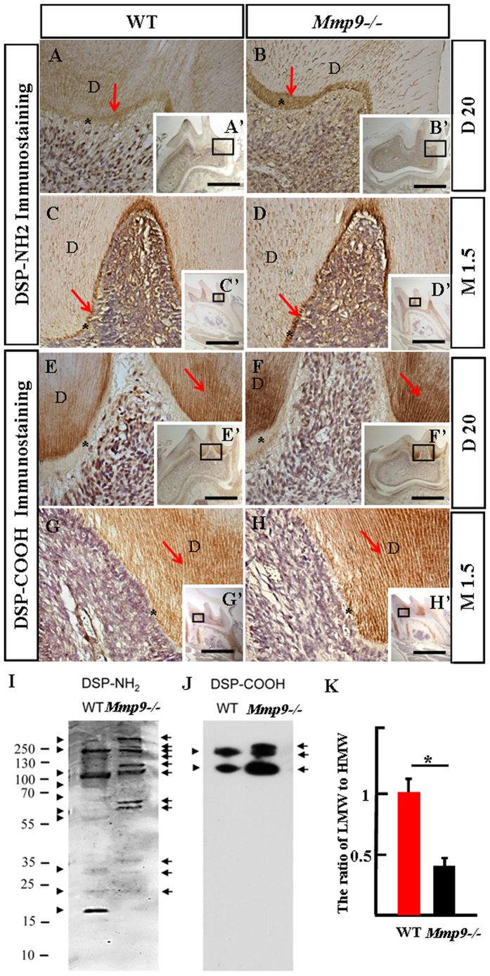
To analyze whether DSP was a substrate of MMP9 in vivo, immunohistochemistry with anti-DSP-NH2 and -COOH specific antibodies were performed in Mmp9−/− first mandibular molars at D20 and M1.5 with wild-type first mandibular molars in parallel as control. Our results showed that immunoreactions for anti-DSP-NH2 antibody were intense in the predentin matrix and odontoblasts, but mild in the mineralized dentin (Fig. 7A–D,A’–D’); for anti-DSP-COOH antibody, immunoreactions were apparently strong in the mineralized dentin, but weak in the predentin and odontoblasts (Fig. 7E–H,E’–H’). DSP signals recognized by both antibodies were more prominent in Mmp9−/− than control molars (Fig. 7A–H,A’–H’).
Figure 7. Immunolocalization and protein profiles of DSP in the Mmp9−/− and the wild-type molars.
(A–H, A’–H’) Immunodistribution of anti-DSP-NH2 antibody was strong in the predentin and the odontoblasts, but weak in the mineralized dentin. In contrast, DSP-COOH fragment(s) was strong in the mineralized dentin, but weak in the predentin and the odontoblasts at D20 and M1.5. (A–H) are higher magnification of the boxes in A’–H’. (I,J) Proteins were isolated from the wild-type and the Mmp9−/− teeth at D15, and Western blots with anti-DSP-NH2 and –COOH antibodies were performed. For normalization, the protein concentration should be measured and same amount of protein was loaded. Different DSP protein profiles were seen in the wild-type and the Mmp9−/− teeth. Arrows and arrowheads show DSP bands from the Mmp9−/− and the wild-type teeth, respectively. (K) The densitometry of anti-DSP-NH2 and -COOH bands of three independent experiments was performed and relative quantification was processed with the ImageJ software. The ratio of the LMW fragments (lower than 95 kDa) to the HMW DSP (higher than 95 kDa) was calculated. Statistical analysis was performed using Student’s t-test. *P < 0.05. In (A–I), *predentin; D, dentin. Scale bars: (A–H) 50 μm, (A’–H’) 1 mm.
To further identify protein profiles of DSP fragments, proteins were isolated from wild-type and Mmp9−/− mouse teeth at D15 and Western blots were conducted using anti-DSP-NH2 and -COOH antibodies. The results showed different expression patterns of DSP in the wild-type and Mmp9−/− teeth. For anti-DSP-NH2 antibody, several high HMW bands (ranging from 95 to over 250 kDa) were seen in the Mmp9−/− but not in wild-type tooth proteins. Several low molecular weight (LMW) DSP bands (lower than 95 kDa) were detected in the wild-type and the Mmp9−/− teeth, but at a higher molecular weight in the Mmp9−/− teeth (Fig. 7I). For anti-DSP-COOH antibody, one additional HMW DSP band was observed in Mmp9−/− teeth but not in wild-type teeth (Fig. 7J). The ratio of the LMW fragments to the HMW DSP was significantly lower in the Mmp9−/− teeth compared to the control (Fig. 7K). Thus, MMP9 activity was responsible for DSP processing in natural dental tissues.
Osteopontin (OPN) has been reported to be localized in tooth tissues including predentin, dentin and RD11. And OPN is known as a substrate of MMP926. Therefore, OPN expression changes in the Mmp9−/− mice from D17 to M7.5 were investigated by immunohistochemistry. OPN showed a higher expression level in the predentin, dentin and RD in the Mmp9−/− molars (Supplementary Fig. 2).
Discussion
Heterogeneous mutations in DSP coding domain are associated with human hereditary dentin defects7. MMP9 and DSP are both highly expressed by the odontoblasts and secreted to the dentin ECM (Fig. 4)11,22. In this study, to gain insights of the role of MMP9 in the tooth development and whether DSP acts as a substrate of MMP9, we investigated the phenotype of Mmp9−/− teeth as well as the processing of DSP by MMP9 in vitro and in vivo. Our results showed that Mmp9−/− mice displayed phenotype similar to human DGI, including severe tooth wear, decreased dentin mineralization, delay of the odontoblast differentiation, and abnormal dentin structures. MMP9 was able to bind to and specifically process DSP into given fragments in vitro. In vivo DSP processing was affected in the Mmp9−/− teeth, which may mainly account for the dentin defects in the Mmp9−/− mice.
Previous studies have reported developmental abnormality of long bone in the mice deficient of Mmp9 and in humans carrying Mmp9 mutations19,20,21. Bone and dentin share many similarities in composition and mechanisms of formation2. Therefore, we hypothesized that dentin formation might be affected by deletion of Mmp9. In this study, we investigated the tooth phenotype of the Mmp9−/− mice by multiple methods including stereomicroscopy, histology, radiography, SEM and NMR. Mmp9−/− molars from M4.5 to M13.5 displayed more severe tooth cusp wear by stereomicroscopy and histology (Fig. 1), suggesting that the resistance to masticatory mechanical strength of Mmp9−/− molars diminished. SEM and radiography experiments did show abnormal appearance of dentin structure, and reduced dentin mineral density in the Mmp9−/− mice. In addition, the enamel in the Mmp9−/− molars was observed abnormal with fissures on the surface and showed reduced mineral density. Meanwhile, larger porosity and pore size were detected in the Mmp9−/− teeth by NMR (Fig. 2). Therefore, reduced mineral content of the enamel and abnormal tooth architecture may explain why Mmp9−/− teeth had lower mechanical resistance to masticatory forces. In this study, we focused on the dentin formation of the Mmp9−/− mice.
In response to severe tooth wear, more amount of RD was formed in Mmp9−/− molars from M4.5 to M13.5 compared to control molars at the corresponding ages (Fig. 1), suggesting that odontoblasts in Mmp9−/− molars are active in secreting dentin matrix to protect themselves from exposure to bacterial environment in oral cavity. This indicates that the capability of odontoblasts to synthesize and secret dentin ECM under masticatory force stimuli was not inhibited by deficiency of Mmp9.
The expression of MMP9 in the early embryonic dental epithelium and mesenchyme suggests a potential role of MMP9 in tooth morphogenesis27. However, Mmp9−/− molars and incisors did not show obvious morphological changes, indicating that MMP9 is dispensable for tooth morphogenesis. The delayed differentiation of odontoblasts in the Mmp9−/− molars (Fig. 3) is consistent with the expression of MMP9 in presecretory and secretory odontoblasts (Fig. 4). Later, MMP9 is present in the predentin and the dentin matrix (Fig. 4)22, suggesting that MMP9 may be involved in the dentin mineralization through processing and degrading dentin ECM, in accordance with which widened predentin and irregular boundary between predentin and dentin were detected in Mmp9−/− molars at D19 (Fig. 3).
The above experiments indicated the necessity of MMP9 activity for odontoblast differentiation and dentin formation, and showed that Mmp9−/− mice exhibited similar tooth phenotype to humans carrying DSP mutations28,29,30. Since DSP is processed into fragments in the odontoblast-like cells and the tooth compartment11,13, further studies were carried out to determine whether MMP9 participated in the DSP processing. Immunostaining experiments confirmed the overlapping distribution of MMP9 and DSP in the odontoblast-like MO6-G3 cells, and in the odontoblasts and dentin ECM of mouse molars (Fig. 4). DSP but not BSA bound to MMP9 in the dose- and time-dependent manner (Fig. 4). The overlapping localization and direct physical interaction between DSP and MMP9 are two prerequisites for processing of DSP by MMP9. Next, in vitro studies revealed that MMP9 selectively cleaved DSP protein into three major fragments (Fig. 5). Enzyme kinetics showed that MMP9 had a high efficiency for processing DSP with a turnover of (kcat/Km) of 146,000 M−1 s−1 (Fig. 6). This value is comparable to other substrates cleaved by MMP931. For in vivo study, Mmp9−/− molars showed elevated expression levels of both DSP NH2-terminal and COOH-terminal fragments by immunohistochemistry (Fig. 7), suggesting that the processing of DSP was reduced in Mmp9−/− molars. Western blot assay was further performed and confirmed that expression patterns of DSP fragments were different between the control and the Mmp9−/− teeth. Several HMW DSP fragments were present in Mmp9−/− teeth but not in control molars and the ratio of the LMW DSP fragments to the HMW DSP was lower in the Mmp9−/− teeth (Fig. 7), assuring that DSP processing in vivo was affected by deletion of Mmp9. DSP is well known as a critical protein for proper dentin formation. Thus, the lack of appropriate DSP processing is likely the most important contributor to the dentin phenotype in Mmp9−/− teeth.
In addition to dentin defects, we revealed severe loss of alveolar bone surrounding Mmp9−/− molar roots, especially in the furcation region of the mandibular molars (Fig. 2 and Supplementary Fig. 1). The alveolar bone phenotype in Mmp9−/− mice is similar to Dspp−/− mice10. Like in dentin, DSPP is also proteolytically processed into DSP and DPP in bone. Processing of DSPP is essential to its biological function in alveolar bone, for disruption of DSPP processing into the given fragments failed to rescue the alveolar bone defect of Dspp−/− mice9,10. Therefore, in addition to dentin, the further cleavage of DSP by MMP9 might also exist in the alveolar bone, which is critical for alveolar bone remodeling. Further studies are needed to verify this possible underlying mechanism of alveolar bone loss in Mmp9−/− mice in the future.
OPN and DSP are both members of SIBLING (Small Integrin-Binding Ligand N-linked Glycoproteins) family. OPN shares many common characteristics with DSP32 and is known as a substrate of MMP926. OPN has been reported in predentin, dentin and RD by our previous study11. As expected, improper processing and degradation of OPN in Mmp9−/− molars was documented by remarkably elevated protein level of OPN (Supplementary Fig. 2). Both in vitro and in vivo studies have shown that OPN is a potent inhibitor of hydroxyapatite crystal formation and growth33,34. However, the teeth of Opn deficient mice were found morphologically normal at the level of light and electron microscopy35. Therefore, compared to DSP, increased OPN protein might play a minor role for the dentin defects in Mmp9−/− molars. The decreased processing of OPN acts as a positive control for the improper processing of DSP in Mmp9−/− teeth.
Taken together, DSP is critical for tooth development, and DSP is a novel target of MMP9 during dentinogenesis. Like other MMPs, MMP9 was originally discovered to function in the breakdown of ECM. However, recent studies have identified intracellular proteins as MMP9 substrates36,37,38. The overlapping prominence of DSP and MMP9 in the cytoplasm of MO6-G3 cells and the existence of multiple LMW DSP fragments in MO6-G3 cell lysate suggest that the cleavage of DSP by MMP9 may be initiated inside odontoblasts11. Further studies are needed to determine the mechanisms of intracellular MMP9 activation and its cleavage of DSP to provide deeper insights into the processing of DSP by MMP9 during dentinogenesis.
Methods
Animal preparation
The protocol of animal use was approved by the Animal Welfare Committee at the University of Texas Health Science Center at San Antonio (UTHSCSA, Protocol No. 05012x). All experiments were performed in accordance with the relevant guidelines and regulations. The Mmp9−/− mice used in this study were originally generated and described by Vu and colleagues19. Genotyping was performed by PCR of genomic DNA extracted from tail snips (Supplementary Fig. 3). The tissue sections from the wild-type and Mmp9−/− mice were immunostained with anti-MMP9 antibody. MMP9 expression was seen in the wild-type tooth tissues, but not in Mmp9−/− mice (Supplementary Fig. 3). At least three mice for each time point were sacrificed and taken for the following analyses.
Stereomicroscopy, X-radiography and low-field nuclear magnetic resonance (NMR)
To compare the overall morphology of wild-type and Mmp9−/− teeth, mice at the ages from M0.5 to M7.5 were put to death. Mandibles at M4.5 and M7.5 were observed under stereomicroscope. Mandibles at M4.5 were incubated in lysis buffer (2 × SSC, 0.2% SDS, 10 mM EDTA, 10 mg/ml proteinase K) for 2 d. After muscles surrounding teeth were digested, the molars were extracted and observed under stereomicroscope.
To compare the mineralization of teeth in Mmp9−/− mice with that in wild-type mice, mandibles from M0.5 to M4.5 were examined using a Faxitron radiograph inspection unit (Field X-ray Corporation, Lincolnshire, IL, USA).
To analyze the structural changes of Mmp9−/− teeth, the extracted first mandibular molars at M1.5 and M2.5 were processed for NMR measurement and compared with control as described previously23.
Histology and immunohistochemistry
To evaluate the histological features of Mmp9−/− teeth, fresh mandibles at ages from D1 to M13.5 were fixed in 4% paraformaldehyde for 1–2 d at 4 °C, and demineralized in 8% EDTA for 1 d to 8 wk depending on age. Then, the tissues were processed for paraffin embedding and 5-μm serial sections were prepared and stained with HE.
To detect the distribution of MMP9, DSP, and OPN in mouse mandibular first molars, immunohistochemistry was performed as previously described11. Primary antibodies included goat anti-MMP9 antibody (Santa Cruz Biotechnology, CA, USA), two rabbit anti-DSP antibodies specific to the NH2 and COOH domains described previously11, and goat anti-OPN antibody (R & D systems, MN, USA). Normal goat or rabbit immunoglobulin G (IgG) (Santa Cruz Biotechnology, CA, USA) was employed as negative control (data not shown).
For double immunofluorescence, mouse odontoblast-like (MO6-G3) cells were grown on glass slides, rinsed with ice-cold phosphate-buffered saline (PBS), and fixed for 10 min on ice with methanol/acetone (1:1). Then, the cells were processed with blocking buffer for 1 h at room temperature (RT), and two primary antibodies were incubated simultaneously overnight at 4 °C (1:200 for rabbit anti-DSP antibody (M-300), 1:50 for goat anti-MMP9 antibody). After washes, slides were incubated with the secondary antibodies conjugated with Alexa Fluo ® 486 green and Alexa Fluo ® 568 red (1:500; Molecular Probes, OR, USA) for 1 h at RT. For nuclear staining, the cells were treated with Hoechst (Sigma-Aldrich, MO, USA).
Scanning electron microscopy (SEM)
Mandibular incisors from Mmp9−/− and wild-type mice were fractured at the level of the labial alveolar crest. The first mandibular molars and the fractured incisor surfaces were fixed in 2% paraformaldehyde and 2.5% glutaraldehyde in 0.1 M cacodylate buffer. After washes, the samples were dehydrated in an ascending alcohol series, allowed to air dry, and then sputter-coated with gold for conventional SEM (JEOL, JSM 6610 LV; JEOL, Inc., Peabody, Mass., USA).
Extraction of proteins from mouse teeth and detection of DSP
Molars from wild-type and Mmp9−/− mice were extracted as described above and tooth proteins were isolated by procedures described previously6. Extracted molars were crushed and incubated with 4 M guanidine HCl solution containing protease inhibitor (Sigma-Aldrich, MO, USA) overnight. The residue was then demineralized with 4 M guanidine HCl, 0.5 M EDTA plus protease inhibitors for 2 d. The solution was dialyzed against 4 M guanidine for 1 d, and against distilled water for another 2 d. Dialyzed solution was lyophilized and dissolved into distilled water. Protein concentrations were then measured.
Same amount of protein from wild-type and Mmp9−/− molars was loaded and subjected to Western blot assay.
Expression and purification of recombinant DSP and MMP9
The cDNA coding for recombinant DSP (rDSP) was subcloned into pGEX-6P1 vector tagged with a NH2-terminal GST (GE Healthcare Biosciences, NJ, USA), termed GST-rDSP. To obtain mutant recombinant MMP9 protein (mut-rMMP9) with ligand-binding property but without catalytic activity, point mutation of the active site Glu402 of proMMP9 to alanine residue was generated as we previously described39. The constructs of GST-rDSP and mut-rMMP9 were transformed to E. coli BL21 (DE3). GST-rDSP and mut-rMMP9 proteins were induced and purified16. A wild-type recombinant mouse MMP9 protein (rMMP9) was purchased (R & D Systems, MN, USA).
Gelatin zymography
To analyze their gelatinolytic activities, rMMP9 and mut-rMMP9 proteins were run on SDS-polyacrylamide gel electrophoresis (SDS-PAGE) containing 0.1% gelatin. The gelatin zymography gel was next washed in 2.5% Triton X-100 with gentle agitation for 1 h at RT, then incubated in the developing buffer (30 mM Tris-HCl, 200 mM NaCl, 5 mM CaCl2 and 1 mM ZnCl2) at 37 °C overnight. Counterstaining with Coomassie Brilliant Blue revealed gelatin degradation as clear bands corresponding to the enzymes against a blue background.
Biotinylation of rDSP and substrate binding assay
For probing protein–protein interactions, rDSP protein at the concentration of 300 μg/ml was dialyzed against 0.1 M NaHCO3 and then reacted with 100 μg/ml Sulfo-NHS (N-hydroxysuccinimido)-LC (long-chain)-biotin (Pierce, Rockford, IL, USA) for 20 min at RT, followed by 2 h at 4 °C. Free biotin was removed by dialysis against 50 mM Tris-HCl and 150 mM NaCl, pH 7.4. To characterize the relative rDSP interaction with MMP9, substrate binding assay was performed as described earlier40. Briefly, 96-microwell plates were coated with 1 μg/well of mut-rMMP9 or bovine serum albumin (BSA) as control overnight at 4 °C and non-specific binding were blocked with 1% BSA (Sigma-Aldrich, MO, USA) for 1 h at RT. After thorough rinses with PBS, the biotinylated rDSP ranged from 0–18 fold molar excesses in PBS with 1% BSA was added into the plates and incubated for 30 min. Bound rDSP was reacted with AP-conjugated streptavidin diluted 1:10,000 in PBS for different time points using 1 mg/ml PNPP (ρ-nitrophenyl phosphate disodium) as substrate (Pierce, Rockford, IL, USA) and quantified at 405 nm (Opsys MR, Dynex, Chantilly, VA, USA). BSA was used as control group and the binding of rDSP to mut-rMMP9 was expressed as relative binding activity compared to the control group. The experiments were performed three times in triplicate.
Processing of rDSP by rMMP9 in vitro
To quantify MMP9 activity, rMMP9 and mut-rMMP9 were reacted with fluorescence-labeled MMP9 substrate {DNP-Pro-Cha-Gly-Cys(Me)-His-Ala-Lys(N-Me-Abz)-NH2} (Calbiochem, Cambridge, MA, USA) as described previously25. In brief, gelatinase assays were carried out in 96-well microplates in total reaction volumes of 200 μl with assay buffer (50 mM Tris/HCl, 200 mM NaCl, 5 mM CaCl2, 20 mM ZnSO4, 0.05 Brij 35, pH 7.6). Reactions were performed at 23 °C and the changes in fluorescence intensity were expressed in relative fluorescent units (RFU) as measured with λexcitation = 365 nm and λemission = 450 nm in a SpectraMAX Gemini XS fluorescent plate reader (Molecular Devices, Sunnyvale, CA, USA).
To assay the role of rMMP9 on rDSP processing, rDSP was incubated alone or in combination with P-aminophenylmercuric acetate (APMA)-activated rMMP9 (enzyme : substrate ratio of 1:20) in reaction buffer (50 mM Tris/HCl, 200 mM NaCl, 5 mM CaCl2, 20 mM ZnSO4, 0.05% Brij 35, pH 7.6). The reactions were carried out at 37 °C for various time intervals and stopped by adding 2 x SDS-PAGE sample buffer containing 2-mercaptoethanol. Samples were boiled for 5 min and subjected to SDS-PAGE and stained with Sypro Ruby dye. To detect DSP fragments in the digestion mixture more specifically, Western blot assay was performed using anti-DSP-NH2 and -COOH antibodies.
Analysis of enzyme kinetics
Kinetics constants Km and kcat were determined from an Eadie-Hofstee plot of initial velocities under multiple turnover conditions31. Mixtures of the rMMP9 (13.6 nM) and substrate rDSP (0.25, 0.5, 1.0, 2.5, 5.0, 10 μM) were incubated in reaction buffer for 30 min at 37 °C. The reaction products were run on SDS-PAGE gel, stained with silver dye, and quantified with ImageJ. All experiments were performed in triplicate.
Mass spectrometry
Twelve μg of purified rDSP were incubated with 0.6 μg of APMA-activated rMMP9 at 37 °C for 1 h in reaction buffer. Products were run on SDS-PAGE gel and visualized by Sypro Ruby dye staining. Then the products were excised from the gel, digested with trypsin, and the peptide mixtures were extracted from the gel as described by Kerkhoff et al.41. MALDI-mass spectrometry was performed using a TofSpec-2E instrument (Micromass, Manchester, UK) provided by Protein Core Facility of UTHSCSA.
Statistical analysis
Quantitative data were presented as means + S. D. from three independent experiments and analyzed with Student’s t test. The differences between groups were statistically significant when P < 0.05. For the Western blot, densitometry of immunoblot bands was collected and relative quantification was processed with the ImageJ.
Additional Information
How to cite this article: Yuan, G. et al. Dentin Sialoprotein is a Novel Substrate of Matrix Metalloproteinase 9 in vitro and in vivo. Sci. Rep. 7, 42449; doi: 10.1038/srep42449 (2017).
Publisher's note: Springer Nature remains neutral with regard to jurisdictional claims in published maps and institutional affiliations.
Supplementary Material
Acknowledgments
We thank Dr. Susan T. Weintraub and Mr. Sammy Pardo from Mass Spectrometry Core Laboratory, UTHSCSA for help with mass spectrometry. This work was supported by NIH grants (DE019892 and HL075360) and grants from the National Natural Science Foundation of China (81670952 and 81570942 and 81420108011).
Footnotes
The authors declare no competing financial interests.
Author Contributions Study design: S.C., Z.C., M.M., K.J.D. Data collection: G.Y., L.C., J.F., G.Y., Q.N., X.X., C.W., M.L. Data analysis: G.Y., S.C., Z.C. Drafting manuscript content: G.Y. Revising manuscript content: G.Y., L.C., J.F., G.Y., Q.N., X.X., C.W., M.L., K.J.D., M.M., Z.C., and S.C. Approving final version of manuscript: G.Y., L.C., J.F., G.Y., Q.N., X.X., C.W., M.L., K.J.D., M.M., Z.C., and S.C. S.C. and Z.C. takes responsibility for the integrity of the data analysis.
References
- Yuan G. et al. The non-canonical BMP signaling and Wnt/β-catenin signaling orchestrate early tooth development. Development 142, 128–139 (2015). [DOI] [PMC free article] [PubMed] [Google Scholar]
- Goldberg M., Kulkarni A. B., Young M. & Boskey A. Dentin: structure, composition and mineralization. Front. Biosci. (Elite Ed) 3, 711–735 (2011). [DOI] [PMC free article] [PubMed] [Google Scholar]
- MacDougall et al. Dentin phosphoprotein and dentin sialoprotein are cleavage products expressed from a single transcript coded by a gene on human chromosome 4. Dentin phosphoprotein DNA sequence determination. J. Biol. Chem. 272, 835–842 (1997). [DOI] [PubMed] [Google Scholar]
- Feng J. Q. et al. Genomic organization, chromosomal mapping, and promoter analysis of the mouse dentin sialophosphoprotein (Dspp) gene, which codes for both dentin sialoprotein and dentin phosphoprotein. J Biol Chem. 273, 9457–9464 (1998). [DOI] [PubMed] [Google Scholar]
- Sreenath T. et al. Dentin sialophosphoprotein knockout mouse teeth display widened predentin zone and develop defective dentin mineralization similar to human dentinogenesis imperfecta type III. J. Biol Chem. 278, 24874–24880 (2003). [DOI] [PubMed] [Google Scholar]
- Suzuki S. et al. Dentin sialoprotein and dentin phosphoprotein have distinct roles in dentin mineralization. Matrix Biol. 28, 221–229 (2009). [DOI] [PMC free article] [PubMed] [Google Scholar]
- Kim J. W. & Simmer J. P. Hereditary dentin defects. J. Dent. Res. 86, 392–399 (2007). [DOI] [PubMed] [Google Scholar]
- Sun Y. et al. Key proteolytic cleavage site and full-length form of DSPP. J. Dent. Res. 89, 498–503 (2010). [DOI] [PMC free article] [PubMed] [Google Scholar]
- Zhu Q. et al. Proteolytic processing of dentin sialophosphoprotein (DSPP) is essential to dentinogenesis. J. Biol. Chem. 287, 30426–30435 (2012). [DOI] [PMC free article] [PubMed] [Google Scholar]
- Gibson M. P. et al. Failure to process dentin sialophosphoprotein into fragments leads to periodontal defects in mice. Eur. J. Oral Sci. 121, 545–550 (2013). [DOI] [PMC free article] [PubMed] [Google Scholar]
- Yuan G., Yang G., Song G., Chen Z. & Chen S. Immunohistochemical localization of the NH(2)-terminal and COOH-terminal fragments of dentin sialoprotein in mouse teeth. Cell Tissue Res. 349, 605–614 (2012). [DOI] [PMC free article] [PubMed] [Google Scholar]
- Yamakoshi Y. et al. Dentin sialophosphoprotein is processed by MMP-2 and MMP-20 in vitro and in vivo. J. Biol. Chem. 281, 38235–38243 (2006). [DOI] [PubMed] [Google Scholar]
- Chen S. et al. Expression and processing of small integrin-binding ligand N-linked glycoproteins in mouse odontoblastic cells. Arch. Oral Biol. 53, 879–889 (2008). [DOI] [PMC free article] [PubMed] [Google Scholar]
- Lee S. Y. et al. Effects of recombinant dentin sialoprotein in dental pulp cells. J. Dent. Res. 91, 407–412 (2012). [DOI] [PubMed] [Google Scholar]
- Ozer A. et al. Domain of dentine sialoprotein mediates proliferation and differentiation of human periodontal ligament stem cells. PloS One. 8, e81655 (2013). [DOI] [PMC free article] [PubMed] [Google Scholar]
- Wan C. et al. The Dentin Sialoprotein (DSP) Domain Regulates Dental Mesenchymal Cell Differentiation through a Novel Surface Receptor. Sci. Rep. 6, 29666 (2016). [DOI] [PMC free article] [PubMed] [Google Scholar]
- Cauwe B. & Opdenakker G. Intracellular substrate cleavage: a novel dimension in the biochemistry, biology and pathology of matrix metalloproteinases. Crit. Rev. Biochem. Mol. Biol. 45, 351–423 (2010). [DOI] [PubMed] [Google Scholar]
- Fanchon S. et al. Involvement of matrix metalloproteinases in the onset of dentin mineralization. Eur. J. Oral Sci. 112, 171–176 (2004). [DOI] [PubMed] [Google Scholar]
- Vu T. H. et al. MMP9/gelatinase B is a key regulator of growth plate angiogenesis and apoptosis of hypertrophic chondrocytes. Cell 93, 411–422 (1998). [DOI] [PMC free article] [PubMed] [Google Scholar]
- Lausch E. et al. Mutations in MMP9 and MMP13 determine the mode of inheritance and the clinical spectrum of metaphyseal anadysplasia. Am. J. Hum. Genet. 85, 168–178 (2009). [DOI] [PMC free article] [PubMed] [Google Scholar]
- Colnot C., Thompson Z., Miclau T., Werb Z. & Helms J. A. Altered fracture repair in the absence of MMP9. Development 130, 4123–4133 (2003). [DOI] [PMC free article] [PubMed] [Google Scholar]
- Niu L. N. et al. Localization of MMP-2, MMP9, TIMP-1, and TIMP-2 in human coronal dentine. J. Dent. 39, 536–542 (2011). [DOI] [PubMed] [Google Scholar]
- Ni Q. & Chen S. Assessment of structural changes of human teeth by low-field nuclear magnetic resonance (NMR). Meas. Sci. Technol. 21, 15803 (2010). [DOI] [PMC free article] [PubMed] [Google Scholar]
- Martins V. L., Caley M. & O’Toole E. A. Matrix metalloproteinases and epidermal wound repair. Cell Tissue Res. 351, 255–268 (2013). [DOI] [PubMed] [Google Scholar]
- Bickett D. M. et al. A high throughput fluorogenic substrate for interstitial collagenase (MMP-1) and gelatinase (MMP9). Anal. Biochem. 212, 58–64 (1993). [DOI] [PubMed] [Google Scholar]
- Takafuji V., Forgues M., Unsworth E., Goldsmith P. & Wang X. W. An osteopontin fragment is essential for tumor cell invasion in hepatocellular carcinoma. Oncogene 26, 6361–6371 (2007). [DOI] [PubMed] [Google Scholar]
- Yoshiba N. et al. Temporospatial gene expression and protein localization of matrix metalloproteinases and their inhibitors during mouse molar tooth development. Dev. Dyn. 228, 105–112 (2003). [DOI] [PubMed] [Google Scholar]
- Xiao S. et al. Dentinogenesis imperfecta 1 with or without progressive hearing loss is associated with distinct mutations in DSPP. Nat. Genet. 27, 201–204 (2001). [DOI] [PubMed] [Google Scholar]
- Rajpar M. H. et al. Mutation of the signal peptide region of the bicistronic gene DSPP affects translocation to the endoplasmic reticulum and results in defective dentine biomineralization. Hum. Mol. Genet. 11, 2559–2565 (2002). [DOI] [PubMed] [Google Scholar]
- Song Y. et al. Phenotypes and genotypes in 2 DGI families with different DSPP mutations. Oral Surg. Oral Med. Oral Pathol. Oral Radiol. Endod. 102, 360–374 (2006). [DOI] [PubMed] [Google Scholar]
- Kridel S. J. et al. Substrate hydrolysis by matrix metalloproteinase-9. J. Biol. Chem. 276, 20572–20578 (2001). [DOI] [PubMed] [Google Scholar]
- Fisher L. W. & Fedarko N. S. Six genes expressed in bones and teeth encode the current members of the SIBLING family of proteins. Connect Tissue Res. 44 (Suppl 1), 33–40 (2003). [PubMed] [Google Scholar]
- Boskey A. L. et al. Osteopontin-hydroxyapatite interactions in vitro: inhibition of hydroxyapatite formation and growth in a gelatin gel. Bone Miner. 22, 147–159 (1993). [DOI] [PubMed] [Google Scholar]
- Sodek J., Ganss B. & Mckee M. D. Osteopontin. Crit. Rev. Oral Biol. Med. 11, 279–303 (2000). [DOI] [PubMed] [Google Scholar]
- Rittling S. R. et al. Mice lacking osteopontin show normal development and bone structure but display altered osteoclast formation in vitro. J. Bone Miner. Res. 13, 1101–1111 (1998), [DOI] [PubMed] [Google Scholar]
- Cauwe B., Martens E., Proost P. & Opdenakker G. Multidimensional degradomics identifies systemic autoantigens and intracellular matrix proteins as novel gelatinase B/MMP-9 substrates. Integr. Biol. (Camb). 1, 404–426 (2009). [DOI] [PubMed] [Google Scholar]
- Cauwe B. et al. Adenylyl cyclase-associated protein-1/CAP1 as a biological target substrate of gelatinase B/MMP-9. Exp. Cell Res. 314, 2739–2749 (2008). [DOI] [PubMed] [Google Scholar]
- Vaisar T. et al. MMP-9 sheds the beta2 integrin subunit (CD18) from macrophages. Mol. Cell Proteomics 8, 1044–1060 (2009). [DOI] [PMC free article] [PubMed] [Google Scholar]
- Feng J. et al. Binding of amelogenin to MMP9 and their co-expression in developing mouse teeth. J. Mol. Histol. 43, 473–485 (2012). [DOI] [PMC free article] [PubMed] [Google Scholar]
- Xu X., Chen Z., Wang Y., Yamada Y. & Steffensen B. Functional basis for the overlap in ligand interactions and substrate specificities of matrix metalloproteinases-9 and -2. Biochem. J. 392 (Pt 1), 127–134 (2005). [DOI] [PMC free article] [PubMed] [Google Scholar]
- Kerkhoff C. et al. Binding of two nuclear complexes to a novel regulatory element within the human S100A9 promoter drives the S100A9 gene expression. J. Biol. Chem. 277, 41879–41887 (2002). [DOI] [PubMed] [Google Scholar]
Associated Data
This section collects any data citations, data availability statements, or supplementary materials included in this article.


