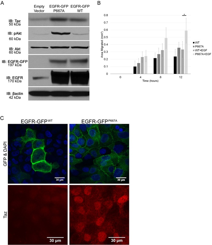
Figure 6. Mislocalization of EGFR drives pro-migratory and survival pathways.

A. Protein lysates were collected from MDCK cells transfected with an Empty Vector, an EGFR-GFPP667A vector, or an EGFR-GFPWT vector, and analyzed by immunoblot using the antibodies: anti-TAZ, anti-pAKT, anti-AKT, anti-EGFR, and anti-βactin. The EGFR-GFP blot indicates the GFP tagged EGFR induced via vector transfection while the EGFR blot indicates all EGFR present. B. EGFR-GFPWT and EGFR-GFPP667A transfected MDCK cells were grown to confluence, scratched, and then observed for wound healing migration in serum free media in either the absence of EGF or in the presence of EGF (20ng/mL). Error bars show ± standard deviation. *P < 0.05. C. MCF12A cells were transfected with EGFR-GFPWT or EGFR-GFPP667A vector, grown on plastic, serum starved overnight, mounted with DAPI and evaluated for localization of EGFR-GFP and TAZ using an anti-GFP and anti-TAZ antibody, respectively.
