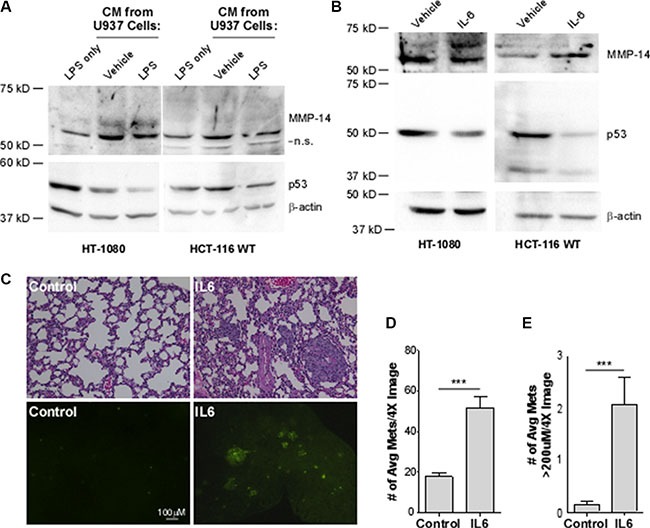
Figure 4. Interleukin-6 decreases p53 levels, leading to a concomitant increase in MMP-14 expression in HT-1080 and HCT-116 WT cells and drives metastasis and growth of HT-1080 cancer cells in vivo.

(A) Western blotting analysis was performed on HT-1080 and HCT-116 p53 wild type (WT) cells in the presence of LPS or with conditioned media (CM) from U937 macrophage cells that were activated with LPS to induce cytokine secretion or with water as vehicle control for 24 hours. While CM from U937 cells increase MMP-14 levels, only after macrophage activation was a decrease in p53 observed using anti-p53 and anti-MMP-14 antibodies. β-actin was used as a loading control. (B) Western blotting analysis was performed on HT-1080 and HCT-116 cells treated with 50 ng/mL recombinant IL-6 or 1% BSA vehicle control for 24 hours followed by Western blotting. A decrease in p53 was observed along with a concomitant increase in endogenous MMP-14 using the corresponding antibodies. β-actin was used as a loading control. (C) H&E staining of paraffin-embedded sectioned lung tissue sections (top panel) and the presence of GFP lesions on the gross lung tissue (bottom panel) indicates increased formation of metastases. The number (D) and size (E) of GFP-positive lesions per image are significantly increased in mice which received IL-6-treated cells (n = 9) compared to control (n = 10). Lesion size was determined using Nikon Imaging Software. ***p < 0.001.
