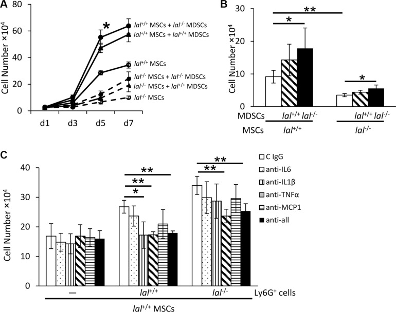
Figure 7. MDSCs partially restored decreased proliferation of lal −/− MSCs.
(A) lal −/− MDSCs stimulated MSC proliferation in the co-culture study. MSCs were seeded at a density of 5 × 10⁴ cells/well in 6-well plates, and lal +/+ or lal −/− MDSCs (2 × 10⁶) were added to the well. The MSC number was counted at d1, d3, d5 and d7 afterwards. (B) lal −/− MDSCs stimulated MSC proliferation in the transwell study. MSCs (2 × 10⁴) were seeded into the lower chamber of transwells plates, while MDSCs (1 × 10⁶) were placed in the upper chamber. After 5 days, the MSC number was counted. (C) Neutralization of cytokines in MSC proliferation. lal +/+ MSCs (2 × 10⁴) were seeded into the lower chamber of transwells, with MDSCs (1 × 10⁶) placed in the upper chamber. MDSCs were treated with 10 μg/ml neutralizing antibody against IL-6, IL-1β, TNF-α, MCP-1 individually, in combination or with control immunoglobulin G at day 1 and day 3. After 5 days, the number of MSCs was counted. In the above experiments, data were expressed as mean ± SD; n = 3~5. *P < 0.05, **P < 0.01.

