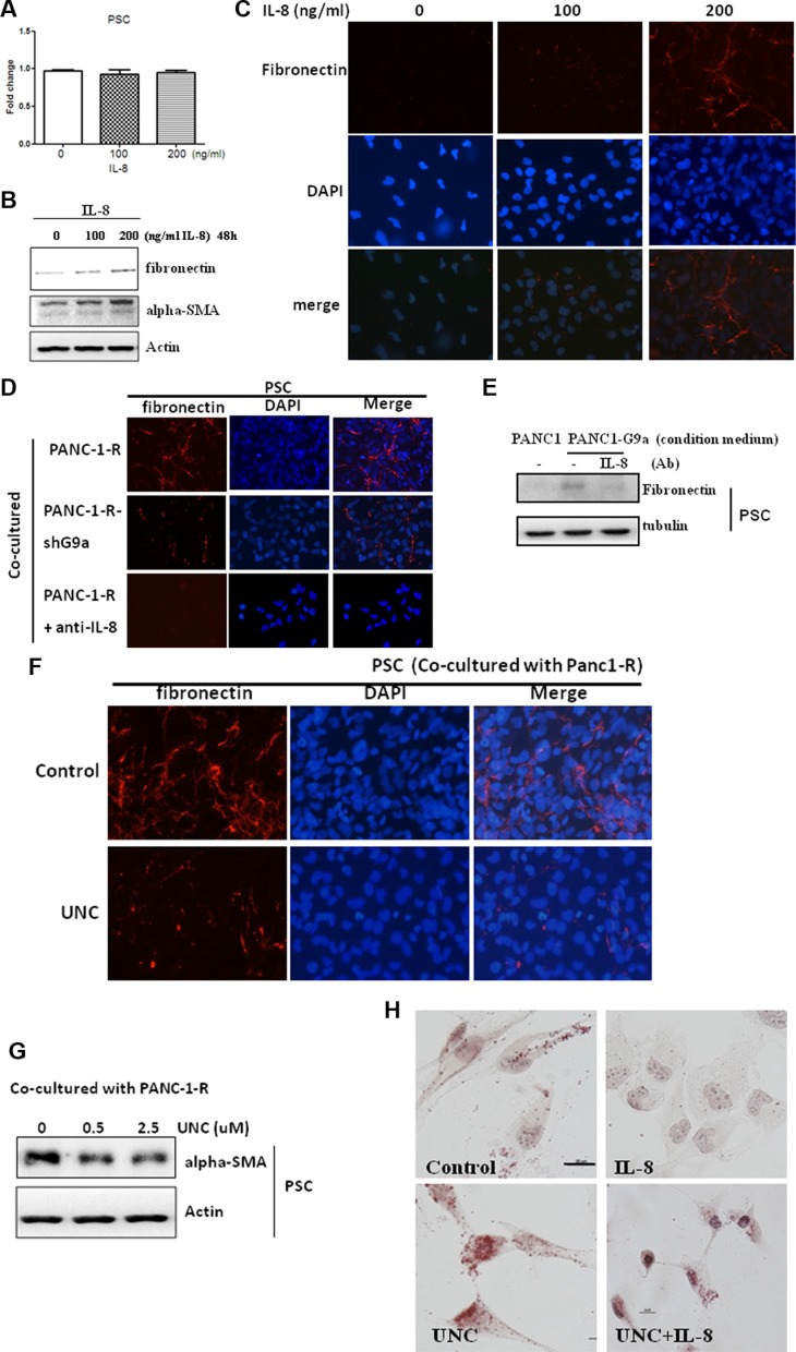
Figure 4. G9a-induced IL-8 promoted PSC activation.
(A) PSC cells were treated with indicated concentrations of recombinant IL-8 for 48 h and cellular proliferation was assessed. (B) PSC cells were treated with indicated concentrations of IL-8 for 48 h, and the protein level of fibronectin and α-SMA were studied by Western blot analysis. (C) PSC cells were treated with indicated concentration of IL-8 for 48 h and the expression and deposition of fibronectin in PSC cells was analyzed by immunofluorescence. (D) PANC-1-R and G9a-depleted PANC-1-R cells were seeded into upper inserts of transwell plates for 24 h and co-cultured with PSC cells for another 48 h. The expression and deposition of fibronectin in PSC cells was analyzed by immunofluorescence. (E) The conditioned medium collected from PANC-1 or G9a-overexpressing PANC-1 cells were pre-incubated with non-immune IgG (—) or IL-8 antibody for 4 h and were used to treat PSC cells. After 48 h, the protein level of fibronectin was investigated by Western blot analysis. (F) PANC-1-R cells were treated without or with UNC0638 (2.5 μM) for 24 h and co-cultured with PSC cells for another 48 h. The expression and deposition of fibronectin in PSC cells was analyzed by immunofluorescence. (G) PANC-1-R cells were treated with indicated concentration of UNC for 24 h and co-cultured with PSC for another 48 h. The expression of α-SMA in PSC cells were analyzed by Western blot. (H) PSC cells were treated with UNC0638 (2.5 μM) or IL-8 (200 ng/ml) for 48 h and the number of lipid droplets was stained by Oil Red dye. Scale bar, 5 micrometer.

