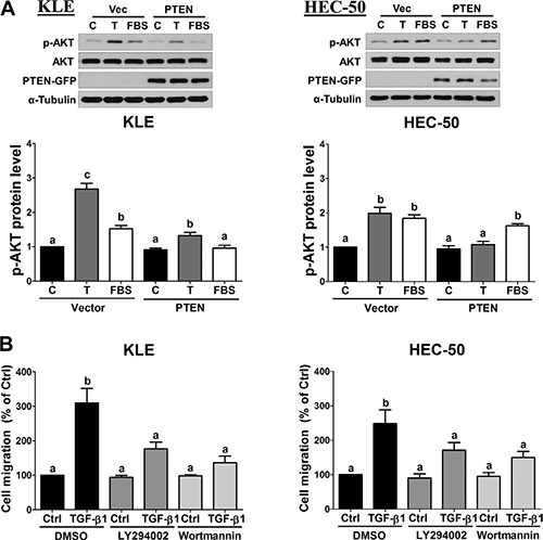
Figure 4. Inhibition of AKT signaling attenuates TGF-β1-stimulated cell migration.

(A) KLE and HEC-50 cells were transfected with 1 μg vector control (Vec; pcDNA-GFP) or vector encoding PTEN (pcDNA-PTEN-GFP) for 48 h and then treated without (C) or with 10 ng/mL TGF-β1 (T) or 10% FBS for a further 24 h. Western blot was used to confirm PTEN-GFP overexpression and to examine the levels of phosphorylated AKT (p-AKT) in relation to its total levels from the same membrane (AKT). (B) KLE and HEC-50 cells were pre-treated with vehicle (DMSO), LY294002 (10 μM) or Wortmannin (1 μM) for 1 h and then treated without (Ctrl) or with 10 ng/mL TGF-β1 for 24 h. After treatment, the levels of cell migration were examined by the transwell migration assay (24 h). Results are expressed as the mean ± SEM of at least three independent experiments and values without common letters are significantly different (P < 0.05).
