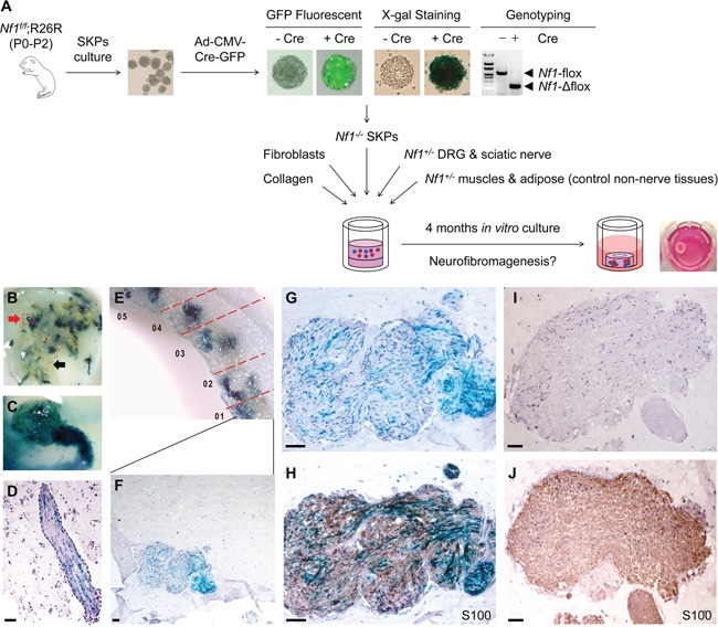
Figure 4. Three-dimensional skin raft culture system for neurofibroma formation in vitro.

Nf1−/− SKPs were co-cultured with Nf1+/− DRGs, sciatic nerves, adipose and muscle tissues in an in vitro raft culture A. Rafts were harvested 4 months after setup for X-gal staining to trace the SKPs that are clustered within nerve tissues (red arrow) but not in muscle/adipose tissues (black arrow), indicating SKPs are neurotropic B. A DRG infiltrated by SKPs C. Histological analysis revealed that SKPs incorporated into nerve tissue D. Many areas within the in vitro raft culture show hyperplasia of LacZ-positive disorganized cells with wavy nuclei E-G. and these areas are positively stained with Schwann cell marker S100 H. In control experiments, nerve tissue did not show any sign of neoplasm I-J. Scale bar = 50 μm.
