Abstract
Schistosomiasis is one of the most important neglected tropical diseases. Despite effective chemotherapeutic treatments, this disease continues to afflict hundreds of millions of people. Understanding the natural intermediate snail hosts of schistosome parasites is vital to the suppression of this disease. A recently identified genomic region in Caribbean Biomphalaria glabrata snails strongly influences their resistance to infection by Schistosoma mansoni. This region contains novel genes having structural similarity to known pathogen recognition proteins. Here we elaborate on the probable structure and role of one of these genes, grctm6. We characterised the expression of Grctm6 in a population of Caribbean snails, and performed a siRNA knockdown of Grctm6. We show that this protein is not only expressed in B. glabrata hemolymph, but that it also has a role in modulating the number of S. mansoni cercariae released by infected snails, making it a possible target for the biological control of schistosomiasis.
Author summary
Schistosomiasis is one of the most prevalent parasitic diseases in the world. Though treatments for schistosomiasis infection exist, there is no vaccine, and reinfection is common in areas where the parasite occurs. One possible way to mitigate schistosomiasis is by controlling the transmission of the parasite larvae from the snails that carry them. Understanding the snail-parasite relationship is essential for the development of new means to interrupt transmission of the parasite from snails to humans. Snails possess immune mechanisms for fighting infection, most of which are based in hemolymph tissue. Here we characterize a novel protein, Grctm6, in a snail host of schistosome parasites. Grctm6 is structurally similar to certain other immune proteins and is present in snail hemolymph. Importantly, we demonstrate that the release of schistosomes by infected snails is exacerbated when this protein is experimentally suppressed in live snails. These results support the suitability of Grctm6 as a possible target for reducing the transmission of this human disease.
Introduction
The World Health Organization (WHO) has estimated that schistosomiasis, a detrimental parasitic helminth disease, affects approximately 258 million people, making it one of the most important parasitic diseases in the world [1, 2]. Millions of people are chemotherapeutically treated for schistosomiasis, but in areas where this parasite is endemic there are high rates of reinfection and persistent debilitating illness. Schistosomiasis-attributed mortality in sub-Saharan Africa alone exceeds 250,000 per year and, with no effective human vaccine, alternative control methods are vital for reducing the burden of this neglected tropical disease [3].
Schistosome miracidia must infect a compatible aquatic snail host in order to produce the cercarial stage that is capable of infecting human hosts. Controlling this intermediate snail host is a primary method for the alternative control of schistosomiasis [4]. Biomphalaria glabrata is the intermediate freshwater snail host of Schistosoma mansoni in the Americas, and has been a target for the successful control of schistosome transmission since the 1950s [5, 6]. Though the initial success of this biological control strategy was limited to Puerto Rico [5], contemporary attempts have expanded to other Caribbean island systems, East Africa (on another snail host), and South America [4–10]. Efforts to control B. glabrata populations have commonly employed the introduction of carnivorous or competitive snail species, but molluscicides have also been heavily exploited [4–12]. Both of these measures can have negative ecological impacts [11]. Despite these consequences, snail control has been shown to be the most successful approach to reduce the prevalence of schistosomiasis, particularly if it is paired with human pharmacological treatment [12]. Recent efforts have begun to focus on determining the relative importance of individual B. glabrata genes on schistosome-infection resistance, with the goal of characterizing snail immune responses to infection, and eventually manipulating snail populations so that they are more naturally resistant to schistosome infection [13–15].
Allelic variation in the Guadeloupe Resistance Complex (GRC), a recently discovered novel gene region in B. glabrata, has been shown to strongly influence Guadeloupean B. glabrata (BgGUA) resistance to Guadeloupean S. mansoni (SmGUA) infection [13]. Allelic variation in this genomic region has an 8-fold effect of infection odds, greater than for any other known snail locus [13, 16, 17]. Resistance is dominant, suggesting a mechanism of parasite recognition and/or clearance by the host, rather than host recognition by the parasite [13]. There are three distinct haplotypes in the GRC region (with 15 coding genes), which we designate R (for the dominant allele that confers increased resistance), S1 and S2 (for the two alleles that confer increased susceptibility; S1 and S2 are equivalent in their effects). The GRC region contains several genes having structural similarity to membrane-bound, pathogen recognition molecules and receptors such as Toll-like receptors and Fc receptors. The region also appears to be under balancing selection, again consistent with a role in pathogen recognition [13]. Determining the functions of these genes, and their potential immunological roles during schistosome infection, is vital for understanding schistosome-infection resistance by this snail species. Given that Biomphalaria species are major intermediate hosts for human schistosomiasis, understanding how schistosome infections can be controlled in these snails may provide insights into ways to proactively limit schistosomiasis transmission. In the present study, we chose one of the GRC genes for in-depth functional analysis: grctm6, which encodes the Guadeloupe Resistance Complex Transmembrane 6 (Grctm6) protein. grctm6 is a particularly compelling candidate locus because the resistant allele at this locus shows high non-synonymous substitution relative to the two susceptibility alleles (particularly in the predicted extracellular domain), susceptibility is not correlated with mRNA levels, and because bioinformatic structural analyses confirms that Grctm6 is a potential candidate for immunological activity due to its predicted transmembrane structure. We report that this gene is expressed at the protein level in hemolymph, and demonstrate that a short interfering RNA (siRNA) knockdown of Grctm6 increased the number of cercariae released into the environment by treated snails.
Materials and methods
Biomphalaria glabrata and Schistosoma mansoni maintenance, lines, and ethics
B. glabrata (BgGUA: “snails”) and S. mansoni (SmGUA: all miracidia or cercariae described) were collected in 2005 in Guadeloupe and maintained as previously described [13, 18]. The SmGUA strain of S. mansoni was cycled through BgGUA and hamsters, and parasite eggs were isolated from rodent livers. BgGUA snails were genotyped based on their GRC locus as previously described [13]. From the outbred BgGUA population we isolated 6 independent, partially-inbred lines that were homozygous at the GRC locus (2 RR, 2 S1S1, and 2 S2S2 lines). We used these lines to verify the baseline resistance (percentage infected) and levels of constitutive expression of grctm6 in each of the three genotypes. All RNAi studies were done on a single RR line. Snails for all experiments were size matched (~7 mm) and housed identically. The Oregon State University Institutional Animal Care and Use Committee, which adheres to Public Health Service Domestic Assurance for humane care and use of laboratory animals (PHS Animal Welfare Assurance Number A3229-01), approved this research as Animal Care and Use Proposal 4360.
In silico predicted protein sequence alignment, domains, and size of Grctm6
Alignment of the protein products of the three alleles of Grctm6 found in BgGUA were calculated previously from RNA-sequencing [13]. We calculated protein molecular weights using Science Gateway (http://www.sciencegateway.org/tools/proteinmw.htm). We examined secondary structure using PSIPRED (http://bioinf.cs.ucl.ac.uk/psipred/). In addition, the signal peptide (http://www.cbs.dtu.dk/services/SignalP/), transmembrane domain (http://www.cbs.dtu.dk/services/TMHMM/), and asparagine glycosylation (http://www.cbs.dtu.dk/services/NetNGlyc/) were predicted using the Center for Biological Sequence Analysis’ prediction servers. Homology searches were performed with DELTA-BLAST (https://blast.ncbi.nlm.nih.gov/) and Pfam (pfam.xfam.org/).
BgGUA and SmGUA infection studies
Parasite challenges were carried out as previously described [13]. In brief, snails were placed in 2 ml of dechlorinated water in individual wells of a 24 well dish containing 10 or 20 miracidia for 24 h, and subsequently transferred into tubs containing 10 snails each to be monitored for infection. All infections were conducted at 1 pm in the afternoon following animal sacrifice at 11 am. Two independent lines of each RR, S1S1, and S2S2 snails were challenged and pooled by genotype for the verification of GRC locus susceptibility. This was done on two separate occasions using a minimum of 30 snails each time (n = 64 S1S1, 68 S2S2, 86 RR). These snails were examined for cercarial shedding, and scored as either infected or uninfected. Starting 5 weeks post challenge, and for 5 subsequent weeks, snails were placed in 2 ml of dechlorinated water in individual wells of a 24 well dish and exposed to light for 3 h beginning at 9 am. For the siRNA experiments, RR snails were treated with enhanced green fluorescent protein, (GFP, Sham injected) or grctm6 oligonucleotides (oligos) and then challenged with miracidia. We challenged using 10 or 20 miracidia, and did two independent trials for each number of miracidia. We used a minimum of 30 snails per trial (Snails that survived for analysis: n = 55 snails using GFP oligos, and n = 37 using grctm6 oligos for 20 miracidial challenges; n = 65 using GFP oligos, and n = 63 using grctm6 oligos for 10 miracidial challenges). These were examined for cercarial shedding as described above [14, 19, 20]. siRNA treated snails were challenged at the beginning of the third day post siRNA injection. Cercariae were enumerated by taking three equal aliquots from a 2 ml sample (or counting all cercariae in a well if density was low). All shedding snails were individually marked with nail polish so that a cumulative count over the 5 week scoring period could be achieved for each snail.
siRNA knockdown of Grctm6
Specific siRNA oligos for grctm6 were designed and produced by Integrated DNA technologies (IDT). Oligos aligned to the more conserved 3′ intracellular domain. Three oligos (GUUAGGACACCGUCAAUU, CACUGCUGACAUUGGCAG, UUUCAUUUGCAUUGCUUG) were suspended according to the manufacturer’s instructions and injected into live ~7 mm RR snails at 2 μg/μl as previously described [14, 19]. In brief, either enhanced GFP IDT control oligos (GFP /Sham) or the grctm6 oligo mixture was suspended in Xfect transfection reagent with nanoparticles (Clontech) according to the manufacturer’s instructions and each snail received a single 10 μl injection distal to the heart [14, 15]. The siRNA-mediated knockdown was assessed at the mRNA level over 4 days (the end of day 0–4), and additionally confirmed by Western blot analysis of protein levels 2–4 days post-injection. Mortality was also compared, within each single day, between GFP control oligos and the grctm6 oligo mixture to ensure that the grctm6 oligo mixture was not inducing additional mortality.
qPCR assessment of grctm6 mRNA transcript levels
Quantitative RT-PCR (qPCR) was used to quantify mRNA transcripts of grctm6 in whole snail lysates of BgGUA snails, and used to detect the extent of siRNA knockdown of grctm6 mRNA in RR snails. Constitutive levels of grctm6 mRNA were assessed in 2 independent homozygous lines of each RR, S1S1, and S2S2 snails and pooled within a genotype (2 RR lines pooled, 2 S1S1 lines pooled, and 2 S2S2 lines pooled). RR snails were also assessed for their grctm6 mRNA levels 0, 1, 2, 3, and 4 days post injection of GFP or grctm6 oligos. In brief, whole snails were snap-frozen in liquid nitrogen, total RNA extracted using the Direct-Zol RNA miniprep kit (Zymo Research) and cDNA synthesized using iScript Reverse Transcriptase Supermix for RT-qPCR (BioRad). Additionally, the head-foot, albumen gland, or hemolymph were removed and snap frozen for tissue analysis of R snails. Hemolymph was collected by head-foot retraction as previously described [14]. qPCR was performed, as previously described [21–23]. All primers were prepared at 300 nM, had a single melt curve, had efficiencies between 90–100%, and were designed or verified using Primer 3 (National Center for Biotechnology Information). grctm6 primers aligned to the more conserved 3′ intracellular domain and showed 100% identity to all three alleles. B-actin (F: 5’-GCTTCCACCTCTTCATCTCTTG -3’; R: 5’-GAACGTAGCTTCTGGACATCTG-3’) was used as an internal control, and did not vary across treatments. grctm6 (F, 5′-TGTTGAGTACGCTGCTGTCAATAAG -3′; R, 5′- ATTCATATCCTTGTTGCTTGGGTCC-3′) was used with the following PCR conditions (in a Applied Biosystems 7500 fast qPCR thermocycler): 95°C for 5 min; 40 cycles of 95°C for 15s and 60°C for 15s. All mRNA levels of GRC lines were normalized to β-actin expression and presented relative to S2 snails. All mRNA levels of oligo injected RR snails were normalized to β-actin expression and presented relative to GFP control samples.
Novel polyclonal anti-Grctm6 antibody production
Rabbit polyclonal Grctm6 antibodies were produced against a peptide epitope (within RR Grctm6, isolated, purified, and validated by Genscript custom antibody services (Genscript). Re-validation of the antibody was performed in house and it was found to be effective at a concentration 1:2000 for western blot detection of synthetically generated Grctm6 peptides and native Grctm6 isolated from hemolymph. Genscript Anti-rabbit IgG secondary HRP conjugate (1:2000) was used for detection.
Western blot analysis of Grctm6
Western blots were used to detect the presence of Grctm6 protein in RR snail hemolymph following siRNA knockdown as previously described with the modifications described below [15]. Snail tissue preps (whole snail, albumen gland, head-foot, hemolymph +/- hemocytes, and hemocytes) were examined from pooled untreated samples from RR snails. Hemolymph preparations were obtained using the head-foot retraction method and either directly added to lysis buffer (Bolt LDS sample and reducing buffer (Thermo)), or cells were removed by centrifugation at 1200g before protein extraction was performed. Unmodified hemolymph produced a consistently detectable band when interrogated with a Grctm6 polyclonal antibody, so this tissue was extracted and used for knockdown analysis. Snail hemolymph was collected, pooled (4–10 snails per sample), and mixed with Bolt LDS sample and reducing buffer (Thermo), and homogenised using a 25G needle before being heated to 95°C for 7 minutes. Tissue samples (whole snail removed from the shell, albumen gland, and head-foot) were homogenized in BOLT LDS sample and reducing buffer using a 18G needle followed by a 25G needle and then treated identically to hemolymph samples. Total protein levels in each sample were quantified using absorbance at 280 nm (Nanodrop, Thermo), additional loading buffer was added to more concentrated samples to ensure all samples had equivalent concentrations of total protein, and 750 ng of total protein was used for each sample per well (500 ng for Fig 3B) [15]. This was the maximum protein concentration that could be used across all samples. Electrophoresis was performed in a 10% pre-cast Bolt Bis-Tris gel for 20 minutes at 200 V. Samples were blotted using a Pierce power blotter cassette (Thermo), and western blot detection was done using an iBind Solutions kit and iBind western device (Thermo). Detection was achieved via Supersignal West Pico luminol solution (Thermo) and chemiluminescence was acquired using a MyECL imager (Thermo). Densitometry was performed post image acquisition using ImageJ software (NIH), and was calculated on the only clear band, which coincided with the predicted 68 kDa size of Grctm6. Three independent blots were run, each inclusive of all 3–5 samples/treatment (5 on Day 3). Relative density normalized to BgActin was calculated independently for each western blot and then averaged as a triplicate for each sample (comparisons within a day only).
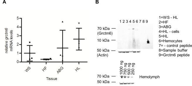
Fig 3. grctm6 mRNA expression and protein detection.
(A) Constitutive grctm6 mRNA levels in resistant whole snail (WS), head-foot (HF), albumen gland (ABG), and hemolymph (HL) lysates (n = 4 samples). HL has significantly elevated levels compared to HF only. (B) Western blot analysis of constitutive Grctm6 protein levels in resistant whole snail after hemolymph was removed (WS- HL), head-foot (HF), albumen gland (ABG), hemolymph with cells removed (HL—cells), whole hemolymph (HL), hemocyte lysates, sample buffer containing no snail tissue, negative control peptide, and Grctm6 peptide (provided by genscript). Equivalent total protein was loaded into each well for experimental samples (500 ng/sample). The band shown is at the ~68 kDa size for Grctm6 (~42 for BgActin loading control), Grctm6 peptides appear lower down on the gel as they are not the full length protein. HL- cells shows lower levels of BgActin protein because most of the actin producing cells were removed by centrifugation. Additionally, hemolymph samples with differing total protein concentrations are shown below. mRNA levels are normalized to β-actin and presented as mean +/- SD relative to WS samples. Significant differences (ln transformed for equal variance, ANOVA, p<0.05) are denoted by asterisks (*).
Statistical analysis
Statistical analyses were completed as indicated, and were generally completed by one-way ANOVA (or unpaired Student’s t-test) with a Tukey post-test unless otherwise specified (p<0.05). If a Barlett’s test (or F-test) for equal variance failed, then data underwent a natural log transformation (ln) before reanalysis. Analyses of the susceptibility of snail populations were done by calculating the Z score (standard score) of the population. Analyses were completed using GraphPad Prism software (La Jolla, CA, USA).
Results
The in silico predicted protein sequence alignment, domains, and size of Grctm6
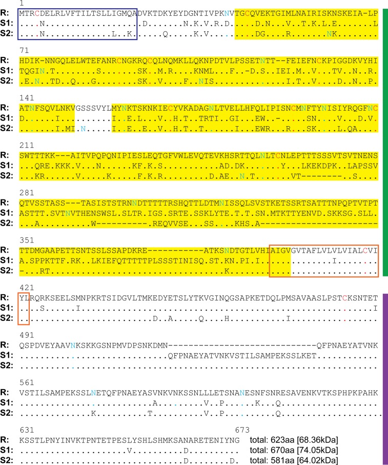
In the GRC region there are seven genes coding for putative membrane spanning proteins that have structural similarities [13]. We examined one of the two genes that were identified by Tennessen et al. [13] as most likely to be responsible for schistosome resistance. Our further analysis of Grctm6 indicates that it is likely a single-pass transmembrane protein of ~68 kDa (Fig 1). The predicted extracellular sequence of the R allele protein differs substantially from those of the two S alleles, but the signal sequence and the transmembrane and cytosolic domains are far less variable between the R and S alleles (Fig 1). This highly variable extracellular domain also has the potential to be glycosylated, which is common in B. glabrata [24]. Interestingly, both the suspected extracellular and intracellular stretches of this protein are long (>200aa) and are likely to support substantial secondary structure (extracellular: 9–11% α helices, 30–39% β strands; intracellular: 6–13% α helices, 7–13% β strands). Therefore, it is likely that both contain unidentified functional domains (Fig 1). However, consistent with previous results [13] we found no significant homology to any known protein domains [13].
Fig 1. grctm6 encodes a single-pass transmembrane protein with an extraordinarily variable extracellular domain.
Alignment of the protein products of the three alleles of grctm6 found in BgGUA. Residues identical to those in the R allele are indicated by dots. The putative signal peptide is shown with a dark blue outlined box. The putative transmembrane domain is shown with an orange box, while the extracellular and intracellular domains are flanked by green and purple bars, respectively. Highly variable regions, defined by 20aa windows containing at least 10 sequence differences, have a yellow background. Cysteines are shown in red. Asparagines predicted to be glycosylated are shown in blue/green.
Variation in resistance to SmGUA among GRC genotypes is not explained by differences in constitutive grctm6 mRNA expression
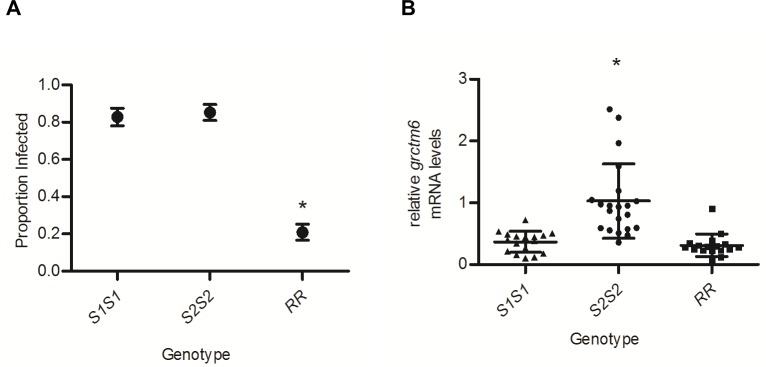
Among our inbred lines, RR snails were ~25% as likely to be infected by SmGUA as either S1S1 or S2S2 snails (p <0.01; Fig 2A ~0.2 vs ~0.8). These same snail lines were examined for mRNA expression of grctm6. RR snails showed no consistent corresponding increase or decrease relative to susceptible snails, although S2S2 snails had ~2–3 fold higher mRNA levels of grctm6 than S1S1 or RR snails (p<0.01; Fig 2B). These findings verify, for our inbred lines, that the constitutive mRNA expression of grctm6 in whole snails does not explain the resistance of the different genotypes at the GRC locus, and that amino acid sequence divergence may be biologically important for this gene’s function [13]. grctm6 mRNA transcripts were detected in all of the snail tissues that were isolated. Transcript levels appear to be slightly elevated in hemolymph, although a statistical difference was only found between hemolymph and the head-foot (p = 0.03; Fig 3A). When Grctm6 protein levels were examined, the only preparations that had consistently detectable Grctm6 protein were from whole hemolymph lysates (Fig 3B). Grctm6 protein was possibly present in isolated hemocytes and cell free hemolymph, but only unmodified hemolymph (hemolymph that received no manipulations prior to protein extraction) produced a consistently detectable band at ~68kDa at various total protein concentrations (Fig 3B). A more sensitive antibody and immunohistological analysis would be required to definitively determine the tissue specific/cellular location of Grctm6. It is puzzling that we were only able to consistently detect Grctm6 in unmodified hemolymph. Perhaps some part of the cell separation protocol modifies or destroys the epitope our antibody binds. It is possible that spinning the cells triggers an intracellular trimming of Grctm6 and loss of the epitope our antibody recognises. It is also possible that, because we are using a novel polyclonal antibody, we were unable to detect smaller amounts of Grctm6 in other tissues because of low sensitivity. Regardless, this is the first evidence that Grctm6 exists at a protein level in any tissue or species of Biomphalaria.
Fig 2. Susceptibility of BgGUA lines is not explained by grctm6 mRNA levels.
(A) The susceptibility of the three different homozygous BgGUA genotypes (S1S1, S2S2, RR) after challenge with 20 miracidia (n = 64, 68, 86 snails respectively). Susceptibility data are presented as the proportion of infected snails +/- standard error of proportions. (B) Constitutive grctm6 mRNA levels (whole snail) of these same homozygous lines (n = 17, 21, 17 snails respectively). Note that the susceptibility differences among the three genotypes does not correlate with expression levels of grctm6. mRNA levels are normalized to β-actin and presented as mean +/- SD relative to levels in S2S2. Significant differences among lines (Susceptibility: Z score of proportion; mRNA levels: ln transformed for equal variance, ANOVA, p<0.05) are denoted by asterisks (*).
siRNA knockdown of grctm6 causes a transient reduction in mRNA transcripts and likely a corresponding reduction in Grctm6 protein levels in the hemolymph of resistant BgGUA
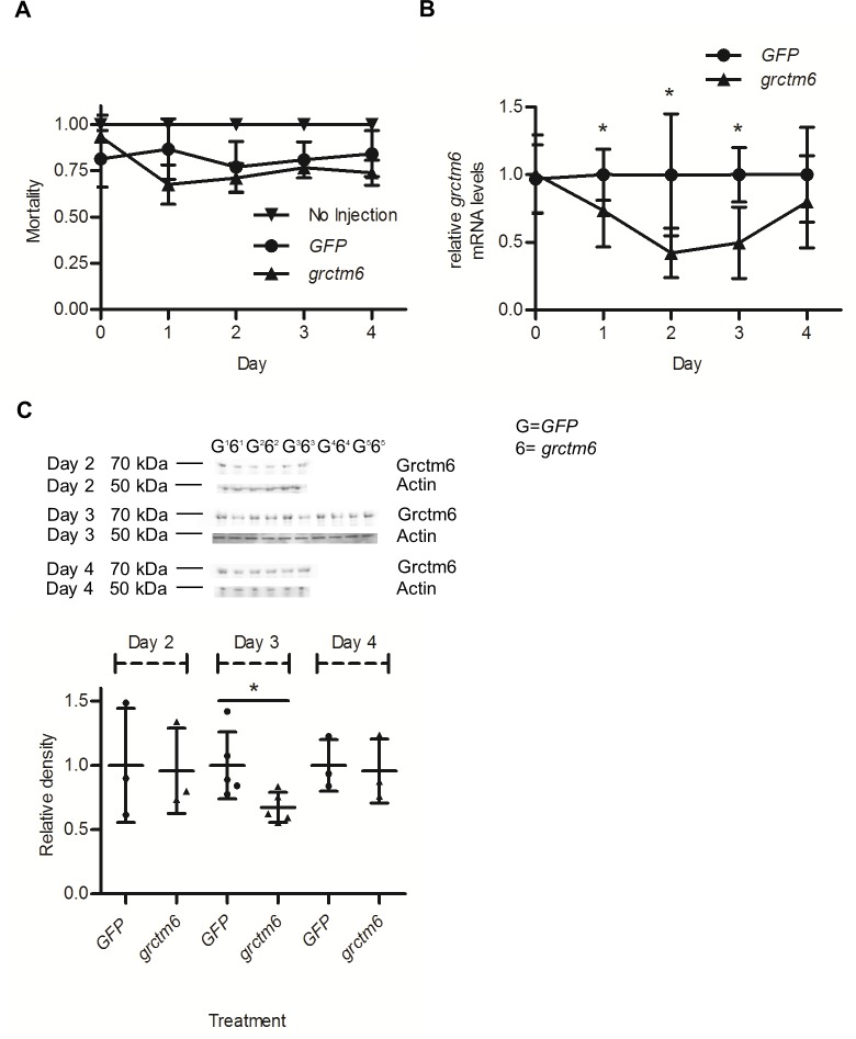
Using RR snails, we knocked down grctm6 mRNA via siRNA. Injections of oligos caused approximately 25–30% mortality over the first days post-injection (Fig 4A). However, our siRNA knockdown of Grctm6 did not increase mortality beyond that of the control (GFP), so the initial dip in survival is likely the result of physical damage to the snail from the injection procedure.
Fig 4. siRNA mediated knockdown of Grctm6.
(A) Mortality of resistant BgGUA snails injected with siRNA taken on 4 separate days post injection (n = 3–6 independent experiments per day). (B) The kinetics of siRNA mediated knockdown of grctm6 mRNA in whole resistant snails after injection compared to GFP siRNA oligos during the first 4 days post-injection (n = 8–10 snails/treatment/day). A reduction of 60% below control was evident by day 3. (C) Western blot analysis of Grctm6 protein levels in resistant hemolymph 2–4 days after injecting snails with grctm6 or GFP siRNA. Equivalent total protein was loaded into each well (750 ng). The band shown in these representative blots is at the ~68 kDa size (~42 for BgActin loading control). Densitometry was preformed using Image J software (Days 2 and 4: n = 3 samples/treatment/day, n = hemolymph pooled from 4–5 individuals; Day 3: n = 5 samples/treatment, n = hemolymph pooled from 6–10 individuals, averaged from three independent Western blots and presented relative to the BgActin loading control for each individual day). A ~30% reduction, below control relative to BgActin, was found in samples taken on Day 3. mRNA levels are normalized to β-actin and relative to GFP oligo injected individuals taken on each individual day (no comparison between days was made). All data are presented as mean +/- SD; significant differences (student t-test, p<0.05) from the GFP control are denoted by asterisks (*).
grctm6 mRNA was significantly reduced by up to ~60% during the first 3 days post siRNA injection in whole snail lysates (p = 0.02; Fig 4B). Given that grctm6 mRNA levels were reduced after 3 days, but normalize by the end of day 4, we examined extent of the protein knockdown surrounding the third day. The amount of Grctm6 protein was significantly reduced by ~30% in the hemolymph 3 days post siRNA injection, but was unmodified on any other day (p = 0.03; Fig 4C). Since we hypothesise that Grctm6 may have some recognition function, we chose to infect snails at the beginning of day 3, given that the protein levels of Grctm6 are reduced from the control on that day, and may be sub-physiological [13].
siRNA knockdown of Grctm6 increases cercarial shedding
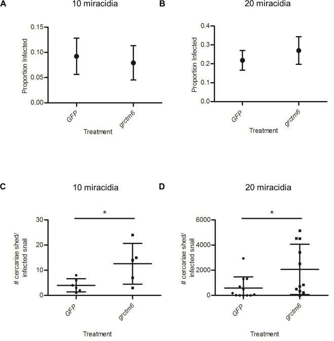
Two independent doses of miracidia were used so that potential changes to schistosome susceptibility were not overlooked by using a single dose of miracidia [18]. Resistant snails treated with grctm6 oligos or GFP oligos exhibited equivalent susceptibility to infection by SmGUA (Fig 5A and 5B). However, grctm6 injected snails had significantly higher (~3–4 fold) cercarial shedding of infected individuals that shed at least one cercariae (p = 0.04, 0.03; Fig 5C and 5D). This pattern was apparent when snails were challenged with either 10 or 20 miracidia, although overall cercarial shedding was ~150-fold lower in snails challenged with 10. This is the first evidence that this protein is directly involved in snail host defense to any pathogen, and specifically to schistosomes.
Fig 5. siRNA knockdown of Grctm6 increases the number of cercariae released by shedding snails but does not modify susceptibility.
Resistant BgGUA snails were treated with GFP or grctm6 oligos and exposed to 10 or 20 miracidia 3 days after injection of oligos. (A-B) Susceptibility over weeks 5–10 post challenge with (A) 10 (n = 65 GFP, 63 snails grctm6) or (B) 20 miracidia (n = 55 GFP, 37 grctm6 snails). (C-D) The total number of cercariae released over a 15 h period (a 3 h period, every week, from weeks 5–10 post exposure) by shedding snails exposed to (C) 10 (n = 6 GFP, 5 grctm6 snails) or (D) 20 miracidia (n = 12 GFP, 11 grctm6 snails). Susceptibility data are presented as the proportion of infected snails +/- standard error of proportions. Cercariae counts are presented as mean +/- SD. Significant differences (Susceptibility: Z score of proportion; cercariae counts: ln transformed for equal variance, student t-test, p<0.05) from the GFP control are denoted by asterisks (*). Note the different scales on the Y-axes in figures for 10-miracidial versus 20-miracidial challenges.
Discussion
In the last decade, there has been a flurry of breakthroughs elucidating snail-schistosome interactions that have exploited a pharmacological, or an RNAi knockdown of a protein of interest in either the snail host or the schistosome [14, 15, 19, 20, 25–36]. Generally, these RNAi knockdowns have targeted proteins that have been relatively well described in other species, or are homologous to another group of defined-immunologic targets. We used this technique to assess the importance of a completely uncharacterized gene/protein, with no known homologs in other species, on snail-schistosome compatibility following its discovery by linkage mapping.
Tennessen et al. [13] described a novel genomic region (the GRC) with alleles that are strongly associated with snail resistance to schistosome infection. Seven transcriptionally expressed, coding GRC genes, designated the grctm loci, are particularly promising candidates in this genomic region. Though these transcripts (including grctm6) bear little resemblance to any known proteins, they share some characteristics common to immunologic membrane bound receptors (single-pass transmembrane proteins with a highly variable extracellular domain, and sequence divergence between disease relevant alleles) [13]. Grctm6 is of particular interest because of its putative structure and because the R allele at the grctm6 locus has high non-synonymous substitution relative to the S alleles. In this study, we verified that GRC genotypes are strongly correlated with resistance and that constitutive transcript variation does not explain resistance differences between the various alleles in BgGUA snails [13]. Previous RNA-Seq data from outbred snails suggested that both S alleles show approximately 2-fold higher expression of grctm6 than R [13]. Our qPCR results on inbred lines indicate that S2, but not S1, has at least 2-fold higher expression than R, supporting the notion that mRNA levels of grctm6 are not a likely explanation for GRC locus-associated resistance in BgGUA (Fig 2B). We also show that Grctm6 is expressed at the protein level in hemolymph (but were unable to determine if it is specific to hemolymph). Although this partial and transient knockdown of Grctm6 did not significantly change the proportion of hosts infected, it did increase cercarial shedding, indicating that Grctm6 has a role in modulating the extent/burden of the schistosome infection in BgGUA. Increasing the number of miracidia used to challenge snails from 10 to 20 has little effect on the proportion of snails infected, but a huge effect on the number of cercariae shed by infected snails (Fig 5). Thus, it is plausible that grctm6 has a role in controlling the number of miracidia that successfully infect the snail, but that we only observed an effect on cercarial shedding because this trait may be more sensitive, than proportion infected, to the number of parasites that successfully established. Alternately, grctm6 may help to regulate some subsequent larval stage leading to cercariogenesis or cercarial release into the environment. Regardless, we have demonstrated that the Grctm6 protein has an important effect on the extent of schistosome infection.
We speculate, based on amino acid sequences, that Grctm6 may be a membrane bound receptor. R and S alleles exhibit substantive sequence divergences (15–45% amino acid differences, Fig 1) in the extracellular region of this protein, which could indicate variation in extracellular domain stabilities, target ligands, and/or binding affinities [24, 37]. The more conserved region of Grctm6 is located in the putative transmembrane and cytoplasmic regions, which could serve to preserve potential outside-in signaling functions of this protein as in other immune receptor proteins [38]. The high expression of Grctm6 in hemolymph, relative to other tissues, (Fig 3) is noteworthy because that is the location of crucial snail-schistosome interactions [39–41]. A rigorous immunological analysis would be required to determine if any of these speculations regarding the mechanistic or immunological role of Grctm6 on parasite infection or immunity are accurate. Although we have clearly shown that this protein influences the numbers of cercariae shed, whether Grctm6 actually functions as an immune receptor, and at what stage in the infection process it acts, remain to be shown. Grctm6 could have negative impacts on any stage of sporocyst growth or development, and further immunohistological and functional analysis are required to determine how Grctm6 is mechanistically involved in schistosome infection.
It is interesting that knocking down Grctm6 expression affected cercarial shedding but not susceptibility (percentage infected). However, this effect was achieved with just a 30% protein knockdown. Perhaps a full knockdown would yield a much stronger phenotype, including an increased susceptibility. A CRISPR/Cas9 knockout would be one way to conclusively test the role of this locus [42]. It would also be pertinent to examine the cellular location of this protein by immunohistochemistry using monoclonal antibodies from recombinant Grctm6. This technique could provide vital future information pertaining to the biological mechanism of Grctm6. We also note that there are six other transmembrane loci in this region, and can’t rule out the possibility that other loci act in concert with grctm6 to produce the observed susceptibility phenotypes.
Here we have shown that a modest reduction in Grctm6 protein levels affects cercarial output, which suggests that this protein may be involved in a pathway which is important during a challenge by S. mansoni. More importantly, this study provides a potentially new type of target for controlling transmission of schistosomes at the snail stage. If, in the future, the resistant allele of genes like grctm6 could be gene-driven into a population of B. glabrata[43], then it is possible that their resistance to schistosomes could be improved without completely immunocompromising the snail. Using these types of modern molecular methods, we would anticipate fewer deleterious ecological consequences than those following contemporary snail control methods, which involve introduced predators/competitors or molluscicides [44, 45]. Even a genetic manipulation that only reduces the number of cercariae shed into the environment could have epidemiological consequences. Further exploration of these genes, their physiological functions, and potential roles for the control of schistosomiasis will be important for combatting this widespread and destructive disease.
Acknowledgments
We would like to thank Clint Sergi, Leeah Whittier, Ryan Wilson, and Ekaterina Peremyslova for their technical support.
Data Availability
All data can be found in the publication itself.
Funding Statement
This work was supported by grants AI109134 and AI111201 from the National Institutes of Health. The funders had no role in study design, data collection and analysis, decision to publish, or preparation of the manuscript.
References
- 1.Schistosomiasis: population requiring preventive chemotherapy and number of people treated in 2010. Wkly Epidemiol Rec. 2012;87(4):37–44. [PubMed] [Google Scholar]
- 2.Schistosomiasis: number of people treated worldwide in 2014. Wkly Epidemiol Rec. 2016;91(5):53–60. [PubMed] [Google Scholar]
- 3.Pearce EJ, MacDonald AS. The immunobiology of schistosomiasis. Nat Rev Immunol. 2002;2(7):499–511. 10.1038/nri843 [DOI] [PubMed] [Google Scholar]
- 4.Martins MC, Silva MC, Silva LR, Lima VL, Pereira EC, Falcao EP, et al. Usnic acid potassium salt: an alternative for the control of Biomphalaria glabrata (Say, 1818). PLoS One. 2014;9(11):e111102 PubMed Central PMCID: PMCPMC4222767. 10.1371/journal.pone.0111102 [DOI] [PMC free article] [PubMed] [Google Scholar]
- 5.Jobin WR, Ferguson FF, Palmer JR. Control of schistosomiasis in Guayama and Arroyo, Puerto Rico. Bull World Health Organ. 1970;42(1):151–6. PubMed Central PMCID: PMCPMC2427502. [PMC free article] [PubMed] [Google Scholar]
- 6.Guimaraes CT, Souza CP, Soares D. Possible competitive displacement of planorbids by Melanoides tuberculata in Minas Gerais, Brazil. Mem Inst Oswaldo Cruz. 2001;96 Suppl:173–6. [DOI] [PubMed] [Google Scholar]
- 7.Prentice MA. Displacement of Biomphalaria glabrata by the snail Thiara granifera in field habitats in St. Lucia, West Indies. Ann Trop Med Parasitol. 1983;77(1):51–9. [DOI] [PubMed] [Google Scholar]
- 8.Pointier JP. Invading freshwater snails and biological control in Martinique Island, French West Indies. Mem Inst Oswaldo Cruz. 2001;96 Suppl:67–74. [DOI] [PubMed] [Google Scholar]
- 9.Pointier JP, Jourdane J. Biological control of the snail hosts of schistosomiasis in areas of low transmission: the example of the Caribbean area. Acta Trop. 2000;77(1):53–60. [DOI] [PubMed] [Google Scholar]
- 10.Nguma JF, McCullough FS, Masha E. Elimination of Biomphalaria pfeifferi, Bulinus tropicus and Lymnaea natalensis by the ampullarid snail, Marisa cornuarietis, in a man-made dam in northern Tanzania. Acta Trop. 1982;39(1):85–90. [PubMed] [Google Scholar]
- 11.Calumpang SM, Medina MJ, Tejada AW, Medina JR. Environmental impact of two molluscicides: niclosamide and metaldehyde in a rice paddy ecosystem. Bull Environ Contam Toxicol. 1995;55(4):494–501. [DOI] [PubMed] [Google Scholar]
- 12.Sokolow SH, Wood CL, Jones IJ, Swartz SJ, Lopez M, Hsieh MH, et al. Global Assessment of Schistosomiasis Control Over the Past Century Shows Targeting the Snail Intermediate Host Works Best. PLoS Negl Trop Dis. 2016;10(7):e0004794 PubMed Central PMCID: PMCPMC4956325. 10.1371/journal.pntd.0004794 [DOI] [PMC free article] [PubMed] [Google Scholar]
- 13.Tennessen JA, Theron A, Marine M, Yeh JY, Rognon A, Blouin MS. Hyperdiverse gene cluster in snail host conveys resistance to human schistosome parasites. PLoS Genet. 2015;11(3):e1005067 PubMed Central PMCID: PMCPMC4361660. 10.1371/journal.pgen.1005067 [DOI] [PMC free article] [PubMed] [Google Scholar]
- 14.Hanington PC, Forys MA, Dragoo JW, Zhang SM, Adema CM, Loker ES. Role for a somatically diversified lectin in resistance of an invertebrate to parasite infection. Proc Natl Acad Sci U S A. 2010;107(49):21087–92. PubMed Central PMCID: PMCPMC3000291. 10.1073/pnas.1011242107 [DOI] [PMC free article] [PubMed] [Google Scholar]
- 15.Pila EA, Gordy MA, Phillips VK, Kabore AL, Rudko SP, Hanington PC. Endogenous growth factor stimulation of hemocyte proliferation induces resistance to Schistosoma mansoni challenge in the snail host. Proc Natl Acad Sci U S A. 2016;113(19):5305–10. PubMed Central PMCID: PMCPMC4868488. 10.1073/pnas.1521239113 [DOI] [PMC free article] [PubMed] [Google Scholar]
- 16.Tennessen JA, Bonner KM, Bollmann SR, Johnstun JA, Yeh JY, Marine M, et al. Genome-Wide Scan and Test of Candidate Genes in the Snail Biomphalaria glabrata Reveal New Locus Influencing Resistance to Schistosoma mansoni. PLoS Negl Trop Dis. 2015;9(9):e0004077 PubMed Central PMCID: PMCPMC4570800. 10.1371/journal.pntd.0004077 [DOI] [PMC free article] [PubMed] [Google Scholar]
- 17.Goodall CP, Bender RC, Brooks JK, Bayne CJ. Biomphalaria glabrata cytosolic copper/zinc superoxide dismutase (SOD1) gene: association of SOD1 alleles with resistance/susceptibility to Schistosoma mansoni. Mol Biochem Parasitol. 2006;147(2):207–10. 10.1016/j.molbiopara.2006.02.009 [DOI] [PubMed] [Google Scholar]
- 18.Theron A, Rognon A, Gourbal B, Mitta G. Multi-parasite host susceptibility and multi-host parasite infectivity: a new approach of the Biomphalaria glabrata/Schistosoma mansoni compatibility polymorphism. Infect Genet Evol. 2014;26:80–8. 10.1016/j.meegid.2014.04.025 [DOI] [PubMed] [Google Scholar]
- 19.Hanington PC, Forys MA, Loker ES. A somatically diversified defense factor, FREP3, is a determinant of snail resistance to schistosome infection. PLoS Negl Trop Dis. 2012;6(3):e1591 PubMed Central PMCID: PMCPMC3313920. 10.1371/journal.pntd.0001591 [DOI] [PMC free article] [PubMed] [Google Scholar]
- 20.Pila EA, Tarrabain M, Kabore AL, Hanington PC. A Novel Toll-Like Receptor (TLR) Influences Compatibility between the Gastropod Biomphalaria glabrata, and the Digenean Trematode Schistosoma mansoni. PLoS Pathog. 2016;12(3):e1005513 PubMed Central PMCID: PMCPMC4807771. 10.1371/journal.ppat.1005513 [DOI] [PMC free article] [PubMed] [Google Scholar]
- 21.Allan ER, Habibi HR. Direct effects of triiodothyronine on production of anterior pituitary hormones and gonadal steroids in goldfish. Mol Reprod Dev. 2012;79(9):592–602. 10.1002/mrd.22066 [DOI] [PubMed] [Google Scholar]
- 22.Allan ER, Tailor P, Balce DR, Pirzadeh P, McKenna NT, Renaux B, et al. NADPH oxidase modifies patterns of MHC class II-restricted epitopic repertoires through redox control of antigen processing. J Immunol. 2014;192(11):4989–5001. 10.4049/jimmunol.1302896 [DOI] [PubMed] [Google Scholar]
- 23.Allan ER, Yates RM. Redundancy between Cysteine Cathepsins in Murine Experimental Autoimmune Encephalomyelitis. PLoS One. 2015;10(6):e0128945 PubMed Central PMCID: PMCPMC4468166. 10.1371/journal.pone.0128945 [DOI] [PMC free article] [PubMed] [Google Scholar]
- 24.Lehr T, Frank S, Natsuka S, Geyer H, Beuerlein K, Doenhoff MJ, et al. N-Glycosylation patterns of hemolymph glycoproteins from Biomphalaria glabrata strains expressing different susceptibility to Schistosoma mansoni infection. Exp Parasitol. 2010;126(4):592–602. 10.1016/j.exppara.2010.06.031 [DOI] [PubMed] [Google Scholar]
- 25.Jiang Y, Loker ES, Zhang SM. In vivo and in vitro knockdown of FREP2 gene expression in the snail Biomphalaria glabrata using RNA interference. Dev Comp Immunol. 2006;30(10):855–66. PubMed Central PMCID: PMCPMC3641767. 10.1016/j.dci.2005.12.004 [DOI] [PMC free article] [PubMed] [Google Scholar]
- 26.Mourao Mde M, Dinguirard N, Franco GR, Yoshino TP. Role of the endogenous antioxidant system in the protection of Schistosoma mansoni primary sporocysts against exogenous oxidative stress. PLoS Negl Trop Dis. 2009;3(11):e550 PubMed Central PMCID: PMCPMC2771906. 10.1371/journal.pntd.0000550 [DOI] [PMC free article] [PubMed] [Google Scholar]
- 27.Knight M, Miller A, Liu Y, Scaria P, Woodle M, Ittiprasert W. Polyethyleneimine (PEI) mediated siRNA gene silencing in the Schistosoma mansoni snail host, Biomphalaria glabrata. PLoS Negl Trop Dis. 2011;5(7):e1212 PubMed Central PMCID: PMCPMC3134429. 10.1371/journal.pntd.0001212 [DOI] [PMC free article] [PubMed] [Google Scholar]
- 28.Bais S, Churgin MA, Fang-Yen C, Greenberg RM. Evidence for Novel Pharmacological Sensitivities of Transient Receptor Potential (TRP) Channels in Schistosoma mansoni. PLoS Negl Trop Dis. 2015;9(12):e0004295 PubMed Central PMCID: PMCPMC4676680. 10.1371/journal.pntd.0004295 [DOI] [PMC free article] [PubMed] [Google Scholar]
- 29.Guidi A, Mansour NR, Paveley RA, Carruthers IM, Besnard J, Hopkins AL, et al. Application of RNAi to Genomic Drug Target Validation in Schistosomes. PLoS Negl Trop Dis. 2015;9(5):e0003801 PubMed Central PMCID: PMCPMC4438872. 10.1371/journal.pntd.0003801 [DOI] [PMC free article] [PubMed] [Google Scholar]
- 30.Hagen J, Young ND, Every AL, Pagel CN, Schnoeller C, Scheerlinck JP, et al. Omega-1 knockdown in Schistosoma mansoni eggs by lentivirus transduction reduces granuloma size in vivo. Nat Commun. 2014;5:5375 PubMed Central PMCID: PMCPMC4243216. 10.1038/ncomms6375 [DOI] [PMC free article] [PubMed] [Google Scholar]
- 31.Valentim CL, Cioli D, Chevalier FD, Cao X, Taylor AB, Holloway SP, et al. Genetic and molecular basis of drug resistance and species-specific drug action in schistosome parasites. Science. 2013;342(6164):1385–9. PubMed Central PMCID: PMCPMC4136436. 10.1126/science.1243106 [DOI] [PMC free article] [PubMed] [Google Scholar]
- 32.Bhardwaj R, Krautz-Peterson G, Skelly PJ. Using RNA interference in Schistosoma mansoni. Methods Mol Biol. 2011;764:223–39. 10.1007/978-1-61779-188-8_15 [DOI] [PubMed] [Google Scholar]
- 33.Stefanic S, Dvorak J, Horn M, Braschi S, Sojka D, Ruelas DS, et al. RNA interference in Schistosoma mansoni schistosomula: selectivity, sensitivity and operation for larger-scale screening. PLoS Negl Trop Dis. 2010;4(10):e850 PubMed Central PMCID: PMCPMC2957409. 10.1371/journal.pntd.0000850 [DOI] [PMC free article] [PubMed] [Google Scholar]
- 34.Rinaldi G, Morales ME, Alrefaei YN, Cancela M, Castillo E, Dalton JP, et al. RNA interference targeting leucine aminopeptidase blocks hatching of Schistosoma mansoni eggs. Mol Biochem Parasitol. 2009;167(2):118–26. PubMed Central PMCID: PMCPMC2705689. [DOI] [PMC free article] [PubMed] [Google Scholar]
- 35.Morales ME, Rinaldi G, Gobert GN, Kines KJ, Tort JF, Brindley PJ. RNA interference of Schistosoma mansoni cathepsin D, the apical enzyme of the hemoglobin proteolysis cascade. Mol Biochem Parasitol. 2008;157(2):160–8. PubMed Central PMCID: PMCPMC4130333. 10.1016/j.molbiopara.2007.10.009 [DOI] [PMC free article] [PubMed] [Google Scholar]
- 36.Geldhof P, Visser A, Clark D, Saunders G, Britton C, Gilleard J, et al. RNA interference in parasitic helminths: current situation, potential pitfalls and future prospects. Parasitology. 2007;134(Pt 5):609–19. 10.1017/S0031182006002071 [DOI] [PubMed] [Google Scholar]
- 37.Moremen KW, Tiemeyer M, Nairn AV. Vertebrate protein glycosylation: diversity, synthesis and function. Nat Rev Mol Cell Biol. 2012;13(7):448–62. PubMed Central PMCID: PMCPMC3934011. 10.1038/nrm3383 [DOI] [PMC free article] [PubMed] [Google Scholar]
- 38.Futosi K, Fodor S, Mocsai A. Neutrophil cell surface receptors and their intracellular signal transduction pathways. Int Immunopharmacol. 2013;17(3):638–50. PubMed Central PMCID: PMCPMC3827506. 10.1016/j.intimp.2013.06.034 [DOI] [PMC free article] [PubMed] [Google Scholar]
- 39.Le Clec'h W, Anderson TJ, Chevalier FD. Characterization of hemolymph phenoloxidase activity in two Biomphalaria snail species and impact of Schistosoma mansoni infection. Parasit Vectors. 2016;9:32 PubMed Central PMCID: PMCPMC4722754. 10.1186/s13071-016-1319-6 [DOI] [PMC free article] [PubMed] [Google Scholar]
- 40.Coustau C, Gourbal B, Duval D, Yoshino TP, Adema CM, Mitta G. Advances in gastropod immunity from the study of the interaction between the snail Biomphalaria glabrata and its parasites: A review of research progress over the last decade. Fish Shellfish Immunol. 2015;46(1):5–16. 10.1016/j.fsi.2015.01.036 [DOI] [PubMed] [Google Scholar]
- 41.Bender RC, Broderick EJ, Goodall CP, Bayne CJ. Respiratory burst of Biomphalaria glabrata hemocytes: Schistosoma mansoni-resistant snails produce more extracellular H2O2 than susceptible snails. J Parasitol. 2005;91(2):275–9. 10.1645/GE-415R [DOI] [PubMed] [Google Scholar]
- 42.Perry KJ, Henry JQ. CRISPR/Cas9-mediated genome modification in the mollusc, Crepidula fornicata. Genesis. 2015;53(2):237–44. 10.1002/dvg.22843 [DOI] [PubMed] [Google Scholar]
- 43.Hammond A, Galizi R, Kyrou K, Simoni A, Siniscalchi C, Katsanos D, et al. A CRISPR-Cas9 gene drive system targeting female reproduction in the malaria mosquito vector Anopheles gambiae. Nat Biotechnol. 2016;34(1):78–83. PubMed Central PMCID: PMCPMC4913862. 10.1038/nbt.3439 [DOI] [PMC free article] [PubMed] [Google Scholar]
- 44.Reardon S. Welcome to the CRISPR zoo. Nature. 2016;531(7593):160–3. 10.1038/531160a [DOI] [PubMed] [Google Scholar]
- 45.Esvelt KM, Smidler AL, Catteruccia F, Church GM. Concerning RNA-guided gene drives for the alteration of wild populations. Elife. 2014:e03401 PubMed Central PMCID: PMCPMC4117217. 10.7554/eLife.03401 [DOI] [PMC free article] [PubMed] [Google Scholar]
Associated Data
This section collects any data citations, data availability statements, or supplementary materials included in this article.
Data Availability Statement
All data can be found in the publication itself.


