Fig. 3.
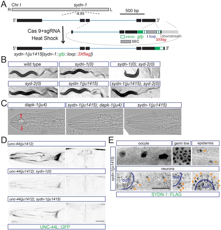
SYDN-1 is expressed in multiple tissues. (A) Experimental design for making gfp::3Xflag knock-in worms of the sydn-1 gene by CRISPR/Cas9 editing. gfp is inserted at the C terminus of sydn-1. The template for C-terminal recombination is indicated. Scale bar: 500 bp. (B) The knock-in allele of sydn-1 mildly enhances syd-2(0) defects. syd-2/α-liprin is essential for synaptogenesis. sydn-1(0) or syd-2(0) single mutants display mildly uncoordinated movement, whereas double mutants display severe paralysis. sydn-1(ju1415) also shows mild uncoordinated movement similar to sydn-1(0). However, sydn-1(ju1415); syd-2(0) double mutants show a less severe locomotor phenotype compared with sydn-1(0); syd-2(0). (C) ju1415 is a loss-of-function allele of sydn-1. Images show head morphology of wild type and mutants. sydn-1(ju1415) suppresses the Mor phenotypes of dapk-1 to the same extent as sydn-1(0). Head region with epidermal defects is labeled by red arrows. (D) UNC-44L::GFP fluorescence from unc-44(ju1412) in sydn-1(ju1415) is reduced compared with wild type, and is noticeably higher than in sydn-1(0). (E) SYDN-1 is widely expressed. Images show immunostaining results with anti-FLAG in sydn-1(ju1415) animals. Immunostaining with anti-FLAG shows expression of SYDN-1::GFP::Flag in nuclei of oocytes, germline, epidermis and neurons. The pharyngeal bulb is labeled in the head image. Nuclear staining of SYDN-1 in epidermal cells or neurons is labeled (orange arrowheads). Scale bar: 20 µm.

