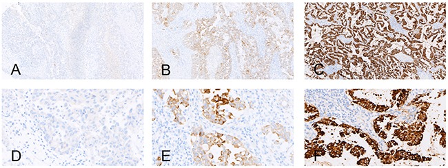
Figure 2. Examples of the immunohistochemical analysis of EML4-ALK.

A, B, and C. show the negative control, positive control and an EML4-ALK (+) case (strong granular cytoplasmic staining) (×100), respectively; D. E. and F. show the negative control, positive control and an EML4-ALK (+) case (strong granular cytoplasmic staining) (×400), respectively.
