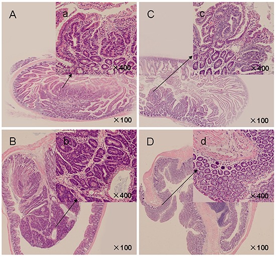
Figure 2. H&E-stained intestinal sections from APCMin/+ mice.

A. Control mice presented crater-shaped adenomatous polyps in small intestines (×100). a' (inset): Adenomatous cells showed enlarged, hyperchromatic, elongated, and crowded dysplastic nuclei (×400). B. Advanced adenomatous polyps with focal high grade dysplasia in colonic polyps of vehicle control mice (×100). b' (inset): Crypt architecture appears complex, nuclei were pleomorphic with frequent mitoses and lack polarization (×400). C. Myricetin-treated small intestinal polyps (×100). c' (inset): Crypt architecture showed decreased dysplastic cells and degree of dysplasia (×400). D. Myricetin-treated colonic polyps (×100). d' (inset): Epithelium presented unremarkable nuclear structure (×400).
