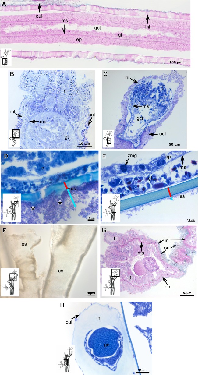
Figure 6. Exoskeletal structure.
(A)–(C) Bougainvillia muscus (Allman, 1863); (D) general internal and external structure of the hydrocaulus, stained with AB + PAS + H; (E)–(F) contracted hydranth, stained with TB. (D)–(H) Bougainvillia rugosa Clarke, 1882. (D)–(E) stained with TB; (A) exoskeleton of the hydrocaulus; E: exoskeleton of the side-branch; (F) general exoskeleton of the polyp; (G) exoskeleton of the hydranth, stained with AB + PAS + H; (H) exoskeleton of mature female gonophore, stained with TB. Cyan-blue line indicates the outer layer of the exoskeleton (=exosarc), red line indicates the inner layer of the exoskeleton (=perisarc), asterisk indicates “perisarc extensions.” Abbreviations: c, coenosarc; ep, epidermis; es, exoskeleton; gct, gastrovascular cavity; gn, gonadal cell cluster; gt, gastrodermis; inl, inner layer; ms, mesoglea; oul, outer layer; t, tentacle; zmg, zymogen glandular cell.

