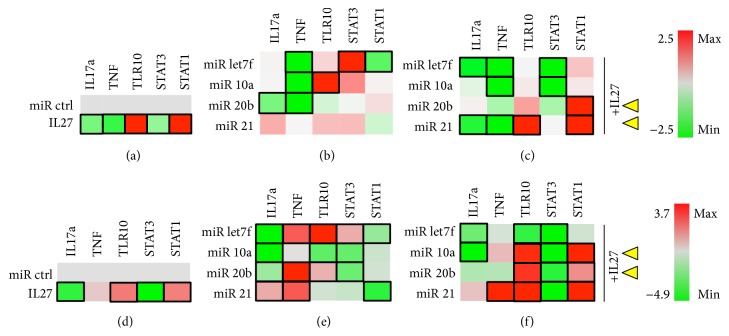
Figure 8.
Jurkat T cells. T cells were grown in the absence (a–c) or presence (d–f) of PMA/Ionomycin stimulus. Cells were cotransfected with miR control or experimental (miRlet7f, miR-10a, miR-20b, or miR-21) and luciferase reporter vectors for detecting IL17a, TNF, and TLR10 expression or STAT3 and STAT1 activity. (a) Effect of IL-27 (50 ng/ml) on luciferase expression. (b) Effect of experimental miRNA on luciferase expression. (c) Effect of miRNA+IL27 combination on luciferase expression. Black rectangle frames (□) highlight the data with significant expression changes (P < 0.05) relative to negative control (miR ctrl); Jurkat T cells cultured in the absence (a–c) or presence of PMA/I (d–f) as per Materials and Methods. Yellow arrowheads, proposed best combinations of miR and IL-27. Color bars, range of fold change in expression, from green (−2.4- or −4.9-fold change or decrease) to red (2.5- or 3.7-fold change or increase).

