Abstract
Patient: Female, 35
Final Diagnosis: Laryngeal sarcoidosis
Symptoms: Hoarseness • stridor
Medication: —
Clinical Procedure: Tracheostomy
Specialty: Otolaryngology
Objective:
Rare disease
Background:
Laryngeal sarcoidosis is a rare extrapulmonary manifestation of sarcoidosis, accounting for 0.33–2.1% of cases. A life-threatening complication of laryngeal sarcoidosis is upper airway obstruction. In this report we describe our experience in the acute and chronic care of a patient who required an emergent tracheostomy, with the aim to provide further insight into this difficult to manage disease.
Case Report:
A 37-year-old African American female with a 10-year history of stage 1 sarcoidosis presented with severe dyspnea. Laryngeal sarcoidosis was diagnosed three years previously, and she remained stable on low-dose prednisone until six months prior to admission, at which time she self-discontinued her prednisone for the homeopathic treatment Nopalea cactus juice. Her physical examination was concerning for impending respiratory failure as she presented with inspiratory stridor and hoarseness. Laryngoscopy showed a retroflexed epiglottis obstructing the glottis with edematous arytenoids and aryepiglottic folds. Otolaryngology performed an emergent tracheostomy to secure her airway and obtained epiglottic biopsies, which were consistent with sarcoidosis. She was eventually discharged home on prednisone 60 mg daily. Following months of corticosteroids, a laryngoscopy showed the epiglottis continuing to obstruct the glottis. The addition of methotrexate to a tapered dosage of prednisone 10 mg daily was unsuccessful, and she remains on prednisone 20 mg daily for disease control.
Conclusions:
Laryngeal sarcoidosis, a rare extrapulmonary manifestation of sarcoidosis, uncommonly presents as the life-threatening complication of complete upper airway obstruction. As such, laryngeal sarcoidosis is associated with significant morbidity and mortality, requiring a high index of suspicion for timely diagnosis and treatment.
MeSH Keywords: Airway Obstruction, Methotrexate, Sarcoidosis, Tracheostomy
Background
Sarcoidosis is a multisystem, chronic granulomatous disease of unknown etiology that predominantly affects the lungs [1]. Typical extrapulmonary manifestations include the heart, eyes, and skin; less frequently described is involvement of the larynx, which accounts for approximately 0.33–2.1% of cases [1,2]. The supraglottic region is most commonly affected, with particular predilection for the epiglottis, arytenoids, and aryepiglottic folds [3]. Patients characteristically present with dysphonia, dyspnea, and dysphagia [3]. We present a rare, life-threatening presentation of laryngeal sarcoidosis in a patient who had upper airway obstruction requiring an emergent tracheostomy. We describe our experience in both the acute and chronic care of our patient to provide further insight into treating this difficult to manage disease for future cases.
Case Report
A 37-year-old African American female with a past medical history significant for stage 1 sarcoidosis presented to our facility with a chief complaint of shortness of breath. Ten years prior, she presented with bilateral uveitis and lupus pernio (LP) on her right ear and was subsequently diagnosed with sarcoidosis. She was successfully treated for these conditions and her sarcoidosis remained quiescent until three years ago, when, at an outside institution, she was discovered to have laryngeal involvement. She was treated with prednisone 60 mg daily for several months and eventually tapered to 10 mg daily. Her sarcoidosis remained well-controlled on this maintenance dose of prednisone until six months prior, at which time she self-discontinued her prednisone in favor of the homeopathic treatment Nopalea cactus juice, which unsubstantially claimed to have anti-inflammatory properties [4]. Subsequently, she developed dyspnea on exertion progressing to dyspnea at rest. During this time her voice became increasingly hoarse and she experienced frequent episodes of difficulty swallowing.
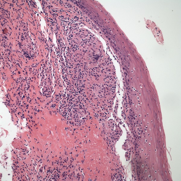
In our emergency department, her physical examination was concerning for respiratory distress as she presented with inspiratory stridor and hoarseness. She also had chronic-appearing, indurated lesions on her right ear. Given that she had signs of upper airway disease for impending respiratory failure, an emergent bedside laryngoscopy was performed, revealing an obstructed airway with the epiglottis retroflexed over the glottis and significant edema in the arytenoids and aryepiglottic folds. She was immediately treated with high-dose intravenous dexamethasone and taken emergently to the operating room (OR) to secure her airway for concern for complete upper airway obstruction, which was confirmed with direct visualization of her larynx in the OR. She was intubated for a surgical airway, and after taking biopsies from the lingual surface of her epiglottis, a #4 cuffed Shiley™ tracheostomy was placed. Her respiratory status immediately stabilized, was extubated, and transferred to our medical intensive care unit. Her tracheostomy was exchanged to a #4 cuffless Shiley on postoperative day 5. During this time she was transitioned from intravenous dexamethasone to prednisone 60 mg daily. Biopsies ultimately revealed non-necrotizing epithelioid granulomas consistent with sarcoidosis (Figure 1). She was prescribed this high-dose prednisone for three months, and after receiving education for self-tracheostomy care, she was discharged home.
Figure 1.

Biopsy of the epiglottis. A squamous lined epithelium with a well formed submucosal non-necrotizing granuloma. The granuloma is relatively distinct from surrounding parenchyma with little adjacent inflammation. 20× objective, H&E stain.
At her subsequent one-month and three-month follow-up visits, she denied any further respiratory issues. Repeat laryngoscopies showed significant improvement in the edema in the arytenoids and aryepiglottic folds, but the epiglottis continued to obscure the glottis despite therapy with high-dose prednisone. Methotrexate was initiated while prednisone was tapered to 10 mg daily; however, this regimen failed and her prednisone dosage was increased to 20 mg daily to reduce edema. Because she declined surgical treatment, she will continue with medical management with immunosuppressive therapy to facilitate eventual de-cannulation.
Discussion
Sarcoidosis of the upper respiratory tract (SURT) is a rare disorder that can have parotid, nasopharyngeal, laryngeal, or tracheal involvement [5]. The most commonly affected structure of the larynx is the epiglottis, which may be in part due to its rich supply of lymphatic vessels [6]. The pathognomonic laryngoscopic findings include a diffusely pale, edematous, and enlarged epiglottis with thickening and rounding of its rim and surrounding supraglottic structures, often described as a “turban-shaped epiglottis [3,5].” Epiglottic biopsies can confirm the diagnosis through the presence of non-caseating granulomas, typically consisting of tubercles composed of epithelioid and giant cells in similar developmental stages [3].
Upper airway obstruction necessitating tracheostomy has been described in 10–20% of cases [7]. Many case reports include significant delays with the initiation of treatment, with a range of 3–24 months from the initial presenting symptom to biopsy confirmed diagnosis [5,7].
Interestingly, an association between lupus pernio (LP) and SURT has been described [5,7]. Although the mechanism remains unclear, concomitant findings of LP and SURT have been reported in up to 54% patients [5]. The specific association between LP and laryngeal sarcoidosis has been described in 6–16% of cases [5]. A finding of LP should herald the possibility of upper airway disease, a potentially life-threatening condition.
Treatment options range from watchful waiting in asymptomatic cases to surgical resection of obstructing lesions [3,5–7]. Asymptomatic patients do not require therapy [5], but close follow-up is needed to monitor for either disease progression necessitating treatment or spontaneous resolution, which occurs in 10% of cases [3]. The treatment of choice is systemic corticosteroids with multiple case series reporting the need for prolonged courses of high doses of prednisone to achieve disease remission [5–7], such as described in our patient. The use of cytotoxic agents in conjunction with low-dose prednisone has been reported, with methotrexate and azathioprine being the two most common agents [8,9]. Because our patient’s preference was to continue medical management to facilitate de-cannulation and had failed treatment with methotrexate, we were considering initiating therapy with azathioprine. Immune modulators, namely cyclosporine, hydroxychloroquine, and infliximab, have also been reported to be used to reduce or obviate the need for systemic corticosteroids [7–9].
The use of intralesional corticosteroid injections, achieved via indirect or direct laryngoscopy, has been shown to be efficacious in patients with well circumscribed disease [3]. In many cases this intervention eliminated the need for systemic corticosteroids, although some patients required multiple injections [3].
Surgical resection of sarcoid lesions is recommended in cases of high-grade airway obstruction [7–10]. An endoscopic approach that applies intralesional corticosteroids with carbon dioxide laser debulking or resection of the lesions has shown efficacy in a small series of patients [10].
For patients who fail medical therapies and are not surgical candidates, low-dose external beam radiation therapy, 3,000 rads applied over six weeks, has been effective in isolated cases [3].
Conclusions
Early diagnosis and treatment is paramount in the management of laryngeal sarcoidosis to avoid upper airway obstruction, a rarely described but life-threatening complication. We described our experience in both the acute and chronic care of our patient to provide further insight into treating this difficult to manage disease for future cases.
Footnotes
Competing interests
The authors declare that they have no competing interests.
References:
- 1.Iannuzzi MC, Rybicki BA, Teirstein AS. Sarcoidosis. N Engl J Med. 2007;357(21):2153–65. doi: 10.1056/NEJMra071714. [DOI] [PubMed] [Google Scholar]
- 2.Tsubouchi K, Hamada N, Ijichi K, et al. Spontaneous improvement of laryngeal sarcoidosis resistant to systemic corticosteroid administration. Respirol Case Rep. 2015;3(3):112–14. doi: 10.1002/rcr2.118. [DOI] [PMC free article] [PubMed] [Google Scholar]
- 3.Dean CM, Sataloff RT, Hawkshaw MJ, Pribikin E. Laryngeal sarcoidosis. J Voice. 2002;16(2):283–88. doi: 10.1016/s0892-1997(02)00099-1. [DOI] [PubMed] [Google Scholar]
- 4.Federal Trade Commission FTC Returns $3 Million to Consumers in Cactus Juice Scam [2015 Press Release] Retrieved from https://www.ftc.gov/news-events/press-releases/2015/05/ftc-returns-3-million-consumers-cactus-juice-scam.
- 5.Sims HS, Thakkar KH. Airway involvement and obstruction from granulomas in African-American patients with sarcoidosis. Respir Med. 2007;101(11):2279–83. doi: 10.1016/j.rmed.2007.06.026. [DOI] [PubMed] [Google Scholar]
- 6.Mrówka-Kata K, Kata D, Lange D, et al. Sarcoidosis and its otolaryngological implications. Eur Arch Otorhinolaryngol. 2010;267(10):1507–14. doi: 10.1007/s00405-010-1331-y. [DOI] [PubMed] [Google Scholar]
- 7.Duchemann B, Lavolé A, Naccache JM, et al. Laryngeal sarcoidosis: A case-control study. Sarcoidosis Vasc Diffuse Lung Dis. 2014;31(3):227–34. [PubMed] [Google Scholar]
- 8.Agrawal Y, Godin DA, Belafsky PC. Cytotoxic agents in the treatment of laryngeal sarcoidosis: a case report and review of the literature. J Voice. 2006;20(3):481–84. doi: 10.1016/j.jvoice.2005.10.010. [DOI] [PubMed] [Google Scholar]
- 9.Baughman RP, Lower EE, Tami T. Upper airway. 4: Sarcoidosis of the upper respiratory tract (SURT) Thorax. 2010;65(2):181–86. doi: 10.1136/thx.2008.112896. [DOI] [PubMed] [Google Scholar]
- 10.Butler CR, Nouraei SA, Mace AD, et al. Endoscopic airway management of laryngeal sarcoidosis. Arch Otolaryngol Head Neck Surg. 2010;136(3):251–55. doi: 10.1001/archoto.2010.16. [DOI] [PubMed] [Google Scholar]


