There are a number of errors in Table 1. The “Application” value for “P38MAPK” in line 4 should read “WB”. The “Dilution” value for “MAPKAPK2” in line 13 should read “1:1000(WB)”. The “Dilution” value for “HSP27” in line 15 should read “1:1000”. Please view the correct Table 1 here.
Table 1. Primary antibodies used for immunodetection.
| Name | Species | Manufacturer | Product number | Application | Dilution |
|---|---|---|---|---|---|
| pEGFR | Rabbit | Cell Signaling | 3777 | WB, IF | 1:1000(WB),1:100(IF) |
| EGFR | Rabbit | Cell Signaling | 4267 | WB, IF | 1:1000(WB),1:100(IF) |
| pP38MAPK | Rabbit | Cell Signaling | 4511 | WB, IF | 1:1000(WB),1:100(IF) |
| P38MAPK | Rabbit | Cell Signaling | 9212 | WB | 1:1000 |
| pJNK | Rabbit | Cell Signaling | 4668 | WB | 1:1000 |
| pJNK | Mouse | Cell Signaling | 9255 | IF | 1:100 |
| JNK | Rabbit | Cell Signaling | 9252 | WB | 1:1000 |
| pERK | Rabbit | Cell Signaling | 4370 | WB, IF | 1:1000(WB),1:100(IF) |
| ERK | Rabbit | Cell Signaling | 9102 | WB | 1:1000 |
| p-c-Jun | Rabbit | Cell Signaling | 3270 | WB, IF | 1:1000(WB),1:100(IF) |
| c-Jun | Rabbit | Cell Signaling | 9165 | WB | 1:1000 |
| pMAPKAPK2 | Rabbit | Cell Signaling | 3007 | WB, IF | 1:1000(WB),1:100(IF) |
| MAPKAPK2 | Rabbit | Cell Signaling | 3042 | WB | 1:1000 |
| pHSP27 | Rabbit | Cell Signaling | 9709 | WB, IF | 1:1000(WB),1:100(IF) |
| HSP27 | Mouse | Cell Signaling | 2402 | WB | 1:1000 |
| BrdU | Mouse | Proteintech | 66241 | IF | 1:500 |
WB, Western blot; IF, immunofluorescence.
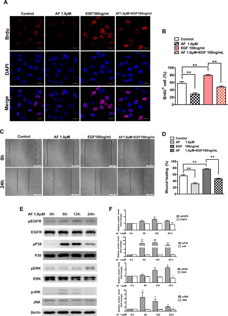
In Fig 5, Fig 5C shows the incorrect image under the “AF 1.0 μM” header for “0h”. Please view the correct Fig 5 here.
Fig 5. Auranofin inhibits EGF-dependent proliferation and migration of ARPE-19 cells.
(A) Immunofluorescence microphotographs of proliferating ARPE-19 cell with BrdU (red) and DAPI (blue) staining after ARPE-19 cells were untreated or treated with AF (1.0 μM) in the absence or presence of EGF (100ng/ml) for 24 hours and then subjected to BrdU labeling for 4 hours, followed by immunostaining with anti-BrdU antibody and DAPI. Scale bar = 20μm. (B) Quantitation data of the number of BrdU+ cells shown in panel A. (C) ARPE-19 cells were subjected to wound healing assay, and then were left untreated or treated with AF (1.0 μM) in the absence or presence of EGF (100 ng/ml) for 24 hours. Scale bar = 100μm. (D) Quantitation of the results shown in panel C. (E) ARPE-19 cells were treated with 1.0 μM AF for 6, 12 and 24 hours. Cell lysates were subjected to Western blot for determination of total and phosphorylated EGFR, P38MAPK, ERK and JNK proteins. β-actin was used as a loading control. (F) Quantitative data of Western blot results shown in panel E from three experiments. The levels of the phosphorylated protein were compared with the control, * P < 0.05, ** P < 0.01. The levels of the total protein were compared with the control, ※P < 0.05, ※※P < 0.01. All data are mean ± SEM.
Reference
- 1.Chen X, Tzekov R, Su M, Hong H, Min W, Han A, et al. (2016) Auranofin Inhibits Retinal Pigment Epithelium Cell Survival through Reactive Oxygen Species-Dependent Epidermal Growth Factor Receptor/ Mitogen-Activated Protein Kinase Signaling Pathway. PLoS ONE 11(11): e0166386 doi:10.1371/journal.pone.0166386 [DOI] [PMC free article] [PubMed] [Google Scholar]


