Since 1970s the evaluation and treatment of bile duct lesions have been accomplished mostly by endoscopic retrograde cholangiopancreatography (ERCP). However, neither ERCP nor other imaging procedures (e.g., endoscopic ultrasound) allow for safe diagnosis of these lesions. Since the emergence of the fiberoptic SpyGlass Direct Visualization System (SDVS) (Boston Scientific, Natick, Massachusetts, USA) in 2006 the sensitivity for detecting cholangiocarcinoma has increased, thereby establishing its superiority over standard ERCP [1]. ERCP-guided cholangiopancreatoscopy with SDVS allows for direct visualization of the bile ducts, tissue sampling and therapeutic maneuvers [1,2]. The impediment of the initial fiberoptic technology has been overcome by the introduction of a new digital SDVS, providing sensitivity and specificity of 90% and 95.8%, respectively, for visual impression of malignancy diagnosis [3].
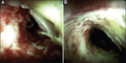
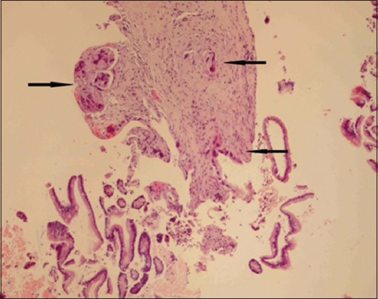
A 51-year-old man was referred to our department due to painless obstructive jaundice for further evaluation with ERCP. He had undergone magnetic resonance cholangiopancreatography (MRCP) which had shown dilation of the common bile duct (CBD diameter ~1.4 cm) and raised suspicion for stenosis in the lower part of CBD. He underwent an ERCP and endoscopic sphincterotomy. A stenosis in the lower part of CBD was observed. Brush cytology was performed, and a stent was placed in CBD. The patient was discharged due to clinical and laboratory improvement (brush cytology was negative for neoplasia). Fifteen days later he was re-admitted for further evaluation and he underwent a second ERCP and digital choledochoscopy with SDVS which showed nodular mucosa with ulceration and mucin containing material in the lower part of CBD (Fig. 1), highly indicative of malignancy. Biopsies obtained under direct visualization from the lesion showed extensive fibrosis and focal infiltration by moderately to poorly differentiated adenocarcinoma (Fig. 2).
Figure 1.

(A and B) Stenosis of the common bile duct presenting nodular mucosa with ulceration
Figure 2.

Biopsy of the common bile duct, showing extensive fibrosis and focal infiltration of the bile duct wall by small foci of moderately to poorly differentiated adenocarcinoma (black arrows). H&E, X200
Biography
Venizeleion General Hospital, Heraklion, Greece
Footnotes
Conflict of Interest: None
References
- 1.Chen YK, Pleskow DK. SpyGlass single-operator peroral cholangiopancreatoscopy system for the diagnosis and therapy of bile-duct disorders: a clinical feasibility study (with video) Gastrointest Endosc. 2007;65:832–841. doi: 10.1016/j.gie.2007.01.025. [DOI] [PubMed] [Google Scholar]
- 2.Theodoropoulou A, Vardas E, Voudoukis E, et al. SpyGlass direct visualization system facilitated management of iatrogenic biliary stricture: a novel approach in difficult cannulation. Endoscopy. 2012;44(Suppl 2 UCTN):E433–E434. doi: 10.1055/s-0032-1325857. [DOI] [PubMed] [Google Scholar]
- 3.Navaneethan U, Hasan MK, Kommaraju K, et al. Digital, single-operator cholangiopancreatoscopy in the diagnosis and management of pancreatobiliary disorders: a multicenter clinical experience (with video) Gastrointest Endosc. 2016;84:649–655. doi: 10.1016/j.gie.2016.03.789. [DOI] [PubMed] [Google Scholar]


