Abstract
Human adenovirus 7 (HAdV-7) strains are a major cause of acute respiratory disease (ARD) among adults and children, associated with fatal pneumonia. An ARD outbreak caused by HAdV-7 that involved 739 college students was reported in this article. To better understand the underlying cause of this large-scale epidemic, virus strains were isolated from infected patients and sequence variations of the whole genome sequence were detected. Evolutionary trees and alignment results indicated that the major capsid protein genes hexon and fibre were strongly conserved among serotype 7 strains in China at that time. Instead, the HAdV-7 strains presented three thymine deletions in the virus associated RNA (VA RNA) II terminal region. We also found that the mutation might lead to increased mRNA expression of an adjacent gene, L1 52/55K, and thus promoted faster growth. These findings suggest that sequence variation of VA RNA II gene was a potential cause of such a severe HAdV-7 infection and this gene should be a new-emerging factor to be monitored for better understanding of HAdV-7 infection.
Introduction
Human adenoviruses (HAdVs) are pathogenic viruses that can usually cause acute respiratory disease (ARD), acute follicular conjunctivitis, haemorrhagic cystitis, gastroenteritis, myocarditis, meningoencephalitis, and even death [1]. Outbreaks of adenovirus infection had been reported worldwide and are recognized as the leading cause of febrile illness and respiratory diseases [2]. Based on serology, genome sequencing, and biological characteristics, HAdVs have been classified into seven species (HAdV-A to G), which include more than 64 serotypes [3]. Different genotypes have been associated with distinct clinical symptoms. For instance, respiratory diseases have been linked mostly to HAdV-B serotypes 3, 7, and 14, and HAdV-C serotypes 1, 2, 5, and 6 [4, 5]. Among these serotypes, HAdV-7 is frequently associated with severe respiratory disease [6]. Numerous reports have covered the consequences of HAdV-7-induced epidemics among children and in crowded spaces. These include febrile pharyngitis and pneumonia accompanied by acute respiratory disease, some of which have been associated with fatal outcomes [7].
The severity of HAdV-7 infection depends on many factors, such as virus virulence, viral transmission environment, or physical condition of the host, and dictates whether occurrence is sporadic vs. epidemic, or mild vs. severe [8–10]. Genetic alterations are the leading cause of changes in the virulence of HAdV-7, as can be seen in the new emerging HAdV-7 strains with genomic variants such as SmaI mutation and VA RNA gene deletions, and could increase the severity of illnesses [11, 12]. A better understanding of viral genetic changes will improve the prediction and prevention of future ARD outbreaks caused by HAdV-7.
Traditionally, capsid hexon and fibre genes have been used to detect genomic variation, as they also determine the HAdV serotypes. However, according to recent reports, the hexon and fibre genes of HAdV-7 were remarkably conserved across time and space [13]. With the application of whole genome sequencing, two HAdV-7 strains identified in separate ARD outbreaks contained a 12-bp deletion in the VA RNA II gene, even though hexon, fibre, and other genes were highly conserved [12]. Nevertheless, there is no clear evidence whether the VA RNA II gene mutations contributed to past HAdV-7 outbreaks.
Here, we report a large-scale outbreak of severe acute respiratory disease associated with HAdV-7 that occurred in Wuhan, China, in 2015. To assess whether this outbreak was related to genetic variation of the VA RNA gene of the incriminated HAdV-7 strain, we analysed the whole genome sequence variation and viral growth of the Wuhan isolates. Knowledge of a link between VA RNA II genetic variation and HAdV-7 virulence may help us understand the function of this gene and take effective measures to control HAdV-7 prevalence.
Materials and methods
Outbreak investigation
An outbreak of ARD was reported in a college in Wuhan city, Hubei Province, between the 20th of January and 21st of February, 2015. Health officials, medical staff, epidemiologists, and laboratory technicians were designated as an epidemic prevention team to investigate and control the outbreak. The names, onset times, clinical symptoms and informed consent for sample collection and subsequent research usage of the students were documented by trained medical staff and epidemiologists. Copies of the case records of the hospitalized students were provided by the corresponding doctors. All the protocols used in these investigations were approved by the Institutional Review Boards of the Centre of Disease Control and Prevention of China, and the Ethics Committee of Wuhan General Hospital.
Sample collection, detection, and serotyping
Nasopharyngeal swabs of the patients were collected using 5-mL collection tubes (Yocon, China). Pure Link Viral RNA/DNA Mini Kits (Invitrogen, Carlsbad, CA, USA) were used to extract nucleic acids. Suspected pathogens, such as adenovirus, coronavirus, parainfluenza virus, influenza A virus, influenza B virus, respiratory syncytial virus A, Bordetella pertussis, Legionella pneumophila, Chlamydophila pneumoniae, Streptococcus pneumoniae, Haemophilus influenzae, and Mycoplasma pneumoniae were detected with Seeplex®RV12 ACE and Seeplex® Pneumabacter detection kits (Seegene, Seoul, Korea). After confirmation of HAdV infection, real-time PCR primers for HAdV serotypes 7, 11, 14, and 55 were used for serotyping (S1 Table).
Virus isolation and gene sequence analysis
The samples of nasopharyngeal swabs were inoculated into A549 cells cultured in DMEM. The viruses were isolated by repeated freeze-thaw and preserved in -80°C. Primer sequences used for the amplification of hexon, fibre and VA RNA genes have been published elsewhere [14]. The whole genome was sequenced using Dideoxyterminator Sanger sequence analysis and primers used were listed in S2 Table. The sequences were aligned with MUSCLE within MEGA6.05 software. Neighbour-joining methods were adopted to construct phylogenetic trees and 1000 bootstrap replicates were used to evaluate their topological accuracy.
Detection of L1 52/55K mRNA
Viral RNA was extracted using an Ultrapure RNA extraction kit (DNase I included, CW0597; KangWei Biotech, China). The primers used for the detection of L1 52/55K mRNA were F: 5’-TGCAAAGAGCTCTAACGGGG-3’ and R: 5’-CTTCCAAGTACTCGCCCTCC-3’. Reference genes VA RNA II and E1 were detected using primers VA-F: 5’-GAGCCAGTGCTGCGTCAA-3’, VA-R: 5’- TTAGAAACGTCGCGGAAA-3’; and E1-F: 5’-CCACCTACGCTGCACGAT-3’, E1-R: 5’- ACCTCTGCCGCTTTCCAC-3’, respectively. KAPA SYBR FAST qPCR Kits (KK4601; Kapa Biosystems, Woburn, MA, USA) were used for amplification.
Virus growth curve
Viruses (1 × 10² genome copies) were propagated in human A549 cells cultured in 12-well plates and were collected 6, 9, 12, 18, 21, 24, 36, 48, and 72 h after infection. The cultures were subjected to repeated freezing and thawing thrice and centrifuged at 12000×g for 10 min. Then, 200 μl of supernatants were used for DNA extraction using Pure Link Viral DNA Mini Kits and virus titres were determined by detecting the number of viral E1 gene copies by real-time PCR. The E1 gene fragment was cloned into the T vector (pEASY-T1 Simple Cloning Kit, CT111-02; TransGen Biotech, Beijing, China) and diluted to 0.5 × 10²–0.5 × 10⁹ copies/μL as standard samples.
Statistical analysis
All experiments were performed at least three times. The data are presented as means ± standard error of the mean (SEM) and were analysed using Student’s t-test. Two-tailed P-values < 0.05 were considered statistically significant.
Results
Epidemiological and clinical characteristics
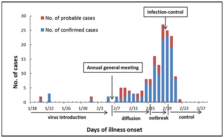
In 2015, an outbreak of acute respiratory disease occurred in a college in Wuhan, China. Of 4113 students, 828 (20.1%) developed the illness and 169 of them were admitted to Wuhan General Hospital between the 20th of January and 21st of February, 2015 (Fig 1). The average age of the hospitalized students was 19 years (between 17 and 24 years). In this study, we took the 169 hospitalized cases for subsequent epidemiological and clinical investigation. The development of the outbreak unfolded as follows. From late January to early February, a few sporadic cases were hospitalized. Then, after an annual college general meeting on the 6th of February, the virus began to spread and led to an outbreak. Control measures, such as isolation, disinfection, and medication, were introduced on the 19th of February. From that point, the number of new infections decreased gradually and after the 23rd of February, no additional patients required hospitalization.
Fig 1. Number of ARD patients admitted to hospital and confirmed cases of HAdV-7 infection in this outbreak.
According to case reports, patients presented typical respiratory infection symptoms, such as fever (92.3%), cough (83.4%), sore throat (62.1%), swelling of tonsils (47.9%), and a runny nose (26.0%). Among them, 92 patients (54.4%) were diagnosed with upper respiratory infection, 77 (45.6%) with pneumonia (Table 1), and 121 (71.6%) presented fever above 39°C. In addition, nearly half (n = 37) of the patients suffering from swollen tonsils (n = 81) presented severely enlarged tonsils. Finally, there were also 17 cases of acute bilateral pneumonia.
Table 1. Characteristics of HAdV infected patients.
| Characteristic | Value (n = 169) | |
|---|---|---|
| Clinical symptom | ||
| Cough | 141 (83.4%) | |
| Sore throat | 105 (62.1%) | |
| Expectoration | 104 (61.5%) | |
| Headache | 48 (28.4%) | |
| Weak | 65 (38.5%) | |
| Pharyngeal lymphoid follicular hyperplasia | 11 (6.5%) | |
| Purulent exudate on the tonsil | 9 (5.3%) | |
| Running nose | 44 (26.0%) | |
| Myalgia | 45 (26.6%) | |
| Diarrhoea | 18 (10.7%) | |
| Fever | 40–41°C | 26 (15.4%) |
| 39–39.9°C | 95 (56.2%) | |
| 38–38.9°C | 30 (17.8%) | |
| <38°C | 5 (3.0%) | |
| Total | 156 (92.3%) | |
| Body function indexes | ||
| WBC count (109 cells/litre) | >10 | 34 (20.1%) |
| <4 | 19 (11.2%) | |
| Lymphocyte % (>0.4%) | 168 (99.4%) | |
| Neutrophil % (>0.7%) | 169 (100%) | |
| Bradycardia (<60 times/min) | 4 (2.4%) | |
| Tachycardia (>100 times/min) | 52 (30.8%) | |
| Tachypnea (>20 times/min) | 6 (3.6%) | |
| Diagnosis | ||
| Upper respiratory infection | 92 (54.4%) | |
| Pneumonia | 77 (45.6%) | |
| Severe cases (admission to intensive care units) | 8 (4.7%) | |
Virus detection and analysis of viral gene sequences
Of the 4113 students, 739 were identified as HAdV-7-positive by real-time PCR. The ratio among hospitalized patients was 76.9% (130 out of 169). Twenty viral strains were isolated from hospitalized positive patients and cultured in the laboratory; after which hexon, fibre, and VA RNA genes were amplified and sequenced. Alignments of these genes of the isolated 20 virus produced 100% similarity.
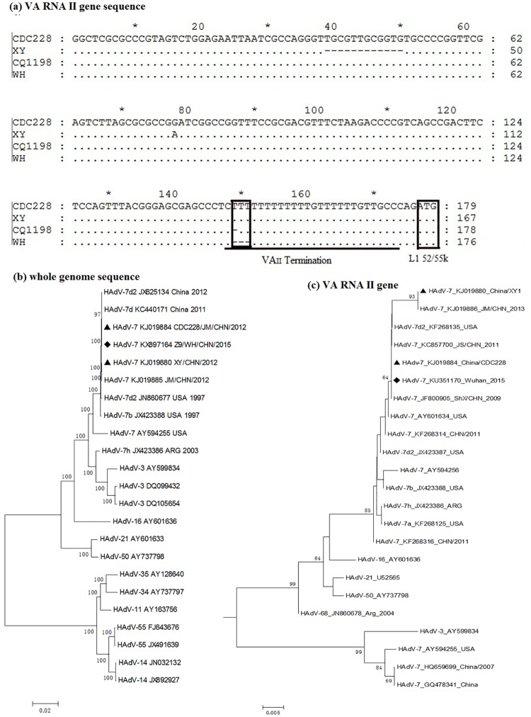
Next, we used the BLASTn tool to find the strains with closest relation to the sequenced hexon, fibre, and VA RNA genes. Both hexon and fibre genes showed 100% identity with the HAdV-7 isolate CQ1198 (GenBank accession number JX625134), which belongs to the HAdV-7d type [10]. The VA RNA gene (GenBank accession number KU351170) also showed the highest identity to this HAdV-7 strain, but it lacked two thymines (T). A comparison of the Wuhan strains with strains CDC228 (KJ019884) and XY1 (KJ019880) previously isolated by our laboratory [12], revealed that the former had three fewer T in the VAII terminal region (Fig 2).
Fig 2. Alignment and phylogenetic analysis of VA RNA II gene and whole genome.
(a) Alignment of VA RNA II gene sequences from different HAdV-7 strains. The empty black box in the VAII region indicates the sites of missing T. The empty black box to the right indicates the start codon of the L1 52/55K gene. Phylogenetic tree corresponding to HAdV-7 (b) whole genome and (c) VA RNA II gene sequences is presented. The Wuhan strain reported in this article is marked by (♦), whereas the reference strains CDC228 and XY1 are indicated by (▲). The scale bar indicates units of nucleotide substitutions per site.
Then, we chose the Z9/WH strain (GenBank No. KX897164) of these Wuhan isolates as representative to make whole-genome sequencing, and the comparison result with strain CDC228 revealed that the Z9/WH strain had some other but insignificant mutations except for VA RNA gene (Table 2).
Table 2. Comparative genomic analysis of the outbreak Z9/WH strain with strain CDC228.
| Region | Location(CDC228) | Gene | Nucleotide Change (amino acid substitution) | |
|---|---|---|---|---|
| CDC228 | Z9/WH | |||
| E1B | 1631 | 20 kDa small T antigen | G(W) | C(C) |
| E2B | 5069 | DNA polymerase | Ta | C |
| 7842 | T(243rdE) | G(A) | ||
| 7882 | G(230thL) | C(V) | ||
| 8057 | G | A | ||
| 8081 | G | A | ||
| L1 | 10748 | VAII | TTT | –b |
| L2 | 15453 | non-coding region | AAAAAA | – |
| E3 | 28601 | 20.3 kDa glycoprotein | A(I) | G(V) |
aThe bases without brackets denote silent mutations.
b“–” denotes deletion mutation.
Phylogenetic tree construction and analysis
Phylogenetic trees based on hexon, fibre, and VA RNA gene sequences were constructed respectively. As all sequences were identical, one sample Wuhan strain (KU351170) was taken as representative. And the Z9/WH strain (GenBank No. KX897164) was chosen for the whole-genome sequence and DNA polymerase gene analysis (Figs A and B in S1 Appendix).
According to the phylogenetic trees, the hexon and fibre genes of the Wuhan strains clustered with the HAdV-7d2 strain CQ1198 (JX625134) and other HAdV-7 strains found in China in recent years. The DNA polymerase gene and whole genome sequence of HAdV-7 found in China in recent years were located within the same branch but with a little difference. And the VA RNA I sequences from all serotype 7 strains clustered together and the sequences were highly conserved except for occasional mutations in the termination region (Figs B and C in S1 Appendix). In contrast, the VA RNA II genes were quite variable even within the same serotype (Fig 2).
Viral L1 52/55K gene expression
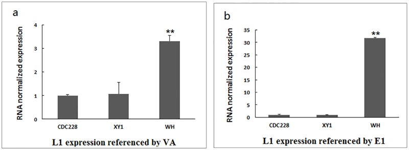
We analysed the expression of the viral gene L1 52/55K using VA RNA and E1 as reference genes. Compared with strains CDC228 and XY1, the representative Wuhan strain displayed substantially higher L1 52/55K expression. L1 52/55K gene expression did not differ between CDC228 and XY1 strains (Fig 3), even though the latter had 12 fewer bases in the VA RNA region than the former [12].
Fig 3. L1 52/55K mRNA expression assay using real-time PCR.
L1 52/55K mRNA expression against (a) VA RNA, n = 3, mean ± SEM, **P = 0.0039 vs. CDC228; and (b) E1, n = 3, mean ± SEM, **P = 0.0098 vs. CDC228. CDC228, XY1, and WH correspond to KJ019884, KJ019880, and Wuhan strains, respectively.
Growth kinetics of viral replication
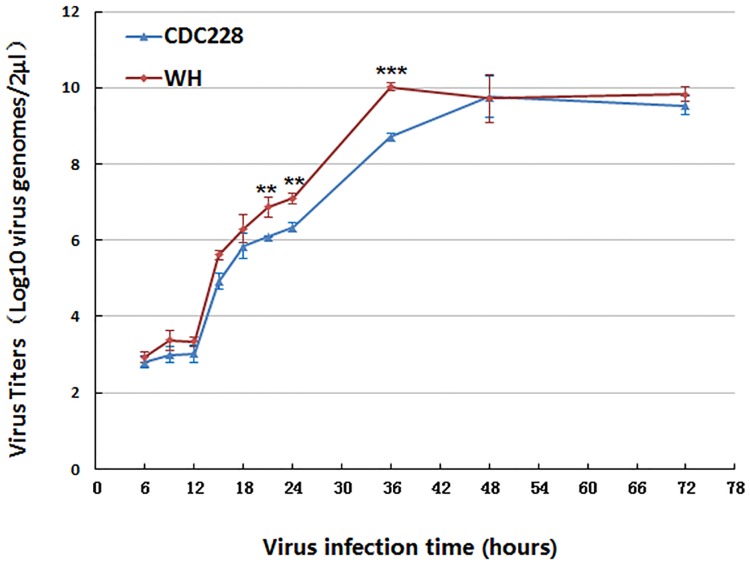
Finally, we compared viral replication kinetics between the CDC228 strain and one of the Wuhan strains (Fig 4). The latter displayed faster growth than CDC228, which contains a complete VA RNA II terminal region.
Fig 4. Growth kinetics of Wuhan and CDC228 strains.
Virus titres were determined in human A549 cells cultured in 12-well plates by detecting virus genomes using real-time PCR (n = 3, mean ± SEM, **P ≤ 0.01, ***P ≤ 0.001 vs. CDC228).
Discussion
Different HAdV serotypes have been shown to be prevalent in different geographic areas [15–18], and different types of HAdV infection have been related to various levels of severity [19–22]. HAdV-B types (HAdV-3 and HAdV-7) can cause respiratory tract infection and result in severe pneumonia and death [7, 23]. Under special conditions, such as fatigue and crowding, HAdV-7 can cause outbreak of acute respiratory disease [6, 24].
Here, we report an outbreak of ARD caused by HAdV-7 among young college students. According to clinical characteristics, this outbreak brought 169 inpatients with poor conditions, like fever above 39°C (71.6%), pneumonia (45.6%) and 8 ICU (intensive care unit) cases. To understand the reason behind this large scale outbreak, genomic variations were detected in our article.
Hexon and fibre capsid proteins are the main antigenic determinants of HAdV serotypes. In a previous work on two other HAdV-7 outbreaks, we found that most genes had few or no mutations besides those in VA RNA genes, which displayed a 12-bp deletion [12]. Therefore, here, we analysed the sequences of hexon, fibre, and VA RNA genes primarily. The hexon, fibre, and VA RNA I genes clustered quite intensely within serotype 7 in the phylogenetic trees, suggesting the conserved prevalence of HAdV-7 in China in recent years. However, the VA RNA II gene had undergone several mutations, even within the same serotype. Besides, the whole genome sequence analysis barely showed any extra mutations, except for two amino acids substitutions in the DNA polymerase gene, which were not located within the functional regions of DNA polymerase [25]. Therefore, more attention should be paid to the VA RNA II gene for future monitoring of HAdV-7 infection.
How the lost T in the VA RNA II terminal region played influence on HAdV-7 is still unknown. Interestingly, the HAdV VA RNA genes are located right in front of the L1 52/55K gene [26], which plays an important role in forming mature, infectious virions [27, 28]. In fact, we show that the start codon of L1 52/55K mRNA is located only two nucleotides after the terminal region of VA RNA II (Fig 2). We first tried to measure the expression ratio between the VA RNA II and L1 52/55K gene of different HAdV-7 strains. However, given the possibility that the mutation in the VA RNA II gene might alter the expression of the gene itself, we chose instead to assess the expression of L1 52/55K against that of E1A. Being the first expressed early gene [29], E1A could reflect the expression of HAdV genes and avoid being influenced by late genes. We show that L1 52/55K mRNA was up-regulated compared with strains CDC228 and XY1, confirming the possibility that deletion in the terminator of VAII could exert an effect on this adjacent gene. The terminal region of VA RNAs is characterized by a cluster of T surrounded by a GC-rich stretch [30]. This region is transcribed by RNA polymerase III [31], which is prone to misreading the termination sequence and read through the next genes [30]. Accordingly, the loss of T in the terminator might destabilize the secondary structure of the terminal stem and increase the chance of continued transcription of L1 52/55K by RNA polymerase III. Considering the role of L1 52/55K protein in promoting the formation of infectious virions, the up-regulation of L1 52/55K might explain the rapid growth of the Wuhan strain and this outbreak. In addition, a previous report suggested that the L1 52/55K gene, which recombined from HAdV-B16, was a reason for the virulence of HAdV-7 strains predominated in China [32]. The WH strain identified in this outbreak shared 100% identity with this reported L1 52/55K gene. Thus, the up-regulated expression of this L1 52/55K gene could lead to increased virulence of WH strain.
In summary, we report here an uncommon outbreak of HAdV-7 among college students in Wuhan, China, in 2015. Sequence analysis revealed several missing T in the VA RNA II terminal region. This mutation, in turn, caused up-regulation of an adjacent gene, L1 52/55K. The latter promoted faster growth of the HAdV-7 strain, thus providing a causal link between the VA RNA II gene and a severe clinical outcome. Taking into account the phylogenetic results on the conserved nature of HAdV-7 hexon and fibre genes in China in recent years, VA RNA genes represent a new determinant factor for HAdV-7 virulence upon infection. However, further work, such as functional verification of the missing T bases by constructing a deletion mutant of strain CDC228 or reverse mutant of the WH strain, should be done to elucidate the function of the VA RNA II gene and its relationship to the increased growth capacity of the HAdV-7 strain.
Supporting information
Fig A. Phylogenetic analysis of HAdV-7 hexon and fibre genes. Fig B. Phylogenetic analysis of DNA polymerase gene and VA RNA I gene of HAdV-7. Fig C. Sequence alignment of HAdV-7 VA genes. Fig D. Sequence alignment of VA II gene of HAdVs.
(DOCX)
(DOCX)
(DOCX)
Data Availability
All relevant data are within the paper and its Supporting Information files.
Funding Statement
This work was supported by the Mega-projects of Science and Technology Research (nos. 2012ZX10004215 and AWS15J006) that received by HBS, who participated in study design and decision to publish. And the National Nature Science Foundation of China (nos. 81373053 and 81371854), and the Beijing Science and Technology Nova program (no. xx2013061) received by SFQ, who participated in study design and decision to publish.
References
- 1.William S. Fields Virology In: David M, editors. Adenoviruses, Lippincott Williams & Wilkins; 2007. pp. 2405–2409. [Google Scholar]
- 2.Yusof MA, Rashid TR, Thayan R, Othman KA, Hasan NA, Adnan N, et al. Human adenovirus type 7 outbreak in Police Training Center, Malaysia, 2011. Emerging Infectious Diseases. 2012;18:852–854. 10.3201/eid1805.110865 [DOI] [PMC free article] [PubMed] [Google Scholar]
- 3.Seto D, Chodosh J, Brister JR, Jones MS. Using the whole-genome sequence to characterize and name human adenoviruses. Journal of Virology. 2011;85:5701–5702. 10.1128/JVI.00354-11 [DOI] [PMC free article] [PubMed] [Google Scholar]
- 4.Echavarria M, Sanchez JL, Kolavic-Gray SA, Polyak CS, Mitchell-Raymundo F, Innis BL, et al. Rapid detection of adenovirus in throat swab specimens by PCR during respiratory disease outbreaks among military recruits. Journal of Clinical Microbiology. 2003;41:810–812. 10.1128/JCM.41.2.810-812.2003 [DOI] [PMC free article] [PubMed] [Google Scholar]
- 5.Washington C, Metzgar D, Hazbon MH, Binn L, Lyons A, Coward C, et al. Multiplexed Luminex xMAP assay for detection and identification of five adenovirus serotypes associated with epidemics of respiratory disease in adults. Journal of Clinical Microbiology. 2010;48:2217–2222. 10.1128/JCM.00029-10 [DOI] [PMC free article] [PubMed] [Google Scholar]
- 6.Tsou TP, Tan BF, Chang HY, Chen WC, Huang YP, Lai CY. Community outbreak of adenovirus, Taiwan, 2011. Emerging Infectious Diseases. 2012;18:1825–1832. 10.3201/eid1811.120629 [DOI] [PMC free article] [PubMed] [Google Scholar]
- 7.Yamamoto D, Okamoto M, Lupisan S, Suzuki A, Saito M, Tamaki R, et al. Impact of human adenovirus serotype 7 in hospitalized children with severe fatal pneumonia in the Philippines. Japanese Journal of Infectious Diseases. 2014;67:105–10. [DOI] [PubMed] [Google Scholar]
- 8.Cui XY, Wen L, Wu ZH, Liu N, Yang CJ, Liu W, et al. Human adenovirus type 7 infection associated with severe and fatal acute lower respiratory illness and nosocomial transmission. Journal of Clinical Microbiology. 2015;53:746–749. 10.1128/JCM.02517-14 [DOI] [PMC free article] [PubMed] [Google Scholar]
- 9.Tang L, Wang L, Tan X, Xu W. Adenovirus serotype 7 associated with a severe lower respiratory tract disease outbreak in infants in Shaanxi Province, China. Virology Journal. 2011;8:23 10.1186/1743-422X-8-23 [DOI] [PMC free article] [PubMed] [Google Scholar]
- 10.Zhao S, Wan C, Ke C, Seto J, Dehghan S, Zou L. Re-emergent human adenovirus genome type 7d caused an acute respiratory disease outbreak in Southern China after a twenty-one year absence. Scientific Reports. 2014;4:7365 10.1038/srep07365 [DOI] [PMC free article] [PubMed] [Google Scholar]
- 11.Calder JA, Erdman DD, Ackelsberg J, Cato SW, Deutsch VJ, Lechich AJ, et al. Adenovirus type 7 genomic-type variant, New York city, 1999. Emerging Infectious Diseases. 2004;10:149–152. 10.3201/eid1001.020605 [DOI] [PMC free article] [PubMed] [Google Scholar]
- 12.Qiu S, Li P, Liu H, Wang Y, Liu N, Li C. Whole-genome sequencing for tracing the transmission link between two ARD outbreaks caused by a novel HAdV serotype 7 variant, China. Scientific Reports. 2015;5:13617 10.1038/srep13617 [DOI] [PMC free article] [PubMed] [Google Scholar]
- 13.Chen M, Zhu Z, Huang F, Liu DL, Zhang TG, Ying D, et al. Adenoviruses associated with acute respiratory diseases reported in Beijing from 2011 to 2013. PloS One. 2015;10:e0121375 10.1371/journal.pone.0121375 [DOI] [PMC free article] [PubMed] [Google Scholar]
- 14.Lin YC, Lu PL, Lin KH, Chu PY, Wang CF, Lin JH, et al. Molecular epidemiology and phylogenetic analysis of human adenovirus caused an outbreak in Taiwan during 2011. PloS One. 2015;10:e0127377 10.1371/journal.pone.0127377 [DOI] [PMC free article] [PubMed] [Google Scholar]
- 15.Sabrina JM, Kurt H, Bjom B, Oyvind K, Svein AN, Samuel M. Prevalence and molecular characterisation of human adenovirus in diarrhoeic children in Tanzania; a case control study. BMC Infectious Diseases. 2014;14:666 10.1186/s12879-014-0666-1 [DOI] [PMC free article] [PubMed] [Google Scholar]
- 16.Pola ND, Katherine CH, Adriana K, Rania S, Amel MNH, Amany ES, et al. Molecular identification of adenoviruses associated with respiratory infection in Egypt from 2003 to 2010. BMC Infectious Diseases. 2014;14:50 10.1186/1471-2334-14-50 [DOI] [PMC free article] [PubMed] [Google Scholar]
- 17.Satoshi H, Saeko M, Kazuo T, Jun K, Tetsuo K. Molecular epidemiology of human adenoviruses D associated with epidemic keratoconjunctivitis in Osaka, Japan, 2001–2010. The Journal of Infectious Diseases. 2013;66:436–438. [DOI] [PubMed] [Google Scholar]
- 18.Punsinee S, Thaweesak C, Chanpim V, Viboonsuk V, Apiradee T, Yong P. Molecular characterization of human adenovirus infection in Thailand, 2009–2012. Virology Journal. 2013;10:193 10.1186/1743-422X-10-193 [DOI] [PMC free article] [PubMed] [Google Scholar]
- 19.Elias H, Daniela H, Tina G, Robert B, Thomas FS, Albert H. A human adenovirus species B subtype 21a associated with severe pneumonia. The Journal of Infection. 2014;69:490–499. 10.1016/j.jinf.2014.06.015 [DOI] [PubMed] [Google Scholar]
- 20.Sun B, He HY, Wang Z, Qu JX, Li XY, Ban CJ. Emergent severe acute respiratory distress syndrome caused by adenovirus type 55 in immunocompetent adults in 2013: a prospective observational study. Critical Care. 2014;18:456 10.1186/s13054-014-0456-6 [DOI] [PMC free article] [PubMed] [Google Scholar]
- 21.Cassir N, Hraiech S, Nougairede A, Zandotti C, Fournier PE, Papazian L. Outbreak of adenovirus type 1 severe pneumonia in a French intensive care unit, September-October 2012. Surveillance and Outbreak Reports. 2014;19:39. [DOI] [PubMed] [Google Scholar]
- 22.Lessa FC, Gould PL, Pascoe N, Erdman DD, Lu X, Bunning ML. Health care transmission of a newly emergent adenovirus serotype in health care personnel at a military hospital in Texas, 2007. The Journal of Infectious Diseases. 2009;200:1759–1765. 10.1086/647987 [DOI] [PubMed] [Google Scholar]
- 23.Gershon A, Edward C, Maureen F, Stanley AP. Outbreak of fatal adenoviral type 7a respiratory disease in a children's long-term care inpatient facility. American Journal of Infection Control. 1986;14:188–190. [DOI] [PubMed] [Google Scholar]
- 24.Dean DE, Xu W, Susan IG, Gray GC, David S, Adriana EK. Molecular epidemiology of adenovirus type 7 in the United States, 1996–2000. Emerging Infectious Diseases. 2002;8:269–277. [DOI] [PMC free article] [PubMed] [Google Scholar]
- 25.Liu H, James HN & Ronald TH. Identification of conserved residues contributing to the activities of adenovirus DNA polymerase. Journal of Virology, 2000, 74: 11681–11689. [DOI] [PMC free article] [PubMed] [Google Scholar]
- 26.Bayar T, Cary W, Robert JS, Thomas S. Adenovirus VAI RNA is required for efficient translation of viral mRNAs at late times after infection. Cell. 1982;31:543–551. [DOI] [PubMed] [Google Scholar]
- 27.Tama BH, David AO, Thomas S. Adenovirus L1 52-and 55-kilodalton proteins are present within assembling virions and colocalize with nuclear structures distinct from replication centers. Journal of Virology. 1992;66:6133–6142. [DOI] [PMC free article] [PubMed] [Google Scholar]
- 28.Perez-Romero P, Gustin KE, Imperiale MJ. Dependence of the encapsidation function of the adenovirus L1 52/55-kilodalton protein on its ability to bind the packaging sequence. Journal of Virology. 2006;80:1965–1971. 10.1128/JVI.80.4.1965-1971.2006 [DOI] [PMC free article] [PubMed] [Google Scholar]
- 29.Joseph RN, Harold SG, Blanchard JM, Michael CW, Jame ED. Regulation of the primary expression of the early adenovirus transcription units. Journal of Virology. 1979;32:727–733. [DOI] [PMC free article] [PubMed] [Google Scholar]
- 30.Daniel FB, Donald DB. Nucleotide sequences in Xenopus 5s DNA required for transcription termination. Cell. 1981;24:261–270. [DOI] [PubMed] [Google Scholar]
- 31.Ma Y, Michael BM. Structure, function, and evolution of adenovirus-associated RNA: a phylogenetic approach. Journal of Virology. 1996;70:5083–5099. [DOI] [PMC free article] [PubMed] [Google Scholar]
- 32.Yu Z, Zeng Z, Zhang J, Pan Y, Chen M, Guo Y, et al. Fatal community-acquired pneumonia in children caused by re-emergent human adenovirus 7d associated with higher severity of illness and fatality rate. Scientific Reports. 2016;6:37216 10.1038/srep37216 [DOI] [PMC free article] [PubMed] [Google Scholar]
Associated Data
This section collects any data citations, data availability statements, or supplementary materials included in this article.
Supplementary Materials
Fig A. Phylogenetic analysis of HAdV-7 hexon and fibre genes. Fig B. Phylogenetic analysis of DNA polymerase gene and VA RNA I gene of HAdV-7. Fig C. Sequence alignment of HAdV-7 VA genes. Fig D. Sequence alignment of VA II gene of HAdVs.
(DOCX)
(DOCX)
(DOCX)
Data Availability Statement
All relevant data are within the paper and its Supporting Information files.


