Abstract
Background
Group 2 and 3 grass pollen allergens are major allergens with high allergenic activity and exhibit structural similarity with the C-terminal portion of major group 1 allergens. In this study, we aimed to determine the crystal structure of timothy grass pollen allergen, Phl p 3, and to study its IgE recognition and cross-reactivity with group 2 and group 1 allergens.
Methods
The three-dimensional structure of Phl p 3 was solved by X-ray crystallography and compared with the structures of group 1 and 2 grass pollen allergens. Cross-reactivity was studied using a human monoclonal antibody which inhibits allergic patients’ IgE binding and by IgE inhibition experiments with patients’ sera. Conformational Phl p 3 IgE epitopes were predicted with the algorithm SPADE, and Phl p 3 variants containing single point mutations in the predicted IgE binding sites were produced to analyze allergic patients’ IgE binding.
Results
Phl p 3 is a globular β-sandwich protein showing structural similarity to Phl p 2 and the Phl p 1–C-terminal domain. Phl p 3 showed IgE cross-reactivity with group 2 allergens but not with group 1 allergens. SPADE identified two conformational IgE epitope-containing areas, of which one overlaps with the epitope defined by the monoclonal antibody. The mutation of arginine 68 to alanine completely abolished binding of the blocking antibody. This mutation and a mutation of D13 in the predicted second IgE epitope area also reduced allergic patients’ IgE binding.
Conclusion
Group 3 and group 2 grass pollen allergens are cross-reactive allergens containing conformational IgE epitopes. They lack relevant IgE cross-reactivity with group 1 allergens and therefore need to be included in diagnostic tests and allergen-specific treatments in addition to group 1 allergens.
Keywords: allergen, conformational IgE epitopes, grass pollen allergy, specific immunotherapy, three-dimensional structure
Approximately 50% of allergic patients are sensitized to allergens from grass pollen which therefore belong to the most frequently recognized allergen sources (1). Due to heavy pollen production and release of allergen-bearing submicronic particles which can intrude in the lower respiratory tract, grasses do not only induce symptoms of allergic rhinoconjunctivitis but can also elicit severe asthma attacks (2, 3). It is therefore not surprising that grass pollen was among the first identified allergen sources (4) and that allergen-specific immunotherapy was conducted for the first time with grass pollen allergens (5). Using protein chemical and molecular approaches, most of the relevant grass pollen allergens have been characterized and their primary structures have been elucidated (6, 7). Group 1, group 2, and group 3 allergens are major grass pollen allergens against which more than 60% of the grass pollen allergic patients are sensitized (7). Since the initial purification of these allergens and the determination of their amino and nucleic acid sequences (8–10), it is known that group 2 and group 3 allergens are highly similar regarding sequence and three-dimensional structure to the C-terminal domain of group 1 allergens (11, 12). The three-dimensional structures of Phl p 2 and Phl p 3 were determined by nuclear magnetic resonance (NMR) and found to be very similar (11, 12). Group 1 allergens belong to a family of evolutionarily closely related proteins, designated as expansins, which are important for the growth of pollen tubes and are thought to originate from common ancestor genes (13). In fact, group 1 and group 2/3 allergens are major components in grass pollen with a percentage weight per total proteins of approximately 0.2 and 5, respectively (14).
Both group 1 and group 2 allergens are major allergens and, according to clinical provocation test studies in allergic patients, are highly allergenic and therefore need to be included in diagnostic tests and allergen-specific forms of treatment (15, 16). Recently, recombinant group 3 grass pollen allergens became available by molecular cloning, but their IgE cross-reactivity with group 2 and group 1 allergens has not yet been studied in detail using individual sera from grass pollen allergic patients (17, 18). Similar as in rye grass (Lolium perenne), group 2 and 3 allergens from timothy grass pollen, Phl p 2, and Phl p 3 exhibit only modest sequence identity and highly divergent isoelectric points (8, 9, 18). Structural similarity and IgE cross-reactivity among group 1, 2, and 3 allergens have not been compared in detail with respect to their potential relevance for diagnosis and specific immunotherapy.
Here, we solved the three-dimensional structure of the timothy grass pollen protein Phl p 3 by high-resolution X-ray crystallography and compared it with the X-ray structures of Phl p 2 (protein data bank, PDB: 1WHO) and with the C-terminal domains of Phl p 1 (PDB: 1N10) and two expansin family proteins, Zea m 1 (PDB: 2HCZ) (19) and YoaJ (PDB: 3D30) (20). Using grass pollen allergic patients’ sera, IgE cross-reactivity of Phl p 3 with group 1 and group 2 allergens was studied by means of IgE inhibition assays. Based on the structural comparisons and the IgE cross-reactivity data, we were able to use the recently developed SPADE algorithm (21) to delineate two major conformational IgE binding sites of Phl p 3 of which one was found to overlap with a major IgE binding site determined from the Phl p 2–IgE complex structure (22). Furthermore, we generated a panel of 24 Phl p 3 variants containing single point mutations to further study allergic patients’ IgE recognition. Our studies identify group 2 and group 3 allergens as a family of highly cross-reactive allergens containing similar conformational IgE epitopes which are immunologically distinct from group 1 allergens despite structural similarity to them. The high structural and immunological similarity of group 2 and 3 allergens indicates that these allergens can replace each other, but not group 1 allergens for diagnosis and specific immunotherapy.
Methods
Protein expression and purification
Phl p 3 was expressed in Escherichia coli (18) and purified from the cytosolic fraction using cation exchange chromatography (GE Healthcare, Uppsala, Sweden) at pH 8.0 and a linear salt gradient from 0 to 0.5 M NaCl. The final purification step was size exclusion chromatography (Superdex 75 HR 10/30; GE Healthcare) in 25 mM Tris pH 8.0, 150 mM NaCl. Recombinant Phl p 1, Phl p 2, Phl p 5, and Bet v 1 made without any N- or C-terminal modifications were purchased (Biomay AG, Vienna, Austria). The immunochemical equivalence of rPhl p 1 and rPhl p 2 with natural group 1 and 2 allergens, respectively, was shown (23). Both rPhl p 2 and rPhl p 3 represented folded nonglycosylated proteins (11, 12, 18). Recombinant Sec c 3 was expressed with a C-terminal His-tag and purified by nickel affinity chromotography to more than 95% purity. Human serum albumin (HSA) was bought from Behring (Marburg, Germany). The recombinant chimeric human Phl p 2-specific mAb containing the variable region of a human IgE Fab on a human IgG1 constant region was purified as described (22). YoaJ was kindly supplied by Paulette Charlier (University of Liège).
Recombinant variants of Phl p 3 containing point mutations of individual residues in the predicted epitopes of Phl p 3 were generated using a site-directed mutagenesis kit (Stratagene, CA, USA). Primers for the point mutants are listed in Table S1 in the Supporting Information. The PCR products were subcloned in the pPROEX-1 vector containing sequences coding for a 6-histidine N-terminal tag and a tobacco etch virus (TEV) cleavage site. Mutations were verified by DNA sequencing of each plasmid construct (Agowa genomics, Berlin, Germany). rPhl p 3 mutants were expressed in E. coli BL21 (DE3) cells in 500 ml liquid Luria-Bertani medium containing 100 mg/l ampicillin. Recombinant proteins were purified by affinity chromatography using His-trap HP columns (GE Healthcare) and size exclusion chromatography using Superdex 75 HR 10/30 (GE Healthcare; see supporting information).
Phl p 3 crystallization and structure determination
For crystallization, recombinant Phl p 3 in 25 mM Tris pH 8.0, 150 mM NaCl, was concentrated to 13.4 mg/ml and polyethylene glycol 3350 was used as precipitant. A Phl p 3 crystal was flash frozen in liquid nitrogen, and a data set collected to 1.8 Å at the X12 beamline (DESY, Hamburg). The structure was solved by molecular replacement (24) using Phl p 2 (PDB: 1WHO, unpublished) as search model. The model was refined to 1.8 Å, containing residues from 1 to 100 for all four molecules. The data (coordinates and diffraction data) were deposited in the protein data bank and were assigned the PDB-ID 3FT1. Details of the structure solution, model building, and refinement are summarized in the supporting information methods section and in Table S2.
Structural analysis and comparisons
The software PROMOTIF (25) was used for identifying prominent secondary structural elements like bulges and well-ordered loops in the protein crystal structures. The Phl p 3 structure was searched against all protein structures in the protein data bank using DALI (26) and VAST (27). FAST (28) and CLUSTALW (29) were used to calculate the root mean square deviation and sequence identity between related structures and their corresponding sequences, respectively. Solvent accessibility values for amino acids in the protein structure were calculated by ASAview (30) using a probe radius of 1.4 Å.
Phl p 3–IgE Fab complex and Sec c 3 Modeling
Sec c 3 from rye (C. Lupinek and R. Valenta, unpublished data) was modeled with Modeller V9.4 (31) using the Phl p 3 crystal structure (Chain A) as template. The Sec c 3 sequence was aligned to the Phl p 3 sequence using T-coffee (32). In each run of Modeller, ten models were generated and the model with least DOPE score was selected. Models were evaluated by the program ERRAT (33). The Phl p 3–IgE Fab complex was modeled based on the Phl p 2–IgE Fab cocrystal structure (PDB: 2VXQ). The Phl p 3 molecule (Chain A) was aligned with Phl p 2 in the Fab complex and docked with the IgE Fab molecule using the RossetaDock server (34).
Epitope prediction using SPADE
To predict IgE epitopes for Phl p 3, a pairwise structural comparison of Phl p 3 was performed using the program SPADE (21) with available structures that exhibit high sequence identity (i.e., Phl p 2, Phl p 1-C, and YoaJ-C, where the latter structures represent the C-terminal domain of the respective proteins). Based on the results of the IgE inhibition experiments, Phl p 2 and Sec c 3 were found to be highly cross-reactive to Phl p 3, while no significant cross-reactivity (CR) was measured to Phl p 1-C. In the absence of a Sec c 3 structure, we based our SPADE prediction solely on the surface similarity between Phl p 3 and Phl p 2, using a formal CR weight of 1.0, and patches with a surface similarity value of 60% or larger were taken into consideration. Prior knowledge of experimentally determined antigen–antibody complexes (35, 36) was used to choose a cut-off value for solvent-accessible surface areas to obtain predicted epitopes within the range of 600 to 1000 Å2.
Circular dichroism (CD) analysis
For secondary structure analysis, CD measurements were performed at room temperature on a Jasco J-715 spectropolarimeter (JASCO Europe s.r.l., Cremella, Italy) using quartz cuvettes with path lengths of 0.2 mm or 1.0 mm. Thermal denaturation was recorded over a temperature range from 25°C to 95°C using a Julabo F25 heating and cooling water bath (Seelbach, Germany) and a scan rate of 60°C/h. All samples were prepared in 10 mM Tris-HCl pH 7.5, 50 mM Na2SO4, 1% glycerol, except for E24A, which was in 50 mM TrisHCl pH 7.5, 0.1 M NaCl.
Antibody and IgE binding assays, IgE inhibition experiments
The IgE binding capacity of purified proteins was determined by a RAST-based assay using nitrocellulose-dotted nondenatured proteins (37) (see supporting information) and by IgE ELISA, and IgE ELISA competition (see supporting information). Binding of the chimeric Phl p 2-specific human IgG1 mAb and of allergic patients’ IgE to purified allergens and variants was also measured by ELISA as described (38, 39).
Results
High-resolution crystal structure of Phl p 3
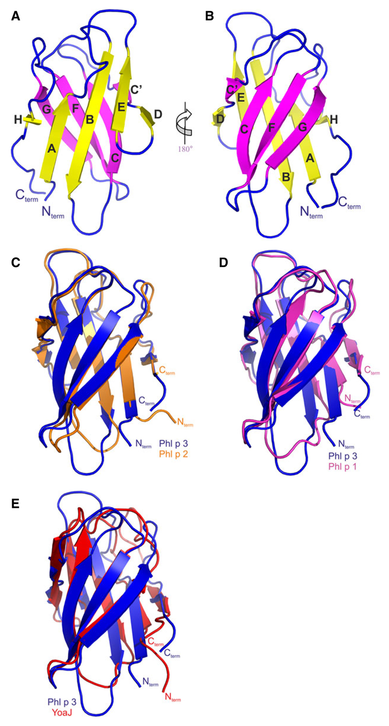
This is the first detailed description of a high-resolution crystal structure of a group 3 pollen allergen. Phl p 3 is a globular β-sandwich protein formed by two closely packed β-pleated sheets which are in face-to-face arrangement, virtually leaving no place for a cavity (Fig. 1A,B). The overall shape of the molecule is ellipsoidal with approximate dimensions 38 × 22 × 23 Å. The front β-sheet (HABED) and the back β-sheet (C’CFG) are separated by a distance of ~15 Å, measured as the distance between the central strands of the front and the back sheet (i.e., strands B and F). Phl p 3 consists of five and four antiparallel beta strands belonging to the front and back β-sheet, respectively. The two β-sheets adopt a twist angle of 30° which is a common feature in immunoglobulin fold structures (40). The overall fold is similar to immunoglobulin h-type fold with an extra C-terminal strand resulting in packing of N-terminal and C-terminal ends close to each other. Apart from Phl p 3 bearing a high structural similarity to Phl p 2 (group 2 allergen) and to the C-terminus of Phl p 1 (group 1 allergen), it also has a close resemblance to the D2 domain of maize expansin EXPB1, which forms part of the proposed glycan-binding stretch (19). The strands are connected by loops of variable sizes which are very well ordered and clearly visible in the electron density. The loops form protruding surface patches with high solvent accessibility due to the twisted beta sheet conformation. Protruding loop regions have been shown to contain conformational epitopes in the hyaluronidase–Fab complex structure (41). The loops connecting strands AB, CC’, EF, and GH form a cap for the hydrophobic core of the protein. The solution structure of Phl p 3 (12) exhibits a close overall structural similarity to the X-ray structure (Table S3), but large deviations at the N-terminal end (due to the His-tag fusion used for the NMR structure) and in the loop regions. The largest differences between the solution structure (model 1 of PDB: 2JNZ) and chain A of the crystal structure occur in the two loops regions AB (residues S12–L17) and EF (residues S57–P60), which actually represent a major part of the IgE epitope 2 (see section ‘Prediction of conformational IgE epitopes’).
Figure 1.
Crystal structure of Phl p 3 and comparison with Phl p 2 and Phl p 1. (A) front view and (B) back view. The strands are labeled A–H according to the Ig fold; the front sheet consists of strands H, A, B, E, and D (yellow), and the back sheet consists of strands C’, C, F, and G (magenta). Connecting loops are shown in blue. (C) Superposition of Phl p 3 (blue) and Phl p 2 (orange), (D) Superposition of Phl p 3 and Phl p 1–C-terminal domain (magenta), (E) Superposition of Phl p 3 and YoaJ–C-terminal domain (red). Structural representations throughout the manuscript were prepared with PyMOL (53).
Structural similarity of Phl p 3 with group 2, group 1 allergens, and expansins
We compared the available high-resolution structures of the group 1, 2, and 3 pollen allergens and of the expansin family. The pairwise sequence alignment showed high similarity (57.3% identity) between the group 2 and group 3 allergens (Fig. 2A, Table S3) and a lower, but still significant similarity to the C-terminal domains of group 1 pollen allergens (Phl p 1 and Zea m 1; 30.5–26.8% identity) and the C-terminal domain of the bacterial expansin YoaJ (20.6% identity). Zea m 1 was included into the structural comparison but not used in the epitope prediction because IgE cross-reactivity between Zea m 1 and Phl p 3 was not analyzed. The Phl p 3 structure determined at 1.8 Å was compared with the group 2 allergen Phl p 2 (PDB: 1WHO, 1.9 Å; Fig. 1C), the group 1 allergen Phl p 1 (PDB: 1N10, 2.9 Å; Fig. 1D) and the bacterial expansin YoaJ from Bacillus subtilis (20) (PDB: 3D3O, 1.9 Å; Fig. 1E). Structural superimposition of the 3D structures of all five proteins yielded root mean square deviations between 1.2 and 1.9 Å using all Cα atoms. The structural alignment of the five different proteins shows a close relation between Phl p 3 and Phl p 2 and a higher deviation between Phl p 3 and the C-terminal domains of expansin proteins (Table S3). Thus, the structural similarity between these proteins correlates well with the sequence identity. The overall alignment of the structures shows a well-conserved Ig-like fold with only minor differences in the loop regions (Fig. 1C–E).
Figure 2.
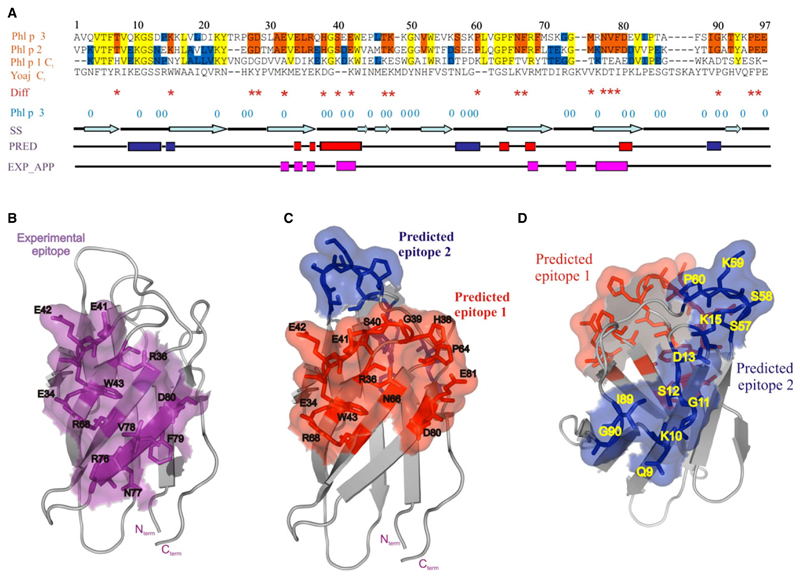
Sequence alignment and epitope prediction: (A) The proteins were aligned using CLUSTALW (54), and the identities between the compared allergens representing group 1, group 2, and group 3 pollen allergens are indicated: yellow (identity in all three groups), orange (identity between group 3 and group 2), and blue (identity between group 1 and group 2 or between group 1 and group 3). Residues with identity between group 2 and group 3, but a nonconservative change in group 1, are marked with an asterisk (*). ‘0’ marks residues in the Phl p 3 structure with a solvent exposure >50%. The secondary structure elements (SS) as observed in the Phl p 3 crystal structure and the predicted epitopes (PRED: Epitope 1 = red boxes, Epitope 2 = blue boxes) and the experimental epitopes (EXP_APP: pink boxes) are aligned to the sequence. (B–D) The epitopes are displayed as semitransparent surface patches, and the residues contained in this epitope are shown in stick representation and labeled. (B) The experimental IgE binding epitope derived from the structure of the Phl p 2–IgE complex and mapped onto the Phl p 3 structure is shown in purple, where only the direct contacts (H-bonds, van der Waals contacts) to the antibody are shown. (C–D), The two epitopes predicted by the program SPADE, based on the structural similarity of Phl p 3 and Phl p 2, epitope 1—red and epitope 2—blue.
Allergic patients’ IgE cross-reacts with group 2 and group 3 allergens but not with group 1 allergens
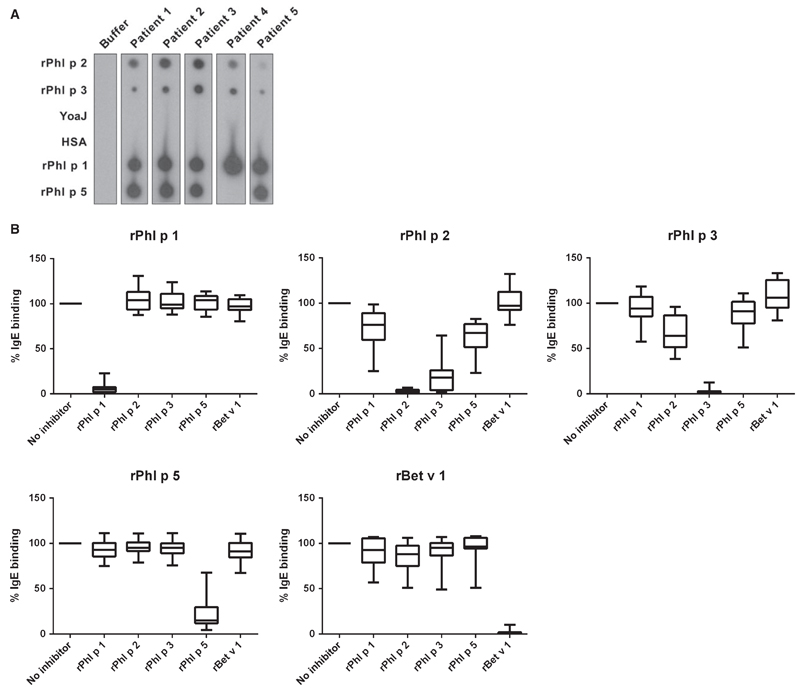
Figure 3A shows IgE reactivities of five representative grass pollen allergic patients with rPhl p 1, rPhl p 2, and rPhl p 3. Each of the sera showed IgE reactivity to group 2 and 3 allergens (i.e., Phl p 2, Phl p 3) and to the group 1 allergen (i. e., Phl p 1) from timothy grass but not with the expansin-like Bacillus subtilis protein YoaJ or the negative control, HSA (Fig. 3A). Four patients reacted also with rPhl p 5, which is considered to be structurally and immunologically unrelated to group 1, 2, and 3 grass pollen allergens (42).
Figure 3.
(A) IgE reactivities of sera from five grass pollen allergic patients (patients 1–5) or buffer alone with dotted recombinant grass pollen allergens [rPhl p 1, 2, 3, 5, an expansin-like protein of Bacillus subtilis (YoaJ)], and, for control purposes, human serum albumin (HSA) are shown. (B) IgE cross-reactivity. Sera from thirteen grass pollen allergic patients were preincubated with grass pollen allergens (Phl p 1, Phl p 2, Phl p 3, and Phl p 5), birch pollen allergen (Bet v 1) or with buffer alone (no inhibitor; x-axes) before exposure to allergens (top: Phl p 1, Phl p 2, Phl p 3, Phl p 5, and Bet v 1). Displayed are allergen-specific IgE reactivities as percentages of binding compared with uninhibited conditions (no inhibitor) in the form of box plots with whiskers representing 10–90 percentiles (y-axes).
Figure 3B and Fig. S1 show the results of IgE cross-reactivity studies performed with sera from 13 grass pollen allergic patients. Despite the structural similarities between Phl p 1, Phl p 2, and Phl p 3, no relevant cross-reactivity was found between Phl p 1 and Phl p 2 as well as between Phl p 1 and Phl p 3. Neither Phl p 2 nor Phl p 3 caused an inhibition of IgE binding to Phl p 1 nor vice versa did Phl p 1 inhibit IgE binding to Phl p 2 or Phl p 3 in a substantial manner (Fig. 3B, Fig. S1). Phl p 3 inhibited IgE binding to Phl p 2 and vice versa, albeit a bit less. We noted some inhibition of IgE binding to Phl p 2 and Phl p 3 by Phl p 5, whereas Bet v 1 did not inhibit IgE binding to any of the grass pollen allergens. Likewise, none of the grass pollen allergens blocked IgE binding to the unrelated birch pollen allergen, Bet v 1. Each of the allergens strongly inhibited IgE binding to itself (Fig. 3B, Fig. S1).
Mapping of a major Phl p 2 IgE epitope to the structures of Phl p 3 and Phl p 1
The structure of a 1 : 1 Phl p 2–IgE–Fab complex has been determined by X-ray crystallography (22). The IgE epitope was a conformational epitope of 876.0 Å2 containing mainly residues on the planar surface of the four-stranded antiparallel β-sheet of Phl p 2. We used the superposition of Phl p 3 and Phl p 2 to map this IgE epitope onto the Phl p 3 structure (Fig. 2B). This epitope exhibits a similar size and largely coincides with epitope 1 predicted with the program SPADE (21). Whereas the Phl p 2–IgE epitope is centered entirely on the β-sheet (C’CFG), the predicted epitope contains additional residues in the loops connecting the strands C–C’ and E–F.
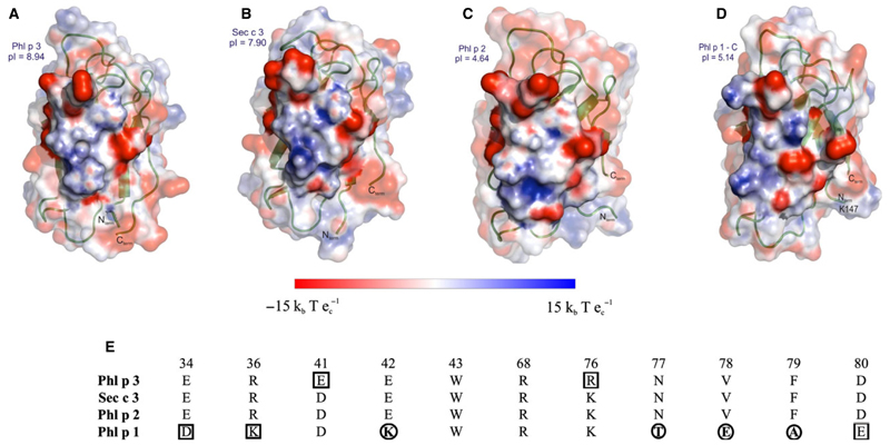
The Phl p 2–IgE epitope was also mapped onto the Sec c 3 structural model and onto the less homologous C-terminal domain of Phl p 1. Furthermore, the electrostatic potentials of all four proteins were calculated and mapped onto the surface, highlighting the experimental IgE epitope (Fig. 4). This comparison shows that the electrostatic charge distribution is highly similar in the epitope area between Phl p 3, Sec c 3, and Phl p 2 in spite of the highly different charge distributions between group 2/3 pollen allergens in general. Phl p 2 is highly acidic (pI 4.6), whereas the compared group 3 allergens are basic (Phl p 3: pI 8.9 and Sec c 3: pI 7.9). The high similarity of the epitope regions between these three cross-reactive allergens is also underlined by the fact that of the eleven residues involved in direct contacts with the human IgE mAb, all are identical except for two conserved amino acid changes between rPhl p 3 and Phl p 2, namely E41 to D39 and R76 to K75, respectively (Fig. 4E). In contrast, mapping of the experimental Phl p 2 epitope onto the Phl p 1–C structure results in a surface patch which exhibits a very different charge distribution resulting from seven changes of the mapped residues, four out of which are nonconservative changes. Two of these lead to a significant change of the local electrostatic potential due to the charge reversal of E42 in Phl p 3 to K186 in Phl p 1 and from V78 to E222, respectively.
Figure 4.
Electrostatic surface potential of the experimental epitope. Electrostatic charge distribution (units ± 15 kbTec−1; kb, Boltzmann’s constant; T, temperature in K; ec, charge of an electron) was calculated for the structures and mapped onto the surface representing the experimental epitope derived from Phl p 2–IgE complex. The experimental epitope area is shown as nontransparent surface while the rest of the surface has a transparency level of 0.5, partly covering the cartoon representation of the proteins. (A) Phl p 3 structure (this work), (B) Sec c 3 model (generated by Modeller based on Phl p 3 structure), (C) Phl p 2 (protein data bank, PDB: 1WHO), (D) the C–terminal domain of Phl p 1 (PDB: 1N10). All structures are oriented according to A. (E) Residues involved in the experimental Phl p 2–IgE epitope by direct contacts (H – bonds and van der Waals contacts) and the equivalent residues in the Phl p 3, Sec c 3 and Phl p 1 structures are listed and numbered according to the Phl p 3 sequence. Differences in the mapped epitope regions are highlighted; conservative changes are boxed and nonconservative changes are circled.
In fact, the human IgE mAb which was used for the determination of the structure of the Phl p 2–IgE complex (22) bound to Phl p 3 and Sec c 3, but not to Phl p 1 (data not shown).
Prediction of conformational IgE epitopes
To predict cross-reactive IgE epitopes, we combined structure-based surface comparison with the obtained immunological cross-reactivity data in the program SPADE (21), which uses the geometrical and physico-chemical similarity of the cross-reactive and noncross-reactive surfaces rather than sequence homology and solvent accessibility as the basis for the predictions. We found high CR between Phl p 3 and Phl p 2, and no CR between Phl p 3 and the proteins belonging to the expansin family (Phl p 1 and YoaJ). Therefore, we have used the crystal structures of Phl p 3 and Phl p 2 with a CR weight of 1.0 between these two proteins. This approach yielded two conformational epitopes which occupy two opposite sites on the Phl p 3 surface (Table 1). The first epitope (Fig. 2C) is predicted to cover a solvent-accessible surface (SAS) area of 922.0 Å2 and contains sequentially distant residues belonging to the β-strands C, C’, F, and G and the connecting loops between these β-sheets (E34, R36, H38, G39, S40, E41, E42, W43, P64, R68, D80, and E81). The second epitope (Fig. 2D) also contains discontinuous sequence stretches comprising the AB loop (connecting strands A and B), the EF loop and two residues from the GH loop (Q9, K10, G11, S12, D13, K15, S57, S58, K59, P60, I89, and G90). This epitope covers a SAS area of 994.0 Å2 and consists almost exclusively of loop regions. Thus, the two predicted epitopes together cover approximately one-third of the whole allergen surface which is 5759.9 Å2. The predicted epitope 1 overlaps with the epitope defined by the human Phl p 2-specific IgE mAb (22), which cross-reacts with Phl p 3 and Sec c 3.
Table 1.
Structural parameters and amino acid residues of the epitopes predicted by the similarity approach
| Epitopes | Epitope 1 | Epitope 2 |
|---|---|---|
| Residues | E34, R36, H38, G39, S40, E41, E42, W43, P64, R68, D80, E81 | Q9, K10, G11, S12, D13, K15, S57, S58, K59, P60, I89, G90 |
| Solvent-accessible area (51) | 922 Å2 | 994.0 Å2 |
| Average accessibility (52) | 56.3% | 66.1% |
| Average excess similarity* | 70.0% | 68.1% |
SPADE (21).
To determine whether the accuracy of the epitope prediction is dependent on the method of structure determination (i.e., crystallography or NMR), we included the minimum-energy NMR models of Phl p 3 and Phl p 2 in the SPADE predictions (Table S5). The comparison of structures from different sources (X-ray vs NMR, free allergen vs complex, etc.) in the presence of conformational flexibility and induced-fit antibody is made possible by two SPADE algorithms, the side-chain standardization and the multilocal backbone superposition (21). Under this premise, the epitope determined experimentally from the Phl p 2-IgE complex (22) was taken as bench mark for the accuracy of the predictions. Using the NMR structures as well as the X-ray structures of Phl p 3 and Phl p 2 in pairwise structural comparisons, we observed that a comparable specificity (correct residues out the total number predicted) could be obtained in both cases. The observed sensitivity (ratio of correctly predicted residues from the experimental epitope) was 9% higher in the case of the X-ray structures (Table S5).
Single point mutations identify the crucial residue for binding a blocking human mAb but have no strong effects on allergic patients’ polyclonal IgE recognition
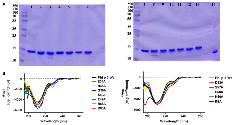
Next, we generated 24 point mutants in the two predicted epitopes. Of the 24 mutants, twelve point mutants (epitope patch 1: E34A, H38A, G39A, S40A, E42A, R68A, and D80A; epitope patch 2: D13A, S57A, S58A, K59A, and I89A) could be purified and were classified as stable according to SDS-PAGE (Fig. 5A). A survey of the fold integrity showed that at room temperature most mutants retained a fold similar to the wt-Phl p 3, except for the construct G39A, which is partly unfolded (Fig. 5B).
Figure 5.
Biophysical characterization of Phl p 3 mutants. (A) Generated mutants of Phl p 3 were loaded on to a 12.5% nonreducing SDS gel. Marked lanes show 1–Phl p 3, 2–E34A, 3–H38A, 4–S40A, 5–G39A, 6–E42A, 7–R68A, 8–D13A, 9–D80A, 10–S57A, 11–S58A, 12–K59A, 13–I89A, and 14–Phl p 2. (B) Circular dichroism spectra of Phl p 3 and the corresponding mutants plotted. x-axes: wavelength range, y-axes: CD signal expressed as mean-residue ellipticity (θmre). The left and right panels correspond to mutations in epitope 1 and epitope 2, respectively.
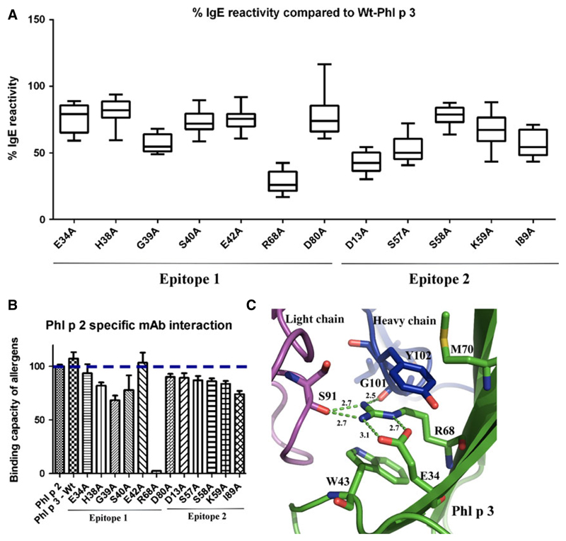
The Phl p 3 derivatives containing single point mutations in epitope patch 1 and 2 were compared with the Phl p 3 allergen regarding IgE reactivity using sera from 15 grass pollen allergic patients (Fig. 6A, Fig. S2). Most of the point mutations had only subtle effects on allergic patients’ IgE binding. Only point mutants R68A (predicted epitope 1), D13A, and S57A (predicted epitope 2) showed a marked mean reduction of ~70%, 60%, and 50% of patients’ IgE reactivity, respectively. The effects of mutations on the binding of the human mAb to Phl p 3 were very clear (Fig. 6B). With the exception of the mutation R68A, which completely abolished the binding of the mAb, none of the other mutations of amino acid residues, neither in epitope patch 1 nor in patch 2, affected its binding (Fig. 6B).
Figure 6.
Effects of single point mutations in epitope patch 1 and 2 on antibody binding. (A) IgE reactivities (y-axis: % IgE binding compared with rPhl p 3) of fifteen grass pollen allergic patients to Phl p 3 mutants (x-axis) as box plots with whiskers representing 10–90 percentiles. (B) Reactivity of Phl p 2, Phl p 3, and Phl p 3 mutants (x-axis) with the Phl p 2-specific mAb determined by ELISA. The y-axis displays the percentages of reactivity to Phl p 2 calculated from the optical density values (means of triplicates ± SD). The dotted horizontal line indicates the binding of Phl p 2. (C) The Phl p 3–IgE Fab complex model depicts the putative interactions of R68 with spatially close residues of Phl p 3 (green), the heavy chain (blue) and the light chain (magenta) of the Fab molecule. Interacting residues are shown in stick presentation and labeled (H-bonds in dashed lines, bond lengths in Å).
Discussion
Here, we determined the high-resolution crystal structure of the major timothy grass pollen allergen Phl p 3 and performed an in-depth structural comparison of Phl p 3 with homologous group 2 and group 1 grass pollen allergens. Furthermore, the IgE recognition of Phl p 3 and its CR with group 2 and group 1 grass pollen allergens was studied. Despite the fact that Phl p 3 and Phl p 2 show only modest sequence homology and a highly divergent isoelectric point (pI Phl p 2: 4.6; pI Phl p 3: 8.9), we found that the three-dimensional structure of both allergens is highly similar and that there is variable cross-reactivity of allergic patients’ IgE between the two allergens. This indicates that group 2 and 3 allergens represent a family of cross-reactive allergens, which may replace each other for diagnosis and allergen-specific immunotherapy. Despite high structural similarity of the C-terminal domain of Phl p 1 with the structures of group 2 and 3 allergens, no relevant IgE CR was observed demonstrating that for diagnosis and allergen-specific immunotherapy group 2 or 3 allergens are needed as independent major allergens in addition to group 1 allergens. The structural comparison of group 2, 3, and 1 allergens indicated that the lack of IgE CR may result from a few amino acid changes occurring in the IgE epitopes, which may introduce considerable local changes in charge, polarity, hydrophobicity, hydrogen binding capacity, or bulkiness of side chains.
As the three-dimensional structures of Phl p 3, Phl p 2, and Phl p 1 as well as experimental data regarding IgE cross-reactivity between these molecules were available, we were able to use a newly developed algorithm, SPADE (21) to predict conformational IgE binding sites. In fact, several programs exist for the prediction of conformational epitopes. There are algorithms based on different principles such as accessible surface area (conformational epitope prediction, CEP) (43), hydrophilicity, surface curvature (ElliPro) (44), or a combination of these (DISCOTOPE) (45). All these approaches use only one structure (i.e., the target allergen itself or a highly homologous protein structure) for the epitope prediction. By contrast, SPADE (21) is based on the structural and immunological comparison of two or more allergens belonging to the same structural family. In this way, topological criteria as well as biophysical properties such as hydrogen bonding, electrostatic surface potential, and hydrophobicity are included in the discrimination process. Using SPADE two cross-reactive IgE epitope-containing areas of 922 and 994 Å2, respectively, could be predicted on Phl p 3 and Phl p 2 (Fig. 2C,D). The size of the predicted areas was well within the range observed for antibody–antigen interactions. The accuracy of the prediction was confirmed by the fact that the first epitope-containing area overlaps to a large extent with the experimental IgE binding site determined by solving the structure of the complex between Phl p 2 and a corresponding human IgE-derived antibody, which strongly inhibited even the polyclonal IgE binding of allergic patients to Phl p 2 (22) (Fig. 3B). SPADE was able to delineate 77% of the experimental epitope area (584.0 Å2 out of 756.8 Å2), which was derived from the Phl p 2–IgE complex by mapping the interface area onto the Phl p 3 structure. The prediction was further confirmed by the fact that the IgE mAb defining epitope 1 cross-reacted with Phl p 3. When the first epitope area was mapped onto group 3, 2, and 1 allergens, a very similar charge distribution was observed between group 3 and group 2 allergens (Fig. 4A–C). Contrarily, the charge distribution was quite different on the equivalent surface patch of the Phl p 1–C-terminal domain due to the changes of two side chains involving charge reversal (E to K and the change from a hydrophobic to a charged amino acid V to E; Fig. 4D). Two other nonconserved changes involve variation in bulkiness and hydrophobicity (F to A and N to T). Therefore, the highly conserved nature of the experimental epitope area 1 between group 3 and group 2 allergens explains the high cross-reactivity between these groups, whereas the significant changes exemplified by the changes in charge distribution may explain the missing cross-reactivity between group 2/3 and group 1 allergens.
The second predicted IgE binding area is a rather elongated surface patch involving several interstrand loops and corresponds to a high extent to the conformation seen in IgG–allergen complexes, for example, Bet v 1–IgG (46), or hyaluronidase-IgG (41). In each of these complexes, the epitope consisted of a loop region of the allergen protruding into the binding site of the variable Fab domain.
We have studied the applicability of SPADE for 3D structures determined by various techniques (X-ray and NMR) and employed NMR models of both Phl p 3 and Phl p 2 for epitope predictions. In fact, given these minimum-energy models, the SPADE prediction result is similar and, with respect to the benchmarking against the experimental epitope, only moderately less accurate than the crystal structure-based prediction. In general, SPADE has been developed mostly on crystal structures (and some minimum-energy NMR models), and therefore, it currently relies on a parameterization optimized for these. However, the program does technically allow for the inclusion of multiple NMR models from the same allergen. An NMR ensemble contains more information than a single representative (minimal-energy or average model) and the consideration of conformational flexibility, as reflected by multiple structural states, might in a certain context describe the dynamic features of epitopes more realistically.
To further study IgE recognition of Phl p 3, we engineered 24 Phl p 3 mutant proteins containing single point mutations in the two IgE epitope-containing areas. The mutants were made to change residues contained in the two predicted epitope-containing areas without changing the overall fold and the spatial arrangement of the other surface-exposed residues. In fact, eleven of the 24 single mutants exhibited a reasonably preserved fold at room temperature and could be purified as intact proteins to study IgE recognition. Although the crystal structure of the Phl p 2-IgE Fab complex indicated that several residues in Phl p 2 which were conserved in Phl p 3 contribute to the direct interaction between the allergen and the IgE Fab, only one mutant, R68A, completely abolished the mIgE binding. In a model of the Phl p 3–mIgE–Fab complex, this residue is placed in the center of the epitope and completely buried by the Fab ligand. R68 forms several important interactions (H-bonds as well as hydrophobic and cation-π-stacking interactions) to both light chain (L_S91) and heavy chain (H_G101, H_Y102) of the IgE–Fab molecule (Fig. 6C), as they were also observed in the Phl p 2–Fab complex (22). In unbound Phl p 3, R68, apart from being involved in crystal packing, forms cation-π interactions with W43 and hydrogen bonds to E34, interactions which are conserved in the Fab complex.
Alterations of allergic patients’ IgE binding to Phl p 3 caused by the single point mutations were less pronounced than the effect of the R68 mutation on the binding of the monoclonal IgE, but the R68 mutation still caused an approximately 70% mean inhibition of allergic patients’ IgE binding. The accuracy of the IgE epitope prediction by SPADE was confirmed by the fact that mutations D13 and S57 in the second predicted area also reduced allergic patients’ IgE binding (mean reduction approximately 60% and 50%, respectively). The other point mutations had little or no effects on allergic patients’ IgE binding. There are at least two explanations for the latter result which are mutually nonexclusive. The first possibility is that due to the polyclonality of the IgE response, allergic patients contain a variety of IgE antibodies, which can bind in close vicinity to each other and thus in high density to the epitope-containing area. This explanation is supported by findings made for several other allergens, demonstrating that there is high density binding of several IgE antibodies toward surface-exposed patches on the allergens (47–50). It is also expected for polyclonal antibodies that they show a variability of cross-reactivity and/or reactivity due to epitope diversity. Another explanation would be that the high-affinity binding of IgE antibodies requires simultaneous interaction with several amino acid residues. In this case, single point mutations may not be sufficient to fully abolish the binding and may rather lead to a decrease in affinity/avidity without complete loss of binding.
Besides analyzing the structure and IgE recognition of two major grass pollen allergen groups required for diagnosis and treatment, our study also highlights a feature of the allergen-IgE interaction. It shows that the allergen–IgE complex formation can tolerate considerable changes in the side-chain chemistry which may have implication for the development of hypoallergenic allergen derivatives for allergen-specific immunotherapy.
Supplementary Material
Additional Supporting Information may be found in the online version of this article
Acknowledgments
We thank Tea Pavkov-Keller and Ulrike Wagner for their helpful comments and discussions, which made the structure solution possible. We are thankful to Winfried Mosler for his technical assistance in protein expression and purification. We acknowledge the EMBL outstation in Hamburg, Germany, for provision of the synchrotron radiation facilities, and we thank Andrea Schmidt for assistance in using beamline X12. We would like to acknowledge Paulette Charlier, Center for Protein Engineering, University of Liege, Belgium for providing the YoaJ protein for the immunological experiments.
Funding
This study was supported by grants F4604, F4605, and F4607 of the Austrian Science Fund (FWF).
Abbreviations
- BSA
bovine serum albumin
- CD
circular dichroism
- CEP
conformational epitope prediction [web server]
- CR
cross-reactivity
- DOPE
discrete optimized protein energy
- ELISA
enzyme-linked immunosorbent assay
- Fab
antigen-binding fragment
- HRP
horse radish peroxidase
- HSA
human serum albumin
- Ig
immunoglobulin
- kDa
kilodalton
- mAb
monoclonal antibody
- NMR
nuclear magnetic resonance
- PBS
phosphate-buffered saline
- PCR
polymerase chain reaction
- PDB
protein data bank
- RMSD
root mean square deviation
- SAS
solvent-accessible surface
- SIT
specific immunotherapy
- SPADE
surface comparison-based prediction of allergenic discontinuous epitopes
- TBST
tris (hydroxymethyl) aminomethane-buffered saline
- TEV
tobacco etch virus
- Tm
melting temperature
Footnotes
Conflicts of interest
RV has received research grants from Biomay AG, Vienna, Austria, Thermofisher, Uppsala, Sweden, from the Austrian Science Fund (FWF) and the European Union. He serves as a consultant for Biomay AG, Vienna and Thermofisher, Uppsala. The other authors of the paper declare no conflicts of interest.
References
- 1.Heinzerling L, Frew AJ, Bindslev-Jensen C, Bonini S, Bousquet J, Bresciani M, et al. Standard skin prick testing and sensitization to inhalant allergens across Europe-a survey from the GALEN network. Allergy. 2005;60:1287–1300. doi: 10.1111/j.1398-9995.2005.00895.x. [DOI] [PubMed] [Google Scholar]
- 2.Suphioglu C, Singh MB, Taylor P, Bellomo R, Holmes P, Puy R, et al. Mechanism of grass-pollen-induced asthma. Lancet. 1992;339:569–572. doi: 10.1016/0140-6736(92)90864-y. [DOI] [PubMed] [Google Scholar]
- 3.Pollart SM, Reid MJ, Fling JA, Chapman MD, Platts-Mills TA. Epidemiology of emergency room asthma in northern California: association with IgE antibody to rye-grass pollen. J Allergy Clin Immunol. 1988;82:224–230. doi: 10.1016/0091-6749(88)91003-2. [DOI] [PubMed] [Google Scholar]
- 4.Blackley CH. Experimental Researches on the Causes and Nature of Catarrhus Aestivus (Hay-Fever or-Hay-Asthma). Publ. cl. Facs. from the 1873 ed. 1959:202. [Google Scholar]
- 5.Noon L. Prophylactic inoculation against hay fever. Lancet. 1911;1:1572–1573. [Google Scholar]
- 6.Thomas WR. The advent of recombinant allergens and allergen cloning. J Allergy Clin Immunol. 2011;127:855–859. doi: 10.1016/j.jaci.2010.12.1084. [DOI] [PubMed] [Google Scholar]
- 7.Andersson K, Lidholm J. Characteristics and immunobiology of grass pollen allergens. Int Arch Allergy Immunol. 2003;130:87–107. doi: 10.1159/000069013. [DOI] [PubMed] [Google Scholar]
- 8.Ansari AA, Kihara TK, Marsh DG. Immunochemical studies of Lolium perenne (rye grass) pollen allergens, Lol p I, II, and III. J Immunol. 1987;139:4034–4041. [PubMed] [Google Scholar]
- 9.Ansari AA, Shenbagamurthi P, Marsh DG. Complete primary structure of a Lolium perenne (perennial rye grass) pollen allergen, Lol p III: comparison with known Lol p I and II sequences. Biochemistry. 1989;28:8665–8670. doi: 10.1021/bi00447a058. [DOI] [PubMed] [Google Scholar]
- 10.Dolecek C, Vrtala S, Laffer S, Steinberger P, Kraft D, Scheiner O, et al. Molecular characterization of Phl p II, a major timothy grass (Phleum pratense) pollen allergen. FEBS Lett. 1993;335:299–304. doi: 10.1016/0014-5793(93)80406-k. [DOI] [PubMed] [Google Scholar]
- 11.De Marino S, Morelli MA, Fraternali F, Tamborini E, Musco G, Vrtala S, et al. An immunoglobulin-like fold in a major plant allergen: the solution structure of Phl p 2 from timothy grass pollen. Structure. 1999;7:943–952. doi: 10.1016/s0969-2126(99)80121-x. [DOI] [PubMed] [Google Scholar]
- 12.Schweimer K, Petersen A, Suck R, Becker WM, Rosch P, Matecko I. Solution structure of Phl p 3, a major allergen from timothy grass pollen. Biol Chem. 2008;389:919–923. doi: 10.1515/BC.2008.102. [DOI] [PubMed] [Google Scholar]
- 13.Cosgrove DJ. Loosening of plant cell walls by expansins. Nature. 2000;407:321–326. doi: 10.1038/35030000. [DOI] [PubMed] [Google Scholar]
- 14.Focke M, Marth K, Flicker S, Valenta R. Heterogeneity of commercial timothy grass pollen extracts. Clin Exp Allergy. 2008;38:1400–1408. doi: 10.1111/j.1365-2222.2008.03031.x. [DOI] [PubMed] [Google Scholar]
- 15.Niederberger V, Stubner P, Spitzauer S, Kraft D, Valenta R, Ehrenberger K, et al. Skin test results but not serology reflect immediate type respiratory sensitivity: a study performed with recombinant allergen molecules. J Invest Dermatol. 2001;117:848–851. doi: 10.1046/j.0022-202x.2001.01470.x. [DOI] [PubMed] [Google Scholar]
- 16.Westritschnig K, Horak F, Swoboda I, Balic N, Spitzauer S, Kundi M, et al. Different allergenic activity of grass pollen allergens revealed by skin testing. Eur J Clin Invest. 2008;38:260–267. doi: 10.1111/j.1365-2362.2008.01938.x. [DOI] [PubMed] [Google Scholar]
- 17.GuerinMarchand C, Senechal H, Bouin AP, LeducBrodard V, Taudou G, Weyer A, et al. Cloning, sequencing and immunological characterization of Dac g 3, a major allergen from Dactylis glomerata pollen. Mol Immunol. 1996;33:797–806. doi: 10.1016/0161-5890(96)00015-6. [DOI] [PubMed] [Google Scholar]
- 18.Petersen A, Suck R, Lindner B, Georgieva D, Ernst M, Notbohm H, et al. Phl p 3: structural and immunological characterization of a major allergen of timothy grass pollen. Clin Exp Allergy. 2006;36:840–849. doi: 10.1111/j.1365-2222.2006.02505.x. [DOI] [PubMed] [Google Scholar]
- 19.Yennawar NH, Li LC, Dudzinski DM, Tabuchi A, Cosgrove DJ. Crystal structure and activities of EXPB1 (Zea m 1), a beta-expansion and group-1 pollen allergen from maize. Proc Natl Acad Sci USA. 2006;103:14664–14671. doi: 10.1073/pnas.0605979103. [DOI] [PMC free article] [PubMed] [Google Scholar]
- 20.Kerff F, Amoroso A, Herman R, Sauvage E, Petrella S, Filee P, et al. Crystal structure and activity of Bacillus subtilis YoaJ (EXLX1), a bacterial expansin that promotes root colonization. Proc Natl Acad Sci USA. 2008;105:16876–16881. doi: 10.1073/pnas.0809382105. [DOI] [PMC free article] [PubMed] [Google Scholar]
- 21.Dall’Antonia F, Gieras A, Devanaboyina SC, Valenta R, Keller W. Prediction of IgE-binding epitopes by means of allergen surface comparison and correlation to cross reactivity. J Allergy Clin Immunol. 2011;128:872–879. doi: 10.1016/j.jaci.2011.07.007. [DOI] [PubMed] [Google Scholar]
- 22.Padavattan S, Flicker S, Schirmer T, Madritsch C, Randow S, Reese G, et al. High-affinity IgE recognition of a conformational epitope of the major respiratory allergen Phl p 2 as revealed by X-ray crystallography. J Immunol. 2009;182:2141–2151. doi: 10.4049/jimmunol.0803018. [DOI] [PubMed] [Google Scholar]
- 23.Niederberger V, Laffer S, Froschl R, Kraft D, Rumpold H, Kapiotis S, et al. IgE antibodies to recombinant pollen allergens (Phl p 1, Phl p 2, Phl p 5, and Bet v 2) account for a high percentage of grass pollen-specific IgE. J Allergy Clin Immunol. 1998;101:258–264. doi: 10.1016/s0091-6749(98)70391-4. [DOI] [PubMed] [Google Scholar]
- 24.Rossmann MG, Blow DM. Detection of sub-units within crystallographic asymmetric unit. Acta Crystallogr. 1962;5:24–32. [Google Scholar]
- 25.Hutchinson EG, Thornton JM. PROMO-TIF-a program to identify and analyze structural motifs in proteins. Protein Sci. 1996;5:212–220. doi: 10.1002/pro.5560050204. [DOI] [PMC free article] [PubMed] [Google Scholar]
- 26.Holm L, Sander C. Dali: a network tool for protein structure comparison. Trends Biochem Sci. 1995;20:478–480. doi: 10.1016/s0968-0004(00)89105-7. [DOI] [PubMed] [Google Scholar]
- 27.Shindyalov IN, Bourne PE. Protein structure alignment by incremental combinatorial extension (CE) of the optimal path. Protein Eng. 1998;11:739–747. doi: 10.1093/protein/11.9.739. [DOI] [PubMed] [Google Scholar]
- 28.Zhu J, Weng Z. FAST: a novel protein structure alignment algorithm. Proteins. 2005;58:618–627. doi: 10.1002/prot.20331. [DOI] [PubMed] [Google Scholar]
- 29.Chenna R, Sugawara H, Koike T, Lopez R, Gibson TJ, Higgins DG, et al. Multiple sequence alignment with the Clustal series of programs. Nucleic Acids Res. 2003;31:3497–3500. doi: 10.1093/nar/gkg500. [DOI] [PMC free article] [PubMed] [Google Scholar]
- 30.Ahmad S, Gromiha M, Fawareh H, Sarai A. ASAView: database and tool for solvent accessibility representation in proteins. BMC Bioinformatics. 2004;5:51. doi: 10.1186/1471-2105-5-51. [DOI] [PMC free article] [PubMed] [Google Scholar]
- 31.Eswar N, Webb B, Marti-Renom MA, Madhusudhan MS, Eramian D, Shen MY, et al. Comparative protein structure modeling using Modeller. Curr Protoc Bioinformatics. 2006;15:5.6.1–5.6.30. doi: 10.1002/0471250953.bi0506s15. [DOI] [PMC free article] [PubMed] [Google Scholar]
- 32.Notredame C, Higgins DG, Heringa J. T-Coffee: a novel method for fast and accurate multiple sequence alignment. J Mol Biol. 2000;302:205–217. doi: 10.1006/jmbi.2000.4042. [DOI] [PubMed] [Google Scholar]
- 33.Colovos C, Yeates TO. Verification of protein structures: patterns of nonbonded atomic interactions. Protein Sci. 1993;2:1511–1519. doi: 10.1002/pro.5560020916. [DOI] [PMC free article] [PubMed] [Google Scholar]
- 34.Lyskov S, Gray JJ. The RosettaDock server for local protein-protein docking. Nucleic Acids Res. 2008;36:W233–W238. doi: 10.1093/nar/gkn216. [DOI] [PMC free article] [PubMed] [Google Scholar]
- 35.Jones S, Thornton JM. Principles of protein-protein interactions. Proc Natl Acad Sci USA. 1996;93:13–20. doi: 10.1073/pnas.93.1.13. [DOI] [PMC free article] [PubMed] [Google Scholar]
- 36.MacCallum RM, Martin AC, Thornton JM. Antibody-antigen interactions: contact analysis and binding site topography. J Mol Biol. 1996;262:732–745. doi: 10.1006/jmbi.1996.0548. [DOI] [PubMed] [Google Scholar]
- 37.Ball T, Edstrom W, Mauch L, Schmitt J, Leistler B, Fiebig H, et al. Gain of structure and IgE epitopes by eukaryotic expression of the major Timothy grass pollen allergen, Phl p 1. FEBS J. 2005;272:217–227. doi: 10.1111/j.1432-1033.2004.04403.x. [DOI] [PubMed] [Google Scholar]
- 38.Madritsch C, Flicker S, Scheiblhofer S, Zafred D, Pavkov-Keller T, Thalhamer J, et al. Recombinant monoclonal human immunoglobulin E to investigate the allergenic activity of major grass pollen allergen Phl p 5. Clin Exp Allergy. 2011;41:270–280. doi: 10.1111/j.1365-2222.2010.03666.x. [DOI] [PubMed] [Google Scholar]
- 39.Niederberger V, Pauli G, Gronlund H, Froschl R, Rumpold H, Kraft D, et al. Recombinant birch pollen allergens (rBet v 1 and rBet v 2) contain most of the IgE epitopes present in birch, alder, hornbeam, hazel, and oak pollen: a quantitative IgE inhibition study with sera from different populations. J Allergy Clin Immunol. 1998;102:579–591. doi: 10.1016/s0091-6749(98)70273-8. [DOI] [PubMed] [Google Scholar]
- 40.Halaby DM, Poupon A, Mornon J. The immunoglobulin fold family: sequence analysis and 3D structure comparisons. Protein Eng. 1999;12:563–571. doi: 10.1093/protein/12.7.563. [DOI] [PubMed] [Google Scholar]
- 41.Padavattan S, Schirmer T, Schmidt M, Akdis C, Valenta R, Mittermann I, et al. Identification of a B-cell epitope of hyaluronidase, a major bee venom allergen, from its crystal structure in complex with a specific Fab. J Mol Biol. 2007;368:742–752. doi: 10.1016/j.jmb.2007.02.036. [DOI] [PubMed] [Google Scholar]
- 42.Vrtala S, Sperr WR, Reimitzer I, Vanree R, Laffer S, Muller WD, et al. Cdna cloning of a major allergen from timothy grass (Phleum-Pratense) pollen – characterization of the recombinant Phl P-V allergen. J Immunol. 1993;151:4773–4781. [PubMed] [Google Scholar]
- 43.Kolaskar AS, Kulkarni-Kale U. Prediction of three-dimensional structure and mapping of conformational epitopes of envelope glycoprotein of Japanese encephalitis virus. Virology. 1999;261:31–42. doi: 10.1006/viro.1999.9859. [DOI] [PubMed] [Google Scholar]
- 44.Ponomarenko J, Bui HH, Li W, Fusseder N, Bourne PE, Sette A, et al. ElliPro: a new structure-based tool for the prediction of antibody epitopes. BMC Bioinformatics. 2008;9:514. doi: 10.1186/1471-2105-9-514. [DOI] [PMC free article] [PubMed] [Google Scholar]
- 45.Andersen PH, Nielsen M, Lund O. Prediction of residues in discontinuous B-cell epitopes using protein 3D structures. Protein Sci. 2006;15:2558–2567. doi: 10.1110/ps.062405906. [DOI] [PMC free article] [PubMed] [Google Scholar]
- 46.Mirza O, Henriksen A, Ipsen H, Larsen JN, Wissenbach M, Spangfort MD, et al. Dominant epitopes and allergic cross-reactivity: complex formation between a Fab fragment of a monoclonal murine IgG antibody and the major allergen from birch pollen Bet v 1. J Immunol. 2000;165:331–338. doi: 10.4049/jimmunol.165.1.331. [DOI] [PubMed] [Google Scholar]
- 47.Fedorov AA, Ball T, Mahoney NM, Valenta R, Almo SC. The molecular basis for allergen cross-reactivity: crystal structure and IgE-epitope mapping of birch pollen profilin. Structure. 1997;5:33–45. doi: 10.1016/s0969-2126(97)00164-0. [DOI] [PubMed] [Google Scholar]
- 48.Flicker S, Steinberger P, Ball T, Krauth MT, Verdino P, Valent P, et al. Spatial clustering of the IgE epitopes on the major timothy grass pollen allergen Phl p 1: importance for allergenic activity. J Allergy Clin Immunol. 2006;117:1336–1343. doi: 10.1016/j.jaci.2006.02.012. [DOI] [PubMed] [Google Scholar]
- 49.Flicker S, Vrtala S, Steinberger P, Vangelista L, Bufe A, Petersen A, et al. A human monoclonal IgE antibody defines a highly allergenic fragment of the major timothy grass pollen allergen, Phl p 5: molecular, immunological, and structural characterization of the epitope-containing domain. J Immunol. 2000;165:3849–3859. doi: 10.4049/jimmunol.165.7.3849. [DOI] [PubMed] [Google Scholar]
- 50.Gieras A, Cejka P, Blatt K, Focke-Tejkl M, Linhart B, Flicker S, et al. Mapping of conformational IgE epitopes with peptide-specific monoclonal antibodies reveals simultaneous binding of different IgE antibodies to a surface patch on the major birch pollen allergen, Bet v 1. J Immunol. 2011;186:5333–5344. doi: 10.4049/jimmunol.1000804. [DOI] [PubMed] [Google Scholar]
- 51.Sanner MF, Olson AJ, Spehner JC. Reduced surface: an efficient way to compute molecular surfaces. Biopolymers. 1996;38:305–320. doi: 10.1002/(SICI)1097-0282(199603)38:3%3C305::AID-BIP4%3E3.0.CO;2-Y. [DOI] [PubMed] [Google Scholar]
- 52.Fraczkiewicz R, Braun W. Exact and efficient analytical calculation of the accessible surface areas and their gradients for macromolecules. J Comput Chem. 1998;19:319–333. [Google Scholar]
- 53.DeLano WL. Use of PYMOL as a communications tool for molecular science. Abstr Pap Am Chem Soc. 2004;228:U313–U314. [Google Scholar]
- 54.Larkin MA, Blackshields G, Brown NP, Chenna R, McGettigan PA, McWilliam H, et al. Clustal W and clustal X version 2.0. Bioinformatics. 2007;23:2947–2948. doi: 10.1093/bioinformatics/btm404. [DOI] [PubMed] [Google Scholar]
Associated Data
This section collects any data citations, data availability statements, or supplementary materials included in this article.


