Abstract
The Old World vulture may carry and spread pathogens for emerging infections since they feed on the carcasses of dead animals and participate in the sky burials of humans, some of whom have died from communicable diseases. Therefore, we studied the precise fecal microbiome of the Old World vulture with metataxonomics, integrating the high-throughput sequencing of almost full-length small subunit ribosomal RNA (16S rRNA) gene amplicons in tandem with the operational phylogenetic unit (OPU) analysis strategy. Nine vultures of three species were sampled using rectal swabs on the Qinghai-Tibet Plateau, China. Using the Pacific Biosciences sequencing platform, we obtained 54 135 high-quality reads of 16S rRNA amplicons with an average of 1442±6.9 bp in length and 6015±1058 reads per vulture. Those sequences were classified into 314 OPUs, including 102 known species, 50 yet to be described species and 161 unknown new lineages of uncultured representatives. Forty-five species have been reported to be responsible for human outbreaks or infections, and 23 yet to be described species belong to genera that include pathogenic species. Only six species were common to all vultures. Clostridium perfringens was the most abundant and present in all vultures, accounting for 30.8% of the total reads. Therefore, using the new technology, we found that vultures are an important reservoir for C. perfringens as evidenced by the isolation of 107 strains encoding for virulence genes, representing 45 sequence types. Our study suggests that the soil-related C. perfringens and other pathogens could have a reservoir in vultures and other animals.
Keywords: Clostridium perfringens, full-length 16S rRNA gene, metataxonomics, operational phylogenetic unit, PacBio, vulture
INTRODUCTION
Animal-associated microbes have received immense attention because of their relevance to human health.1 The majority of human pathogens are of animal origin and especially have wild animals as host reservoirs.2, 3 Studying the microbiome of wild animals will provide valuable information to deliver an advance alert of or prevent putative infection outbreaks. Vultures are nature's most successful scavengers, feeding on the carcasses of dead animals without suffering any apparent illness effect. They perform an important task in removing rotting carcasses that could otherwise be a source of disease. However, they may also constitute a reservoir of pathogens that could be widely disseminated. Sky burial is practiced on the Qinghai-Tibet Plateau, and in Bhutan, Nepal and some parts of India.4 Old World vultures are attracted to the ritual and play a major role in removing the carcasses from the ritual sites. Our driving hypothesis was that vultures could acquire and disseminate pathogens from dead animals, such as Marmota himalayana, a major reservoir of Yersinia pestis on the Qinghai-Tibet Plateau.5
16S rRNA sequencing is the most widely used technique for microbial identification and diversity analysis. Metataxonomics is defined as the use of high-throughput 16S rRNA amplicon sequencing along with phylogenetic analysis to characterize the entire microbiota of a given sample.6 The major problem with most of the previous studies is that they rely on the use of platforms, such as Illumina or Roche 454, that generate only partial 16S rRNA gene sequences, mostly from hypervariable regions of no more than 600 bp in length and in general <400 bp. The PacBio platform offers the possibility to sequence almost full-length 16S rRNA gene amplicons, increasing the information content by at least 3 ×, which is necessary for an accurate phylogenetic inference.7 Despite some restrictions of PacBio caused by its higher error rate compared with other platforms,8 our approach minimizes their effect, as it does not rely on just clustering sequences at a given identity threshold in operational taxonomic units (OTUs). We use the operational phylogenetic unit (OPU) strategy that is based on phylogenetic inference after a treeing approach, which, among other benefits, diminishes the influence of sequence errors and indels.9, 10 The additional advantage of using almost full-length amplicon sequences is that the phylogenetic inference can be done after a de novo tree reconstruction instead of just the addition of partial sequences using the parsimony algorithm.7 This is probably the most accurate way to analyze the diversity based on 16S rRNA gene amplicons.
MATERIALS AND METHODS
Vulture sampling
The vultures were live-captured in Qinghai Province, China. All animals were anesthetized, sampled using rectal swabs and released. The species of vultures were identified by mitochondrial cytochrome c oxidase I or b sequence analysis, amplified from the feathers.11, 12 The animal welfare practices associated with this study were approved by the Wildlife Protection Agents of Qinghai Province.
High-throughput 16S rRNA amplicon sequencing by PacBio and Illumina MiSeq systems
Genomic DNA was extracted from rectal swabs using the QIAamp Fast DNA Stool Kit (Qiagen, Hilden, Germany). For the PacBio sequencing, the almost complete region of the bacterial 16S rRNA gene was amplified using the universal primer set 27F (5′-AGA GTT TGA TCC TGG CTC AG-3′) and 1492R (5′-GNT ACC TTG TTA CGA CTT-3′).13 PCR was performed using Ex Taq DNA polymerase, and the parameters for amplification were as follows: initial denaturation for 5 min at 94 °C; 25 cycles of denaturation at 94 °C for 30 s, annealing at 52 °C for 30 s, and extension at 72 °C for 1 min 30 s; and finally an extension step at 72 °C for 7 min. Sequencing was conducted on a PacBio RS II platform at TianJin Biochip Corporation, China. The primary 16S rRNA sequences generated were filtered using the SMRT Portal (version 2.3.0; www.pacb.com/devnet/). To ensure that the barcoded reads are correctly assigned to their original samples, a minimum barcode score of 22 was selected to achieve 99.5% accuracy. Data containing ambiguous bases was removed, primer sequences and adapters were excised from the filtered reads, and sequences outside of the 10–1490 nucleotide positions were trimmed.
For the Illumina MiSeq sequencing, the PCR amplification of the V3-V4 region of the bacterial 16S rRNA was performed using universal primers 341F (5′-CCT ACG GGN GGC WGC AG-3′) and 805R (5′-GAC TAC HVG GGT ATC TAA TCC-3′).14 The cycling parameters for amplifying V3-V4 using Ex Taq DNA Polymerase were as follows: initial denaturation for 5 min at 94 °C; 25 cycles of denaturation at 94 °C for 30 s, annealing at 55 °C for 30 s and elongation at 72 °C for 30 s; and a final step at 72 °C for 7 min. Sequencing was conducted using a paired-end 2 × 300 bp cycle run on an Illumina MiSeq platform at Berry Genomics Co., Ltd, Beijing, China. The image analysis, base calling and error estimation were performed using Illumina Real-Time Analysis software. The paired-end reads produced by Illumina Miseq were merged using FLASH.15
Qiime was applied for further filtering.16 Chimeras were removed using the UCHIME algorithm and using RDP gold as a reference database.17, 18 Rarefaction curves using PAST19 software, version 1.82b, and Good's coverage were calculated as previously indicated.9 The microbiome raw sequence data of this study have been deposited in the NCBI Sequence Read Archive (SRA) with the accession code SRP082183.
Operational taxonomic unit and operational phylogenetic unit analysis strategy
The USEARCH pipeline was used to cluster the 16S rRNA sequences of both sequencing platforms into OTUs with a threshold set at 98.7% identity.20 The most frequent read of each OTU was selected as a representative to be added to the LTP123 database (The All-Species Living Tree Project)21 and aligned using the SINA tool (SILVA Incremental Aligner).22 The aligned sequences were inserted into the default tree using the Parsimony tool implemented in the ARB software package.23 The resulting insertions were manually inspected to recognize all representative sequences affiliating closely with either type strain sequences or clearly within a genus lineage. All sequences that remained unaffiliated were added to the SILVA SSURef_NR_123 (Silva Reference Non Redundant) database and inserted into the default tree.22 Approximately three closest relative sequences representing uncultured organisms were selected for each independent lineage made by the OTU representatives and exported to the LTP123 database.
A subset of sequences containing (i) all PacBio OTU representative sequences, (ii) the selection of the reference type strains and the SILVA REF123 recruited sequences, and (iii) the neighbor-joining supporting sequence data set was used to reconstruct a neighbor-joining tree.24 The reconstruction was performed using a 30% conservational filter to avoid phylogenetic noise. The partial Illumina MiSeq sequences obtained and the downloaded Roche 454 sequences of the New World vulture study25 were posteriorly added with the Parsimony tool to the final tree. The reconstruction was manually inspected, and each unique lineage including OTU representatives and at least one reference sequence was recognized as an OPU.10, 26 The sequences that were identical or nearly identical (>98.7% identity) to the type strain sequences were considered members of the same species. When the OPU represented an independent lineage within a genus, it was considered a new species of this taxon. Unique lineages affiliated with outside known genera were considered uncultured lineages of known families, orders or classes. The sequencing error rate was determined for each sample as previously decribed.8 Insertion and deletion rates were obtained by comparing the final alignment of all representative selected sequences with the corresponding reference sequence in the LTP123.
Microbiome-guided culturing
The species with putative medical significance as suggested by the microbiome analysis, such as C. perfringens, were attempted to be isolated from fecal swabs and analyzed for virulence genes and genetic diversity.27, 28, 29
RESULTS
Use of the almost full-length 16S rRNA gene for OPU assignment
The PacBio platform rendered a total of 89 412 raw 16S rRNA sequence reads for the nine vultures, with an average of 9935±1182 each. After quality filtering and the removal of chimeras, a total of 54 135 (64.1%) high-quality reads were retained, with an average 6015±1058 per vulture, ranging from 3914 to 7501 reads. The sequences were on average 1442±6.9 bp in length. The sequences were clustered into 4223 OTUs at 98.7% identity, with an average of 469±257 OTUs per vulture (Table 1). The representative sequences for each OTU were used for the OPU design, in which an OPU is the smallest monophyletic group of sequences containing OTU representatives together with the closest reference sequence, including the sequence of a type strain when possible.9 The 4223 OTUs were classified into 314 OPUs, with 78±49.6 per vulture (Table 1). The amplification for the V3-V4 region using the Illumina MiSeq system rendered a total of 826 772 high-quality reads, with a mean of 91 864±23 335 per vulture, with average of 443±3.1 bp in length. Using a threshold of 98.7% identity, an average of 71±26.2 OTUs per vulture was obtained. The selected sequences of each OTU were affiliated with 126 OPUs, with an average of 31±14.4 per vulture (Table 1).
Table 1. Quality, richness and diversity estimation of the 16S rRNA sequencing by PacBio and Illuminaa.
| Sample | PacBio | Illumina | ||||||||||||||
|---|---|---|---|---|---|---|---|---|---|---|---|---|---|---|---|---|
| Reads | Length (bp) | Q20 (%) | Q30 (%) | OTUs | OPUs | Shannon | Coverage | Reads | Length | Q20 (%) | Q30 (%) | OTUs | OPUs | Shannon | Coverage | |
| Am1 | 3914 | 1456±31.83 | 99.06 | 98.20 | 270 | 40 | 2.685 | 0.996 | 107 049 | 446±10.19 | 99.48 | 97.54 | 77 | 46 | 3.13 | 0.999999 |
| Am2 | 6911 | 1438±29.82 | 99.03 | 98.18 | 1041 | 180 | 2.885 | 0.989 | 113 973 | 440±8.86 | 99.54 | 97.81 | 95 | 53 | 1.35 | 0.999997 |
| Am3 | 5379 | 1445±31.49 | 99.02 | 98.16 | 380 | 52 | 2.761 | 0.994 | 43 454 | 440±11.29 | 97.55 | 91.28 | 16 | 12 | 1.72 | 0.999980 |
| Gb1 | 5524 | 1433±28.74 | 99.08 | 98.25 | 321 | 46 | 1.459 | 0.995 | 109 183 | 442±8.07 | 99.48 | 97.57 | 68 | 21 | 1.58 | 0.999996 |
| Gb2 | 6210 | 1437±30.25 | 99.19 | 98.46 | 518 | 128 | 2.977 | 0.990 | 107 243 | 444±8.47 | 99.51 | 97.62 | 108 | 40 | 2.00 | 1 |
| Gb3 | 5613 | 1440±33.21 | 99.18 | 98.38 | 284 | 70 | 1.864 | 0.994 | 100 036 | 441±6.00 | 99.55 | 97.8 | 71 | 19 | 1.54 | 1 |
| Gh1 | 6288 | 1437±30.77 | 99.05 | 98.22 | 346 | 30 | 1.368 | 0.999 | 98 151 | 441±5.03 | 99.55 | 97.83 | 79 | 17 | 1.64 | 0.999999 |
| Gh2 | 6795 | 1446±32.17 | 99.16 | 98.33 | 341 | 51 | 2.054 | 0.997 | 73 172 | 448±11.31 | 99.52 | 97.6 | 51 | 33 | 1.93 | 0.999984 |
| Gh3 | 7501 | 1445±29.77 | 99.07 | 98.23 | 722 | 101 | 3.288 | 0.994 | 74 511 | 447±9.92 | 99.44 | 97.42 | 72 | 39 | 2.98 | 0.999997 |
| Average | 6015±1057.91 | 1442±6.90 | 99.09 | 98.27 | 469±257.09 | 78±49.59 | 2.371 | 0.994 | 91 864±23 335.08 | 443±3.11 | 99.29 | 96.94 | 71±26.17 | 31±14.40 | 1.99 | 0.999995 |
| Total | 54 135 | ND | 314 | 826 772 | ND | 126 | ||||||||||
Abbreviations: not done, ND; operational phylogenetic unit, OPU; operational taxonomic unit, OUT.
The good coverage was calculated on OPUs.
Trimming and quality control rendered similar percentage yields, with 62.6%±8.4% and 49.0%±13.8% for PacBio and Illumina, respectively (Supplementary Table S1). In accordance with the rarefaction analysis, the 314 OPUs detected by PacBio did not enter saturation, whereas for the 126 MiSeq OPUs, the curves were well-saturated (Supplementary Figure S1). The error rates generated by the PacBio sequencing were slightly higher than those of Illumina MiSeq (Supplementary Figure S1). Three of the four most abundant OPUs (OPU107, OPU123 and OPU236), gathering 57.9% of the total reads and summing to 1540 representative OTU sequences, were selected for a deeper analysis of the insertion and deletion rates compared with the corresponding type strain reference sequence in the LTP123.21 For PacBio (Supplementary Table S2), the insertion and deletion rates were 0.4%±0.96 and 2.37%±1.25, respectively, accounting for mean inserted and deleted nucleotides in each sequence of 5.9±14.3 and 33.3±17.2. For the Illumina MiSeq system, the insertion and deletion rates were 0.06%±0.23 and 0.1%±0.17, respectively, accounting for mean inserted and deleted nucleotides in each sequence of 0.29±1 and 0.44±0.77. As the phylogenetic inferences in each case were done using a 30% conservational filter, the influence of the insertions was negligible and that of the deletions was also low.
Fecal microbiome profile at high taxon levels
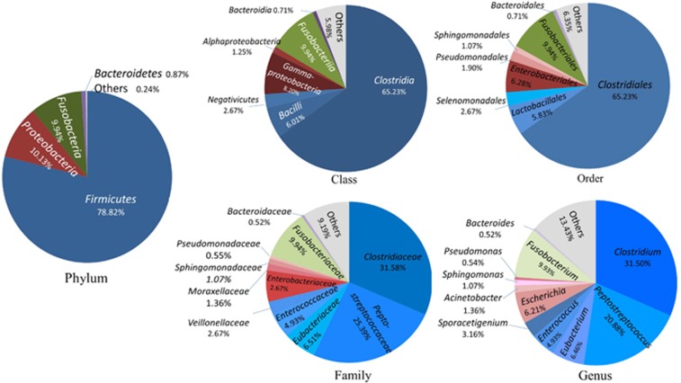
The 314 OPUs obtained with the PacBio approach were affiliated with 13 phyla, 25 classes, 37 orders, 66 families and 89 genera (Figure 1). Only two of the 13 phyla were detected in all vultures, namely, Firmicutes and Proteobacteria. Four of the 25 classes were detected in all vultures, namely, Clostridia, Gammaproteobacteria, Bacilli and Alphaproteobacteria. Four of the 37 orders were detected in all vultures, namely, Clostridiales, Enterobacteriales, Lactobacillales and Sphingomonadales. Six of the 66 families were detected in all vultures, namely, Clostridiaceae, Peptostreptococcaceae, Eubacteriaceae, Sphingomonadaceae, Peptoniphilaceae and Lactobacillaceae. Seven of the 89 genera were detected in all vultures, namely, Clostridium, Peptostreptococcus, Peptoniphilus, Eubacterium, Sporacetigenium, Sphingomonas and Lactobacillus (Figure 1).
Figure 1.
Microbiome profile at phylum, class, order, family and genus categories of the Old World vultures. Phylum: of 13 phyla classified, only four with >0.1% total reads are displayed. Class: of 25 classes classified, only seven with >0.5% total reads are displayed. Order: of 37 orders classified, only eight with>0.5% total reads are displayed. Family: of 66 families classified, only 11 with>0.5% total reads are displayed. Genus: of 89 genera classified, only 11 with >0.5% total reads are displayed.
Species composition of the fecal microbiomes
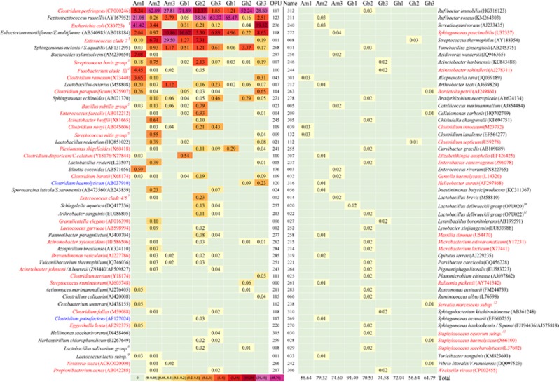
Of the 314 OPUs detected, 102 (32%) were identified as members of known species, as they were very closely affiliated (>98.7% identity) or exhibited an identical sequence to their respective type strain (Figure 2). In some cases, due to the high identity between type strain sequences, an accurate identification was not possible, and the OPU was identified by the distinct members of the clade, such as OPU001, identified as Enterococcus clade 7, including several species;30 and OPU108 and OPU268, identified as Eubacterium moniliforme/E. multiforme and Sphingomonas melonis/S. aquatilis, respectively.
Figure 2.
Abundance heat map of the 102 known species detected and their medical significance. The abundances are shown as percentage of reads in each vulture, and highlighted with a color scale. Species on the list of medically significant bacteria are highlighted in red. The species in blue were reported to be isolated from diseased animals. The species in black have no information to be associated with human or animal disease. The species Gyps himalayensis, Gypaetus barbatus and Aegypius monachus were abbreviated as Gh, Gb and Am. The number following the abbreviation was the number for a given sampled vulture. #: OPUs including several species (for example, 1OPU236, E. coli or Shigella).
The 102 known species gathered between 57.6% and 91.4% of the reads in each vulture and 73.0% of the total reads for all vultures. The abundances of individual species in individual vultures varied significantly, ranging from 0.002% to 71.9% reads (Figure 2). Only four characterized species were present in all vultures, namely, C. perfringens (OPU107), Peptostreptococcus russellii (OPU123), E. moniliforme/E. multiforme (OPU108), and S. melonis/S. aquatilis (OPU268). In addition, there were only five species with >1% total reads, i.e., C. perfringens (OPU107; 1.2%–71.9%), P. russellii (OPU123; 0.05%–65.5%), Escherichia coli/Shigella (OPU236; 0.04%–41.42%, but absent in Am3 and Gh1), E. moniliforme/E. multiforme (OPU108; 0.22%–16.6%) and the Enterococcus clade 7 (0.1%–29.5% Figure 2; Supplementary Table S3).
Of the 102 known species, 45 were considered medically significant bacteria or had been reported to be responsible for outbreaks or infections in humans, as evidenced by literature searches for the combination of the given species name and outbreak31 (Figure 2), and ranged from three to 25 in the individual vultures. The analyses evidenced outbreak causative agents including Plesiomonas shigelloides (OPU241), C. perfringens (OPU107), C. septicum (OPU112), C. tertium (OPU111), C. barati (OPU109), Bordetella petrii (OPU263), Brevundimonas vesicularis (OPU285), Gemella haemolysans (OPU032), Granulicatella elegans (OPU006), Propionibacterium acnes (OPU217), Serratia marcescens (OPU238), Ralstonia pickettii (OPU261), Sphingomonas paucimobilis (OPU272), Staphylococcus haemolyticus (OPU028), and S. saccharolyticus (OPU029), among others.31
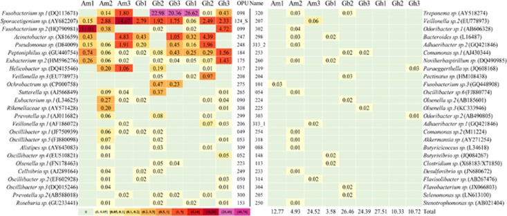
Of the 314 OPUs, 50 affiliated with 34 known genera (Figure 3) but represented independent lineages with identity values <98.7% from any type strain within each respective genus as an indication that they may represent new unclassified species. Therefore, we considered them yet to be described species. These 50 new species accounted for between 3.6% and 27.5% of the reads in the individual vultures and 15.9% of the total reads for all vultures (Figure 3; Supplementary Table S4). Only Sporacetigenium sp. (OPU124_S) and Peptoniphilus sp. (OPU184) were detected in all nine vultures. Of the 34 genera with putative new species, 23 have species of pathogens that caused human infections or have been isolated from clinical specimens, such as Acinetobacter (OPU247), Alistipes (OPU309), Bacteroides (OPU298), Butyrivibrio (OPU148), Clostridium (OPU113), Comamonas (OPU253), Desulfovibrio (OPU233), Eubacterium (OPU090), Fusobacterium (OPU098), Helicobacter (OPU317), Ochrobactrum (OPU275), Odoribacter (OPU302), Olsenella (OPU223), Oscillibacter (OPU049), Paraeggerthella (OPU219), Peptoniphilus (OPU184), Prevotella (OPU299), Pseudomonas (OPU248), Selenomonas (OPU205), Stenotrophomonas (OPU250), Sutterella (OPU265), Treponema (OPU320), Veillonella (OPU206).
Figure 3.
Abundance heatmap of the 50 yet to be described bacterial species.
Of the 314 OPUs, 161 were considered unknown new lineages of uncultured representatives, which represented new branches affiliating outside any known genus but representing putative unclassified new lineages different from the known genera. These summed to between 0.4% and 33.0% of the reads of each individual vulture (Supplementary Table S5) and 11.0% of the total reads for all vultures. However, none of those unknown new lineages of uncultured representatives was detected in all nine vultures studied. In addition, OPU287 was identified as a chloroplast.
Top 10 most abundant species and two-component model
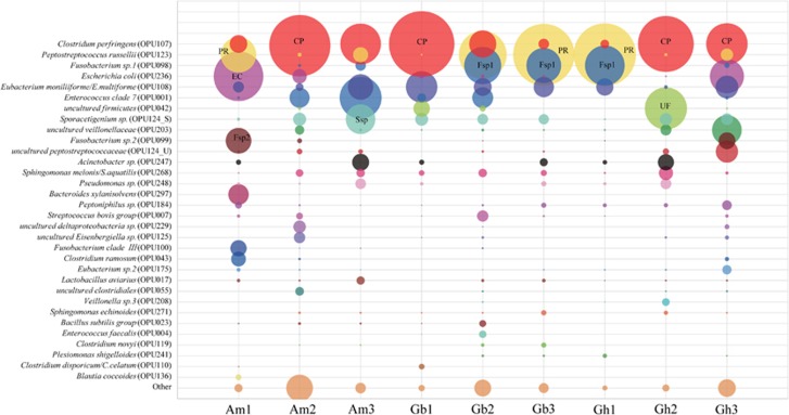
The top ten most abundant OPUs of each vulture summed to a total of 32 OPUs, covering between 88.1% and 99.3% of their respective total reads (Figure 4, Supplementary Tables S3–S5). Only six OPUs were shared by all vultures, namely, C. perfringens (OPU107), P. russellii (OPU123), E. moniliiforme/E. multiforme (OPU108), Sporacetigenium sp. (OPU124_S), S. melonis/S. aquatilis (OPU268) and Peptoniphilus sp. (OPU184). It was remarkable that the two OPUs (C+P) C. perfringens (OPU107) and P. russellii (OPU123) accounted for 51.7% of the total reads, ranging from 26.3% to 71.9% in each individual sample. The OPUs with total reads higher than 1% also included Fusobacterium sp. 1 (OPU098), E. coli/Shigella (OPU236), Eubacterium moniliiforme/E. multiforme (OPU_108), Enterococcus clade 7 (OPU001), uncultured Firmicutes (OPU042), Sporacetigenium sp. (OPU124_S), uncultured Veillonellaceae (OPU203), Fusobacterium sp. 2 (OPU099), uncultured Peptostreptococcaceae (OPU124_U) and Acinetobacter sp. (OPU247) (Figure 4).
Figure 4.
Abundances of the top ten most abundant OPUs. The abundances are shown as percentages of reads in each vulture. A total of 32 OPUs were collected as being the top 10 within the set of all vultures. The others indicated the rest of the OPUs for a given vulture.
Comparison between the PacBio and Illumina MiSeq approaches
Using the Illumina MiSeq platform, we detected a total of 126 OPUs of which only 44 (35%) were identified as known species, representing 43.9% of the total reads (Supplementary Tables S3–S6). The combination of the two approaches summed a total of 355 OPUs. Of these, the majority (87.5% or 83.1% of the total reads, respectively; Supplementary Figure S2) affiliated with OPUs common to the two approaches. Actually, the two methods produced equivalent relative abundances for all shared taxa. Only OPU123 (P. russellii) was underrepresented in the Illumina approach, a fact that is most likely related to amplification biases. Of the 355 OPUs, 229 (64.5%) were only detected by PacBio, accounting for 12.5% of the total reads. Finally, 41 OPUs were only detected by Illumina MiSeq, accounting for 16.8% of its total reads (Supplementary Figure S2, Supplementary Table S6).
Microbiome profiles between Old World and New World vultures
We compared our data with those generated for the New World vultures.25 The 466 824 sequences of 435 bp in length affiliated with 105 OPUs (Supplementary Figure S3). The Old and New World vultures shared 40 OPUs, which accounted for 66.7 and 85.3% of the total reads, respectively. Only 65 OPUs were detected in the New World vultures (Supplementary Figure S3). The five most abundant OPUs shared by all vultures were C. perfringens (OPU107), P. russellii (OPU123), E. coli/Shigella (OPU236), Sporacetigenium sp. (OPU124_S), and Fusobacterium sp. 2 (OPU099). The five most abundant species only detected in Old World vultures were Fusobacterium sp. 1 (OPU098), E. moniliiforme/E. multiforme (OPU108), Enterococcus clade 7 (OPU001), uncultured Firmicutes (OPU042) and uncultured Veillonellaceae (OPU203) (Supplementary Figure S3).
Microbiome-guided culturing of C. perfringens
The species-level microbiome analysis indicated that the deadly pathogen C. perfringens was the most abundant commensal member in the intestinal flora of all vultures. Therefore, the specific isolation of the members of this species was conducted. A total of 107 strains of C. perfringens were isolated from the rectal swabs of the nine vultures (Supplementary Figure S4). All strains were identified by full-length 16S rRNA sequence analysis and tested positive for the major lethal alpha-toxin gene by the PCR method, which was then confirmed by sequencing. The toxin gene beta2 toxin gene (cpb2) was found in fifteen isolates belonging to two sequence types (STs), while other toxin genes were tested negative. The multi-locus sequence typing (MLST) approach based on eight genes grouped the 107 strains into 45 sequence types, including alpha-toxin gene (plc), D-alanine-D-alanine ligase gene (ddlA), deoxyuridine triphosphatase gene (dut), glycerol kinase gene (glpK), deoxyguanylate kinase gene (gmk), recombinase gene (recA), superoxide dismutase gene (sod) and triose phosphate isomerase gene (tpiA) (Supplementary Table S7).27 Twenty-seven STs contained a single strain. There were three predominant STs, namely, ST33, ST41, and ST45, containing 18, 11 and six strains, respectively. The neighbor-joining reconstruction based on the concatenated MLST sequences showed that the strains from the vultures were distributed into four groups, different from the avian isolates.
DISCUSSION
Metataxonomic analysis with almost full-length 16S rRNA gene sequencing generated by the PacBio system allowed the precise identification of the microbiome composition up to the species category.9, 10, 26 Our study evidenced the benefits of using this approach as it (i) produces an almost complete gene sequence of the amplicons and (ii) seems to render higher-diversity yields of especially the low-abundance microbiota in comparison with the Illumina MiSeq approach. The fact that the rates of insertion and deletion were low, together with the capabilities to use general filters to remove phylogenetic noise23 and the de novo reconstruction based on phylogenetic inferences, provide the OPU recognition and identification with high confidence. The combination of PacBio and Illumina MiSeq data revealed the recognition of 355 OPUs that could be assumed to represent different individual species.10 The PacBio revealed approximately 65% and Illumina MiSeq approximately 12% OPUs that were not reciprocally detected. However, the coincident taxa detected represented >83% of the reads in both cases, and the relative proportions were generally equivalent. The Illumina MiSeq rendered much a lower resolution in revealing the low-abundance microorganisms, despite the number of sequences being approximately 15x higher. Using the metataxonomic approach, we could assign approximately 72.9% of the total reads to known species and 15.9% to yet to be described species. Actually, of the top 10 most abundant microbes in each individual vulture, approximately 72.2% of the total reads could be readily identified as members of known species and 15.3% as yet to be described species.
In this study, an especially remarkable finding was the unexpected abundance and predominance of C. perfringens, a well-known pathogen causing enteric diseases and gas gangrene.32 C. perfringens had been already detected as prevalent in healthy dogs by means of culture-dependent approaches.33 To compare our results with the New World vulture's microbiome,25 we applied the OPU approach to the available sequences and followed the same strategy as for other Roche 454 datasets with partial sequences.9, 10 Approximately 40 OPUs were shared by the two sample sets, with sequence contributions of >66% in the Old World and >85% in the New World. The shorter sequences, together with the OTU strategy used in the original study, restricted the accurate identification to only the genus level.25 However, we identified 88% as members of known species or new species within known genera. Within the shared microbiome, C. perfringens was again one of the most relevant species. Although an accurate species identification was not achieved, Roggenbuck already identified gene coding for the tissue-degrading enzymes and toxins of C. perfringens. The New World vultures seem to have a similar pattern of two components to that observed in our data set (C+P). In both cases, C. perfringens is one of the key players.25
The abundance of C. perfringens in all vultures yet studied brought us to believe that the relationships between C. perfringens and vultures could be mutualistic and not pathogenic. Therefore, both the New and Old World vultures in the USA and China seem to be important animal reservoirs for this pathogenic species. We also analyzed lineages of C. perfringens isolates associated with distinct disease presentations. These 107 isolates were identified into three evolutionary lineages: Lineage I, Gangrene, bovine hemorrhagic enteritis (β2+), antibiotic-associated diarrhea, retail meats (non-pathogenic); Lineage II, Human peritonitis and septicemia; and Lineage III, Bovine and equine hemorrhagic enteritis (β2-) (data not shown).34 An estimated 9.4 million cases of foodborne diseases occur each year in the US, and one million (10%) of them have been related to C. perfringens poisoning.35 Therefore, this finding has significance in public health, which was reinforced by the isolation of 107 strains from the nine vultures encoding for the virulence gene and the surprising diversity of STs and evolutionary lineages.
Most gastrointestinal infections by C. perfringens are caused by the contamination of food sources by humans or by animal feces. However, C. perfringens is ubiquitous in the soil and, thus, can be a source of gastrointestinal diseases36, 37 and gas gangrene, which was common in the combat injuries of soldiers well into the 20th century.37 Although we have no direct evidence of soilborne food poisoning or gas gangrene, our results indicate that the vulture's fecal deposits could be a means of dispersion of pathogenic bacteria, especially C. perfringens due to its high prevalence. In this regard, it was remarkable that both the C. perfringens isolates in this study and those recovered from the soil were predominantly type A.36
It seems that vultures could be an animal reservoir of many additional emerging bacterial pathogens, as from the 102 species identified in Old World vultures, 45 have been isolated from patients or were reported as responsible for outbreaks.31 It should be noted that both R. pickettii and S. paucimobilis have been recognized as emerging pathogens in hospital settings as causative agents for respiratory infections or meningitis. Very interestingly, both were isolated from soil samples.38, 39, 40 The results provide an almost complete inventory of the potentially zoonotic known bacteria in vultures.
In short, our metataxonomic approach offers a species-precise identification for microbiome profiling. It could be used for a pre-screening of putative pathogens for routine implementation in future large-scale epidemiological studies, for the culture-independent etiological investigation of infectious disease outbreaks, and as a culture-guidance methodology.
It should be noted that some of the Old World vultures on the Tibet-Qinghai Plateau are on the list of state-protected wildlife, including Gypaetus barbatus, Gyps himalayensis and Aegypius monachus. This wildlife protection regulation limited the number of vultures that could be sampled.
Acknowledgments
This work was supported by grants from the National Natural Science Foundation of China (81290340 and 81290345) and by a grant (2016YFC1201903) from the Ministry of Science and Technology, China. RRM was supported by the Spanish Ministry of Economy project CLG2015_66686-C3-1-P, and by European Regional Development Fund (FEDER) funds.
Footnotes
Supplementary Information for this article can be found on the Emerging Microbes & Infections website (http://www.nature.com/emi)
Supplementary Material
References
- Woolhouse M, Gaunt E. Ecological origins of novel human pathogens. Crit Rev Microbiol 2007; 33: 231–242. [DOI] [PubMed] [Google Scholar]
- Mazet JA, Wei Q, Zhao G J et al. Joint China-US call for employing a transdisciplinary approach to emerging infectious diseases. Ecohealth 2015; 12: 555–559. [DOI] [PMC free article] [PubMed] [Google Scholar]
- Kahn RE, Clouser DF, Richt JA. Emerging infections: a tribute to the one medicine, one health concept. Zoonoses Public Health 2009; 56: 407–428. [DOI] [PubMed] [Google Scholar]
- Lu X, Ke D, Zeng X, Gong G, Ci R. Status, ecology, and conservation of the Himalayan griffon Gyps himalayensis (Aves, Accipitridae) in the Tibetan plateau. Ambio 2009; 38: 166–173. [DOI] [PubMed] [Google Scholar]
- Ge P, Xi J, Ding J et al. Primary case of human pneumonic plague occurring in a Himalayan marmot natural focus area Gansu Province, China. Int J Infect Dis 2015; 33: 67–70. [DOI] [PubMed] [Google Scholar]
- Marchesi JR, Ravel J. The vocabulary of microbiome research: a proposal. Microbiome 2015; 3: 31. [DOI] [PMC free article] [PubMed] [Google Scholar]
- Yarza P, Yilmaz P, Pruesse E et al. Uniting the classification of cultured and uncultured bacteria and archaea using 16S rRNA gene sequences. Nat Rev Microbiol 2014; 12: 635–645. [DOI] [PubMed] [Google Scholar]
- Schloss PD, Jenior ML, Koumpouras CC, Westcott SL, Highlander SK. Sequencing 16S rRNA gene fragments using the PacBio SMRT DNA sequencing system. PeerJ 2016; 4: e1869. [DOI] [PMC free article] [PubMed] [Google Scholar]
- Mora-Ruiz MR, Font-Verdera F, Orfila A, Rita J, Rossello-Mora R. Endophytic microbial diversity of the halophyte Arthrocnemum macrostachyum across plant compartments. FEMS Microbiol Ecol 2016; 92: fiw145. [DOI] [PubMed] [Google Scholar]
- Vidal R, Ginard D, Khorrami S et al. Crohn associated microbial communities associated to colonic mucosal biopsies in patients of the western Mediterranean. Syst Appl Microbiol 2015; 38: 442–452. [DOI] [PubMed] [Google Scholar]
- Seibold I, Helbig AJ. Evolutionary history of New and Old World vultures inferred from nucleotide sequences of the mitochondrial cytochrome b gene. Philos Trans R Soc Lond B Biol Sci 1995; 350: 163–178. [DOI] [PubMed] [Google Scholar]
- Kerr KCR, Stoeckle MY, Dove CJ, Weigt LA, Francis CM. Hebert PDN. Comprehensive DNA barcode coverage of North American birds. Mol Ecol Notes 2007; 7: 535–543. [DOI] [PMC free article] [PubMed] [Google Scholar]
- Weisburg WG, Barns SM, Pelletier DA, Lane DJ. 16S ribosomal DNA amplification for phylogenetic study. J Bacteriol 1991; 173: 697–703. [DOI] [PMC free article] [PubMed] [Google Scholar]
- Klindworth A, Pruesse E, Schweer T et al. Evaluation of general 16S ribosomal RNA gene PCR primers for classical and next-generation sequencing-based diversity studies. Nucleic Acids Res 2013; 41: e1. [DOI] [PMC free article] [PubMed] [Google Scholar]
- Magoc T, Salzberg SL. FLASH: fast length adjustment of short reads to improve genome assemblies. Bioinformatics 2011; 27: 2957–2963. [DOI] [PMC free article] [PubMed] [Google Scholar]
- Bokulich NA, Subramanian S, Faith JJ et al. Quality-filtering vastly improves diversity estimates from Illumina amplicon sequencing. Nat Methods 2013; 10: 57–59. [DOI] [PMC free article] [PubMed] [Google Scholar]
- Edgar RC, Haas BJ, Clemente JC, Quince C, Knight R. UCHIME improves sensitivity and speed of chimera detection. Bioinformatics 2011; 27: 2194–2200. [DOI] [PMC free article] [PubMed] [Google Scholar]
- Wang Q, Garrity GM, Tiedje JM, Cole JR. Naive Bayesian classifier for rapid assignment of rRNA sequences into the new bacterial taxonomy. Appl Environ Microbiol 2007; 73: 5261–5267. [DOI] [PMC free article] [PubMed] [Google Scholar]
- Hammer Ø, Harper DA, Ryan PD. PAST: paleontological statistics software package for education and data analysis. Palaeontol Electr 2001; 4: 1–9. [Google Scholar]
- Edgar RC. UPARSE: highly accurate OTU sequences from microbial amplicon reads. Nat Methods 2013; 10: 996–998. [DOI] [PubMed] [Google Scholar]
- Yarza P, Ludwig W, Euzeby J et al. Update of the All-Species Living Tree Project based on 16S and 23S rRNA sequence analyses. Syst Appl Microbiol 2010; 33: 291–299. [DOI] [PubMed] [Google Scholar]
- Quast C, Pruesse E, Yilmaz P et al. The SILVA ribosomal RNA gene database project: improved data processing and web-based tools. Nucleic Acids Res 2013; 41: D590–D596. [DOI] [PMC free article] [PubMed] [Google Scholar]
- Ludwig W, Strunk O, Westram R et al. ARB: a software environment for sequence data. Nucleic Acids Res 2004; 32: 1363–1371. [DOI] [PMC free article] [PubMed] [Google Scholar]
- Munoz R, Yarza P, Rosselló-Móra R. Harmonized phylogenetic trees for the Prokaryotes. In: Rosenberg E, DeLong EF, Stackebrandt E, Thompson F (eds). The Prokaryotes: Actinobacteria. Springer-Verlag Berlin Heidelberg. 2014, 1–3. [Google Scholar]
- Roggenbuck M, Baerholm Schnell I, Blom N et al. The microbiome of New World vultures. Nat Commun 2014; 5: 5498. [DOI] [PubMed] [Google Scholar]
- Mora-Ruiz MR, Font-Verdera F, Diaz-Gil C et al. Moderate halophilic bacteria colonizing the phylloplane of halophytes of the subfamily Salicornioideae (Amaranthaceae). Syst Appl Microbiol 2015; 38: 406–416. [DOI] [PubMed] [Google Scholar]
- Jost BH, Trinh HT, Songer JG. Clonal relationships among Clostridium perfringens of porcine origin as determined by multilocus sequence typing. Vet Microbiol 2006; 116: 158–165. [DOI] [PubMed] [Google Scholar]
- Baums CG, Schotte U, Amtsberg G, Goethe R. Diagnostic multiplex PCR for toxin genotyping of Clostridium perfringens isolates. Vet Microbiol 2004; 100: 11–16. [DOI] [PubMed] [Google Scholar]
- Chalmers G, Bruce HL, Hunter DB et al. Multilocus sequence typing analysis of Clostridium perfringens isolates from necrotic enteritis outbreaks in broiler chicken populations. J Clin Microbiol 2008; 46: 3957–3964. [DOI] [PMC free article] [PubMed] [Google Scholar]
- Lebreton F, Willems RJL, Gilmore MS. Enterococcus diversity, origins in nature, and gut colonization. In: Gilmore MS, Clewell DB, Ike Y, Shankar N (eds). Enterococci: From Commensals to Leading Causes of Drug Resistant Infection [Internet]. Boston: Massachusetts Eye and Ear Infirmary, 2014. [PubMed] [Google Scholar]
- Woo PC, Teng JL, Yeung JM, Tse H, Lau SK, Yuen KY. Automated identification of medically important bacteria by 16S rRNA gene sequencing using a novel comprehensive database, 16SpathDB. J Clin Microbiol 2011; 49: 1799–1809. [DOI] [PMC free article] [PubMed] [Google Scholar]
- Silva RO, Lobato FC. Clostridium perfringens: A review of enteric diseases in dogs, cats and wild animals. Anaerobe 2015; 33: 14–17. [DOI] [PubMed] [Google Scholar]
- Goldstein MR, Kruth SA, Bersenas AM, Holowaychuk MK, Weese JS. Detection and characterization of Clostridium perfringens in the feces of healthy and diarrheic dogs. Can J Vet Res 2012; 76: 161–165. [PMC free article] [PubMed] [Google Scholar]
- Rooney AP, Swezey JL, Friedman R, Hecht DW, Maddox CW. Analysis of core housekeeping and virulence genes reveals cryptic lineages of Clostridium perfringens that are associated with distinct disease presentations. Genetics 2006; 172: 2081–2092. [DOI] [PMC free article] [PubMed] [Google Scholar]
- Scallan E, Hoekstra RM, Angulo FJ et al. Foodborne illness acquired in the United States—major pathogens. Emerg Infect Dis 2011; 17: 7–15. [DOI] [PMC free article] [PubMed] [Google Scholar]
- Li J, Sayeed S, McClane BA. Prevalence of Enterotoxigenic Clostridium perfringens Isolates in Pittsburgh (Pennsylvania) Area Soils and Home Kitchens. Appl Environ Microbiol 2007; 73: 7218–7224. [DOI] [PMC free article] [PubMed] [Google Scholar]
- Baumgardner DJ. Soil-related bacterial and fungal infections. J Am Board Fam Med 2012; 25: 734–744. [DOI] [PubMed] [Google Scholar]
- Ryan MP, Pembroke JT, Adley CC. Genotypic and phenotypic diversity of Ralstonia pickettii and Ralstonia insidiosa isolates from clinical and environmental sources including High-purity Water. Diversity in Ralstonia pickettii. BMC Microbiol 2011; 11: 194. [DOI] [PMC free article] [PubMed] [Google Scholar]
- Labarca JA, Trick WE, Peterson CL et al. Multistate nosocomial outbreak of Ralstonia pickettii colonization associated with an intrinsically contaminated respiratory care solution. Clin Infect Dis 1999; 29: 1281–1286. [DOI] [PubMed] [Google Scholar]
- Coppotelli BM, Ibarrolaza A, Del Panno MT, Morelli IS. Effects of the inoculant strain Sphingomonas paucimobilis 20006FA on soil bacterial community and biodegradation in phenanthrene-contaminated soil. Microb Ecol 2007; 55: 173–183. [DOI] [PubMed] [Google Scholar]
Associated Data
This section collects any data citations, data availability statements, or supplementary materials included in this article.


