. 2017 Jan 30;6:e20991. doi: 10.7554/eLife.20991

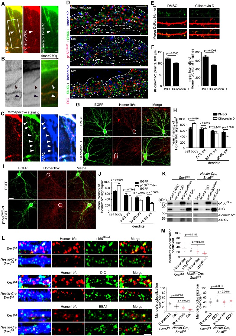
Figure 6. SNX6 is required for motility of Homer1b/c vesicles in dendritic shaft and their association with dynein−dynactin.

(A–C) Dynamic behavior of mCherry-SNX6 and mEmerald-Homer1c in distal dendrite. The last frame of motile SNX6-, Homer1c-positive puncta (arrowhead) in dendrite (A) with the respective kymograph of boxed area (B) is shown. A retrospective staining of MAP2 after live imaging (C) illuminates dendrite identity. (D) Superresolution images of DIV18 neurons immunostained with antibodies to SNX6, Homer1b/c, and p150Glued or DIC. Shown are representative images of dendrites outlined with dashed lines. White arrowheads indicate overlapped signals. (E) DIV14 neurons were transfected with construct expressing EGFP, treated with DMSO or Ciliobrevin D on DIV16 for 2 hr and immunostained with antibodies to Homer1b/c. (F) Quantification of puncta number per 100 μm dendrite length and mean intensity in spines for Homer1b/c in (E) (mean ± SEM, n = 30, N = 3). (G) Same as (E), shown are representative confocal images of EGFP-expressing neurons. Dashed lines outline the cell bodies. (H) Quantification of Homer1b/c distribution in the cell body and dendrites in (G) (mean ± SEM, n = 30, N = 3). (I) DIV14 neurons were transfected with construct overexpressing EGFP or p150Glued-N-EGFP, fixed on DIV16 and immunostained with antibodies to Homer1b/c. Dashed lines outline the cell bodies. (J) Quantification of Homer1b/c distribution in the cell body and dendrites in (I) (mean ± SEM, n = 30, N = 3). (K) Membrane fractions from mouse brain lysates were subjected to immunoisolation with antibodies to p150Glued or DIC coupled to Dynabeads Protein G, respectively. Shown are immunoblots probed with antibodies to SNX6, p150Glued, DIC, and Homer1b/c. (L) Hippocampal neurons cultured from Snx6fl/fl and Nestin-Cre; Snx6fl/fl mice were transfected with pLL3.7.1 on DIV14, fixed on DIV17 and immunostained with antibodies to Homer1b/c and p150Glued, DIC or EEA1. Shown are representative confocal images of dendritic segments. (M) Quantification of colocalization in (L) from 45 dendritic segments of 15 neurons (mean ± SEM, N = 2. Total length of dendrites: Snx6fl/fl/Nestin-Cre; Snx6fl/fl: 1247 μm/1058 μm for p150Glued; 1264 μm/1301 μm for DIC; 1244 μm/1291 μm for EEA1). Bars, 2 μm in (A), (D), (E) and (L), 20 μm in (G) and (I).

DOI: http://dx.doi.org/10.7554/eLife.20991.014

Figure 6.

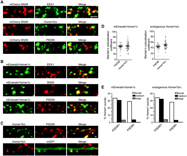
Figure 6—figure supplement 1. Overexpressed mCherry-SNX6 and mEmerald-Homer1c recapitulate the distribution of endogenous proteins in hippocampal neurons.

Figure 6—figure supplement 1.

(A) Hippocampal neurons in dissociated culture were transfected with construct expressing SNX6 on DIV14, digitonin extracted and fixed on DIV16 and immunostained with antibodies to EEA1, Homer1b/c, or PSD95. Shown are the representative confocal images. (B) Hippocampal neurons in dissociated culture were transfected with construct expressing mEmerald-Homer1c on DIV14, digitonin extracted and fixed on DIV16 and immunostained with antibodies to EEA1, SNX6, or PSD95. (C) Hippocampal neurons in dissociated culture were fixed and stained with antibodies to Homer1b/c and PSD95 on DIV16, or transfected with construct expressing membrane-bound GFP (mGFP) on DIV14, digitonin extracted and fixed on DIV16 and immunostained with antibodies to Homer1b/c. (D) Quantification of colocalization between Homer1 and PSD95 in (B) and (C) (mEmerald-Homer1c, 26 dendritic segments of 13 neurons; endogenous Homer1b/c, 22 dendritic segments of 11 neurons). (E) Quantification of size distribution of PSD95-positive (+) or negative (−) Homer1 puncta in dendrites from (B) and (C). Sizes of puncta: small (0.04–0.3 μm2), medium (0.3–0.8 μm2) and large (0.8–2.0 μm2). Numbers of puncta quantified: mEmerald-Homer1c, 672 from 13 neurons; endogenous Homer1b/c, 502 from 11 neurons. Bar: 2 μm.
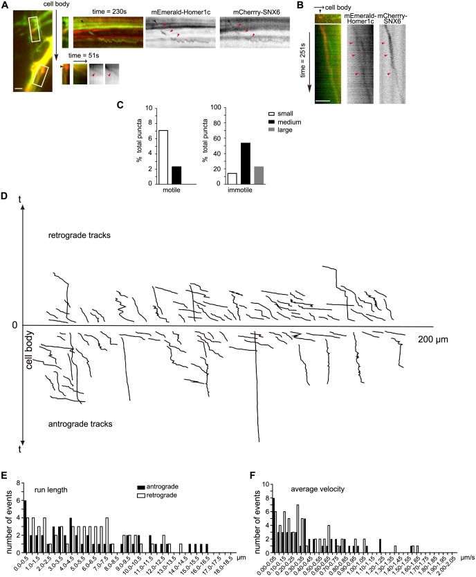
Figure 6—figure supplement 2. SNX6-, Homer1c-positive puncta move bidirectionally in the dendritic shaft.

Figure 6—figure supplement 2.

(A) SNX6-, Homer1c-positive puncta in proximal dendrite move away from the cell body. Shown are the first frames of motile SNX6-, Homer1c-positive puncta in boxed areas (Left panel, black arrow indicates the relative orientation of the cell body) in proximal dendrite and the respective kymographs (right panels, black arrowheads indicate the initial position and red arrowheads indicate the trajectory of vesicles). (B) SNX6-, Homer1c-positive puncta in distal dendrite moves towards the cell body. Shown are the first frame of a motile SNX6-, Homer-positive structure (top left panel) in distal dendrite and the kymograph (all other panels). Bars: 2 μm. (C) Quantification of the size distribution of motile and immotile SNX6-, Homer1c-positive puncta in dendrites (1128 puncta from 31 neurons). Shown are the percentage of small (0.14‒0.3 μm2), medium (0.3‒0.8 μm2) and large puncta (0.8‒2.8 μm2) in the motile and immotile populations, respectively. (D) The positions of all motile SNX6 and Homer1c-positive puncta as a function of time (the trajectory of each vesicle) were aligned at their starting positions away from the cell body (106 vesicles from 31 neurons). (E and F) Histograms of frequency distribution for run lengths (E) and average velocities (F) of anterograde and retrograde motile SNX6-, Homer1b/c-positive puncta.