Supplemental Digital Content is available in the text.
Keywords: exercise, heart failure
Abstract
Background:
Small studies have suggested that high-intensity interval training (HIIT) is superior to moderate continuous training (MCT) in reversing cardiac remodeling and increasing aerobic capacity in patients with heart failure with reduced ejection fraction. The present multicenter trial compared 12 weeks of supervised interventions of HIIT, MCT, or a recommendation of regular exercise (RRE).
Methods:
Two hundred sixty-one patients with left ventricular ejection fraction ≤35% and New York Heart Association class II to III were randomly assigned to HIIT at 90% to 95% of maximal heart rate, MCT at 60% to 70% of maximal heart rate, or RRE. Thereafter, patients were encouraged to continue exercising on their own. Clinical assessments were performed at baseline, after the intervention, and at follow-up after 52 weeks. Primary end point was a between-group comparison of change in left ventricular end-diastolic diameter from baseline to 12 weeks.
Results:
Groups did not differ in age (median, 60 years), sex (19% women), ischemic pathogenesis (59%), or medication. Change in left ventricular end-diastolic diameter from baseline to 12 weeks was not different between HIIT and MCT (P=0.45); left ventricular end-diastolic diameter changes compared with RRE were −2.8 mm (−5.2 to −0.4 mm; P=0.02) in HIIT and −1.2 mm (−3.6 to 1.2 mm; P=0.34) in MCT. There was also no difference between HIIT and MCT in peak oxygen uptake (P=0.70), but both were superior to RRE. However, none of these changes was maintained at follow-up after 52 weeks. Serious adverse events were not statistically different during supervised intervention or at follow-up at 52 weeks (HIIT, 39%; MCT, 25%; RRE, 34%; P=0.16). Training records showed that 51% of patients exercised below prescribed target during supervised HIIT and 80% above target in MCT.
Conclusions:
HIIT was not superior to MCT in changing left ventricular remodeling or aerobic capacity, and its feasibility remains unresolved in patients with heart failure.
Clinical Trial Registration:
URL: http://www.clinicaltrials.gov. Unique identifier: NCT00917046.
Current guidelines recommend exercise training as an adjunctive therapy in patients with chronic heart failure.1 A universal agreement on exercise prescription does not exist; thus, an individualized approach, including behavioral characteristics, personal goals, and preferences, is recommended.2,3 At present, moderate continuous endurance exercise is the best described and established form of training because of its well-demonstrated efficacy and safety.2 This advice is based mainly on a large multicenter exercise intervention trial (HF-ACTION [Heart Failure: A Controlled Trial Investigating Outcomes of Exercise Training]) with 2331 patients with heart failure, which observed a moderate reduction of symptoms, improvement of exercise capacity, and a reduction of hospital readmissions for heart failure.4
Exercise of high submaximal intensity performed in intervals of 1 to 4 minutes, also called high-intensity interval training (HIIT), has been tested in a small study of patients with heart failure with reduced ejection fraction, showing that HIIT was superior to moderate continuous training (MCT) in improving exercise capacity, quality of life, endothelial function, and left ventricular diameter and ejection fraction.5 The results were better than those observed in previous studies and meta-analyses of patients with chronic heart failure.6,7 They also prompted discussions of whether HIIT should be included in standard care of patients with chronic heart failure.
This background formed the basis for a larger randomized controlled multicenter trial, the SMARTEX Heart Failure Study (Study of Myocardial Recovery After Exercise Training in Heart Failure), to test the hypothesis that HIIT is superior to MCT with regard to improvement of left ventricular dimensions and exercise capacity.
Methods
Study Design
The SMARTEX Heart Failure Study is an investigator-initiated randomized controlled clinical trial conducted at 9 European centers (Antwerp, Copenhagen, Leipzig, Luxembourg, Munich, Stavanger, Trondheim/Levanger, Veruno, and Ålesund) between June 2009 and July 2014. The final patient was randomized July 1, 2013, and had the 52-week follow-up on July 22, 2014. The Clinical Trials database registration reports 268 patients enrolled in the Web case report form database. However, 7 randomizations were error entries during initial testing and demonstration of the database. Thus, the correct number of patients randomized was 261. The trial was approved by the Regional Committee for Medical and Health Ethics of Central Norway and by national and local committees where required. Informed written consent was obtained from all participants. Details of rationale, design, methods, sample size, randomization, and organization have previously been published.8 Data management and statistical analyses were performed by the coordinating center with oversight by the steering committee (Ø.E., M.H., A.L., E.P.), whose members had full access to all data and vouch for the accuracy and completeness of data and analyses.
Patients and Interventions
Patients were enrolled from outpatient heart failure clinics, referrals to cardiac rehabilitation, public announcements, and screening of eligible patients in hospital registries. Eligible patients with symptomatic (New York Heart Association class II–III), stable, pharmacologically optimally treated chronic heart failure were randomized 1:1:1 to a 12-week program of HIIT, MCT, or recommendation of regular exercise (RRE), stratified by study center and pathogenesis (ischemic versus nonischemic). Stratification by center was performed to avoid bias from unobserved treatment differences, and stratification by pathogenesis was performed to allow possible post hoc analysis of the influence on left ventricular end-diastolic diameter (LVEDD) changes. Exercise training protocols have been described elsewhere.5,8 Briefly, HIIT and MCT had 3 supervised sessions per week on a treadmill or bicycle. HIIT included four 4-minute intervals aiming at 90% to 95% of maximal heart rate separated by 3-minute active recovery periods of moderate intensity. HIIT sessions lasted 38 minutes including warm-up and cool-down at moderate intensity. MCT sessions aimed at 60% to 70% of maximal heart rate and lasted 47 minutes. HIIT and MCT sessions were estimated to obtain similar energy expenditures.9 Patients randomized to RRE were advised to exercise at home according to current recommendations and attended a session of moderate-intensity training at 50% to 70% of maximal heart rate every 3 weeks.8 In all 3 groups, there were no supervised training sessions after the 12-week interventions, but the investigators had telephone contact with the participants every 4 weeks to register clinical events and to encourage physical activity.
Clinical Assessments
Screening procedures and clinical assessments before and after exercise interventions were performed at local study centers as previously described.8 Briefly, medical history, anthropometrics, physical examination including fasting blood sampling, quality-of-life questionnaires, cardiopulmonary exercise testing, and echocardiography were performed, in addition to prespecified substudies.
Echocardiography data were acquired according to standard operation procedures of the study, stored digitally, and transferred as DICOM (digital imaging and communications in medicine) files or raw data to the core laboratory in Trondheim, Norway. Analyses were performed by 1 of 2 expert echocardiographers (A.S. and H.D.) blinded to group assignment but not always to time point of assessment on EchoPAC SW (version BT 11–13; GE Ultrasound, Horten, Norway). LVEDD was measured at the tip of the mitral leaflet in the 2-dimensional parasternal long-axis view.10 Repeatability was tested by Bland-Altman analyses of the first 25 baseline assessments between the 2 investigators. There was no bias (0.3 mm), and the coefficient of variation was 4.1%.
Cardiopulmonary exercise testing was performed with standard equipment for indirect calorimetry in an incremental protocol until exhaustion on either a treadmill or a bicycle ergometer, depending on exercise training equipment. The protocol comprised a 10- or 20-W increase in workload ≈1 minute, starting at 20 or 40 W, respectively. For comparisons per patient, the baseline, 12-week, and 52-week tests were performed with the same protocol. The mean of the 3 highest 10-second consecutive measurements was identified as peak oxygen uptake (Vo2peak). Respiratory quotient and other related values are reported from this time point. Cardiopulmonary exercise testing personnel were not blinded to assignment to intervention group, but analysis was performed separately by an independent investigator (P.B.).
End Points
The primary end point was comparison of groups with respect to change in LVEDD from baseline to the 12-week assessment by echocardiography. Key secondary end points were change in left ventricular ejection fraction and Vo2peak; the latter was also considered a measure of training effect. Safety was assessed by the rate of serious adverse events (SAEs) defined as all-cause and cardiovascular death, worsening heart failure requiring hospitalization or intensified diuretic treatment, atrial and ventricular arrhythmias, unstable angina, inappropriate implantable cardioverter-defibrillator shocks, and other events leading to hospital admission or clinical evaluation. Events were considered training related when occurring during or within 3 hours of supervised exercise training sessions. An independent blinded end-point committee (J.K., R.H., and S.G.) classified all events. Quality of life was assessed with the Kansas City Cardiomyopathy Questionnaire, the Hospital Anxiety and Depression Scale, the Global Mood Scale, and the Type D Scale 14. Physical activity was assessed with the International Physical Activity Questionnaire (7-day short form), but its reliability and validity have not been established in patients with heart failure, so the data have not been included. The measurements were taken at baseline, at 12 weeks immediately after the intervention, and at follow-up 52 weeks from the start of the training program.
Statistical Analysis
Power calculations for the main end point (comparison of the groups with respect to change in LVEDD from baseline to 12 weeks) have been detailed in a previous article on rationale and design.8 We estimated that a total number of 200 patients, randomized 1:1:1 between RRE, MCT, and HIIT, would be sufficient to detect a reduction of LVEDD of 3.0 mm between HIIT and MCT and 5.0 mm between HIIT and RRE. Calculations were based on LVEDD of 70 mm, coefficient of variance of 0.04, statistical power of 0.90, and value of P=0.05, adjusted for 3 comparisons by the Bonferroni correction. Unless otherwise specified, data are presented as median with 95% confidence interval of the median because many variables were nonnormally distributed or as observed numbers with percentages.
The main end-point analysis was prespecified as mixed-models linear regression with robust standard errors, with 12-week values used as outcome and baseline values used as adjustment variables, and included adjustments for center, ischemic or nonischemic pathogenesis, and height. Model fit was checked by residual plots, and estimated contrasts are presented with 95% confidence intervals and P values corrected for 3 pairwise comparisons with the Scheffé method. For comparisons including the 52-week data, similar analyses were performed. A 2-sided value of P≤0.05 was considered statistically significant.
To monitor adherence to training intensity, heart rate and workload were recorded during training sessions, and average training intensity was calculated as percentage of maximal heart rate at baseline. For HIIT and MCT patients, regression models including average percentage of maximal heart rate during training (continuous variable or lowest versus highest quartile), atrial fibrillation (yes versus no), or smoking (present versus former/never) were developed.
The study was not powered to assess differences in safety or clinical events; therefore, SAEs were not a prespecified end point.8 However, safety is an important concern in this population, especially when performing exercise. With acknowledgment of the limitations of post hoc analysis,11 χ2 tests for cardiovascular, noncardiovascular, and total SAEs during the training intervention period and during follow-up were performed with no corrections for multiple testing. Statistical analyses were performed with Stata (version 13.1, StataCorp, College Station, TX).
Results
Patient Population and Adherence to Intervention
After initial exclusions and withdrawals, 231 patients were included in HIIT, MCT, or RRE. Nine dropped out because of SAEs, and 7 withdrew or were lost to follow-up (Figure 1). Two hundred fifteen patients were assessed after 12 weeks and were included in the intention-to-treat analysis reported here. Median adherence to supervised training was 35 (34–36) sessions of 36 possible in HIIT and MCT and 4 (3–4) of 4 in RRE. Eight patients completed <24 of 36 exercise sessions, leaving 207 patients included in the per-protocol analysis that yielded equivalent results (data not shown).
Figure 1.
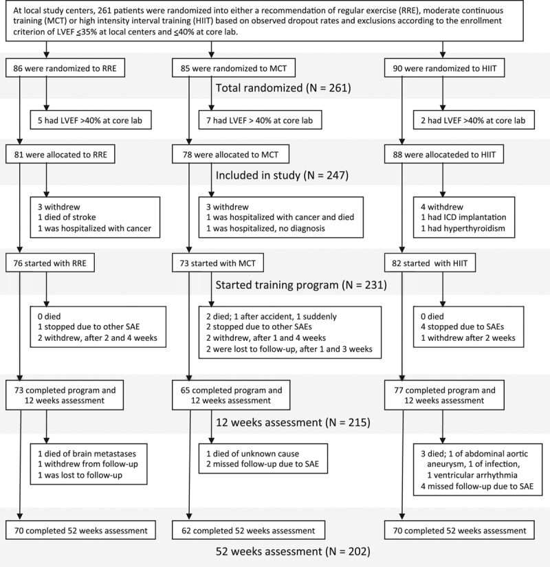
Study enrollment, randomization, and follow-up. Enrollment was stopped when it was estimated that at least 200 patients would complete the 12-week assessments according to protocol. Two hundred fifteen patients came to follow-up assessments and were included in the intention-to-treat analysis; 207 of these were included in per-protocol analysis. Two hundred two patients came to the 52-week assessments and fulfilled the criterion of having completed either echocardiography or cardiopulmonary exercise testing. LVEF indicates left ventricular ejection fraction; and SAE, serious adverse event.
Baseline characteristics were similar in all groups, although more RRE patients had a history of hypertension (Table 1). Median age was 60 years (interquartile range [IQR] 53–70 years); 71% were in New York Heart Association class II, and the rest were in class III. All patients were considered to be on optimal medical treatment. Only 19% of the patients were women (Table 1). Median left ventricular ejection fraction at baseline was 29% (IQR, 24%–34%), and median Vo2peak was 17.1 mL·kg−1·min−1 (IQR, 14.2–20.3 mL·kg−1·min−1) with no difference between groups at baseline (Table 2 and Tables I and II in the online-only Data Supplement).
Table 1.
Patient Characteristics at Baseline
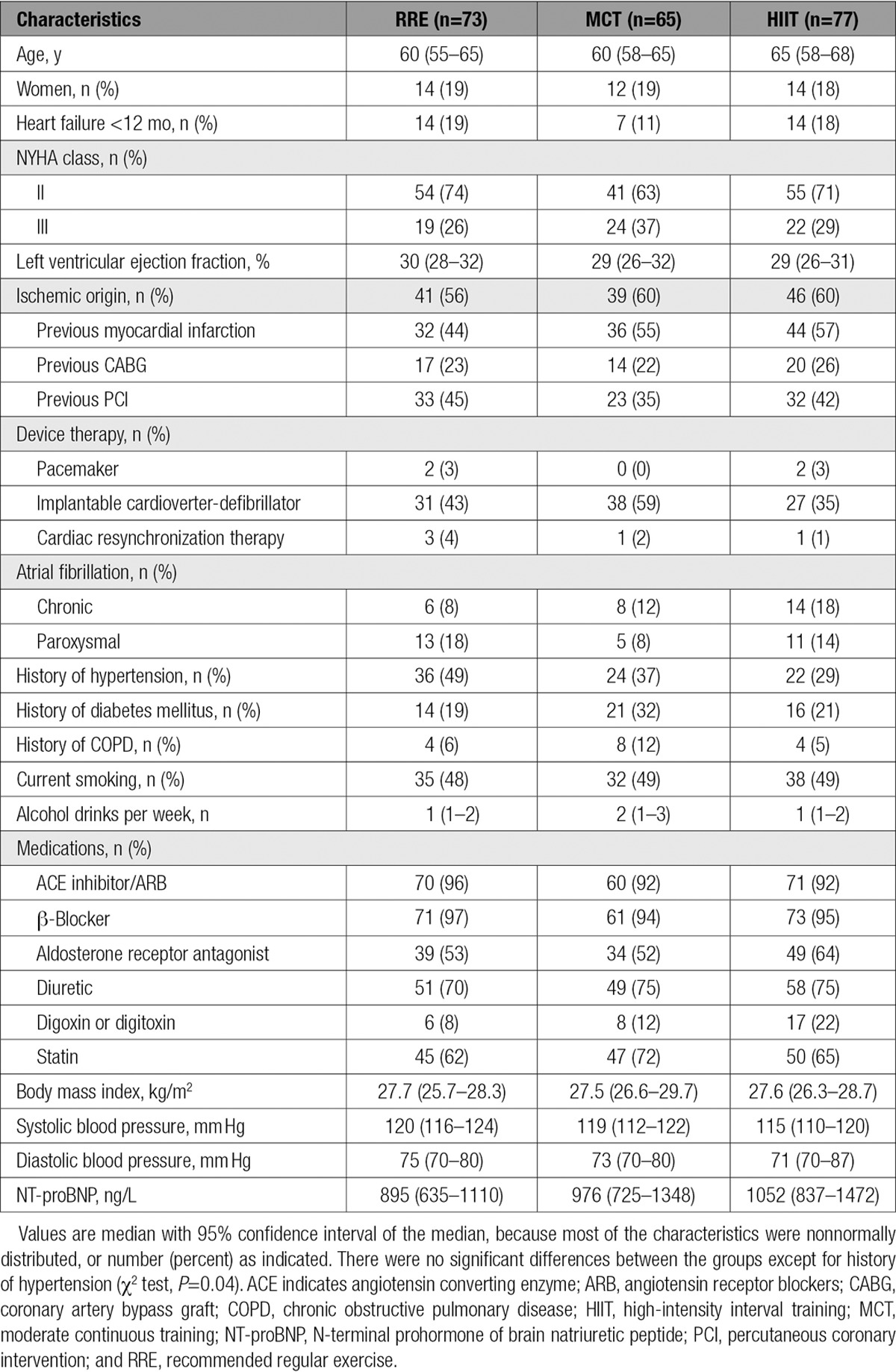
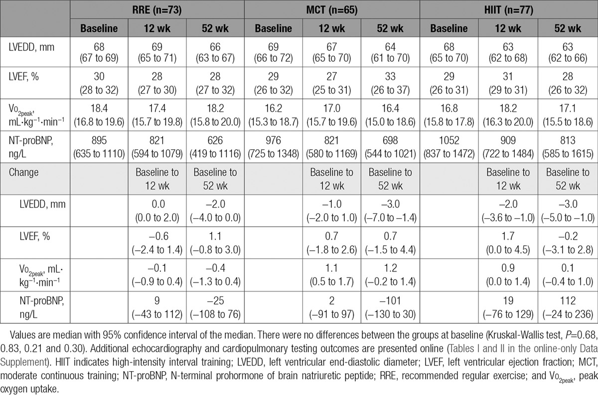
Table 2.
Main Echocardiography and Cardiopulmonary Testing Measures at Baseline, 12 weeks, and 52 Weeks With Unadjusted Changes
Training Intensity Compared With Protocol Targets
Heart rate and workload were monitored at all centers during the supervised training sessions (Figure 2). Average heart rate during sessions remained unchanged in the 12 weeks of supervised training, indicating constant relative exercise intensity during interventions (Figure 2A). Workload during intervals in the HIIT group was consistently 33 W (IQR, 24–42 W; P<0.001), higher than during continuous exercise in MCT (Figure 2B). Median relative training intensity based on maximal heart rate was 90% (IQR, 88%–92%) in HIIT and 77% (74%–82%) in MCT. Thus, the difference in training intensity was only 10% (8%–13%) with adjustment for center and pathogenesis (Figure 2C) compared with the protocol target difference of 27.5%. The training records showed that 51% of the patients in the HIIT group exercised at a lower intensity than prescribed, whereas 80% of those in the MCT group trained at a higher intensity than the protocol target (Figure 2D).
Figure 2.
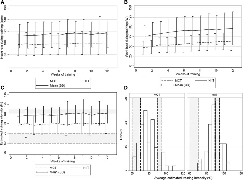
Training intensity during the 12-week intervention. A, Heart rate during training. Average heart rate during the 12-week intervention, estimated as weekly mean (SD) during moderate continuous training (MCT) and during the last 2 minutes of high-intensity interval training (HIIT). Constant difference between groups: 16 bpm (10–22 bpm; P<0.001). B, Workload. Average workload estimated as for heart rate. Difference between groups: 33 W (24–42 W; P<0.001). C, Training intensity. Average relative training intensity (percentage of maximal heart rate) estimated as for heart rate: HIIT, 90% (88%–92%); MCT, 77% (74%–82%); difference, 10% (8%–13%; P<0.001). Some of the variability in estimated training intensity probably results from variation in maximal heart rate. Comparing baseline and follow-up assessments in individual patients revealed differences that seemed randomly distributed and independent of intervention group, center, and whether the patients had sinus rhythm or atrial fibrillation (data not shown). Shaded areas mark boundaries of prescribed training intensity: HIIT, 90% to 95%; MCT, 60% to 70%. D, Training intensity on target. Distribution of average training intensity during the 12-week intervention; MCT, left histogram; HIIT, right histogram. Shaded areas mark boundaries for prescribed training intensity. Fifty-one percent of HIIT patients exercised below their prescribed training intensity, and 80% of MCT patients exercised above theirs. Density scales the height of the bars so that the sum of their areas equals 1.00.
Echocardiography and Cardiopulmonary Exercise Testing
Table 2 presents the crude within-group changes in main results from baseline to 12 and 52 weeks. Results of the prespecified primary analyses, the adjusted between-group differences in changes in LVEDD from baseline to 12 weeks (primary end point), are given in Table 3. Change of LVEDD in HIIT was not significantly different from that in MCT (−1.2 mm; −3.6 to 1.2 mm; P=0.45) but larger than in RRE (−2.8 mm; −5.2 to −0.4 mm; P=0.02), whereas the change in MCT was not significantly different from the change in RRE (−1.6 mm; −4.2 to 1.1 mm; P=0.34; Table 3). There were no other significant differences in echocardiographic measurements or in prohormone of brain natriuretic peptide (Table 3 and Table I in the online-only Data Supplement).
Table 3.
Main Outcomes
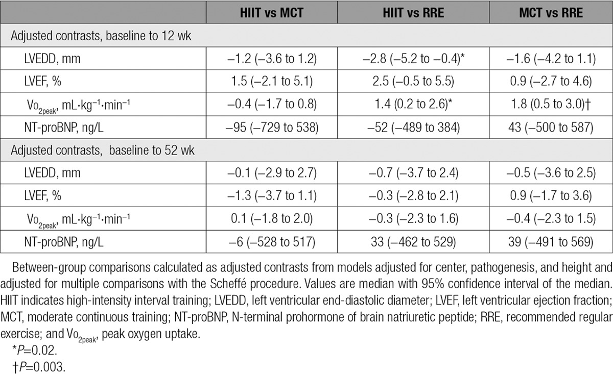
Change in Vo2peak in HIIT was not significantly different from MCT (−0.4 mL·kg−1·min−1; −1.7 to 0.8 mL·kg−1·min−1; P=0.70) but was 1.4 mL·kg−1·min−1 (0.2−2.6 mL·kg−1·min−1; P=0.02) larger than in RRE. Change in Vo2peak was 1.8 mL·kg−1·min−1 (0.5−3.0 mL·kg−1·min−1; P=0.003) larger in MCT compared with RRE (Table 3 and Table II in the online-only Data Supplement). There were no differences in respiratory quotient between groups at Vo2peak at baseline, 12 weeks, or 1 year, indicating similar levels of effort during testing (Table II in the online-only Data Supplement). At the 1-year follow-up, there were no differences in primary or secondary end points between the groups (Table 3 and Tables I–III in the online-only Data Supplement).
Sensitivity analyses exploring factors that might have influenced the changes of LVEDD in response to exercise did not identify predictors of response. Change of LVEDD in HIIT and MCT was not associated with average percentage of maximal heart rate during supervised training sessions when added to the regression model (P=0.52) or when used to substitute for intervention group (P=0.24). Likewise, findings were similar when comparing patients in the highest and lowest quartiles of achieved percentage of maximal heart rate during training and when using these quartiles as a categorical variable. Change in LVEDD was also not associated with atrial fibrillation (P=0.22). An alternative model for LVEDD changes from baseline to 12 weeks excluding patients with atrial fibrillation gave results comparable to the results from the model with all patients, albeit with slightly larger effect sizes for HIIT versus RRE (−3.7 mm; −6.7 to −0.8 mm; P=0.009) and for HIIT versus MCT (−2.0; −4.9 to 0.9 mm; P=0.23). Smoking was not significantly associated with change in LVEDD (P=0.26). There was no difference in exercise intensity assessed as percentage of maximal heart rate during sessions between centers (P=0.61) or between training on treadmill (89%; 82%–92%) versus bicycle (85%; 83%–88%; P=0.20), whether HIIT and MCT were analyzed jointly or separately (data not shown).
Quality of Life
There were no within-group or between-group differences in the quality-of-life measures Kansas City Cardiomyopathy Questionnaire, Hospital Anxiety and Depression Scale, Global Mood Scale, or Type D Scale 14 at baseline, 12 weeks, or 52 weeks (Table III in the online-only Data Supplement).
Serious Adverse Events
There were no statistically significant differences between groups in total number of patients with SAEs or cardiovascular SAEs during the 12-week intervention, although SAEs were numerically higher in HIIT, followed by MCT and RRE (Table 4). During the follow-up period from week 13 to 52, there was a possible trend (uncorrected P=0.10) for more patients admitted to hospital with cardiovascular events in HIIT (n=19) and RRE (n=17) compared with MCT (n=8), mainly because of fewer admissions for heart failure worsening in MCT (Table 4). This was also reflected in the 52-week total number of patients with SAEs: HIIT, 32 (39%); RRE, 26 (34%); and MCT, 18 (25%; P=0.16). The corresponding number of fatal events at 52 weeks was as follows: HIIT, 3; RRE, 1; and MCT 3.
Table 4.
Serious Adverse Events
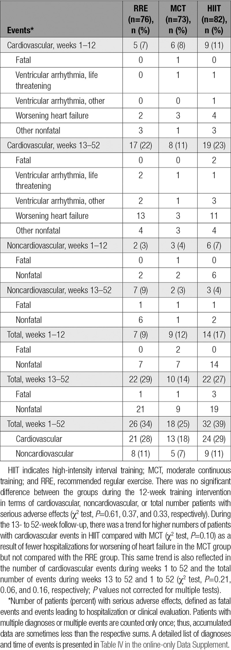
Details of diagnoses and time of events, including multiple diagnoses or multiple admissions in single patients, are reported in Table IV in the online-only Data Supplement. Three events occurred during or within 3 hours of supervised exercise in the HIIT group. One patient had ventricular arrhythmia with cardiac arrest during supervised exercise in week 1, was successfully resuscitated, and stopped the exercise program. This patient had refused cardioverter-defibrillator implantation before inclusion. Another patient had inappropriate implantable cardioverter-defibrillator discharge unrelated to arrhythmia during supervised exercise in week 12 and stopped the exercise program. A third patient experienced dizziness within 3 hours after supervised exercise, with no detectable cardiovascular cause, and continued the exercise program without any reoccurrences.
Discussion
The present study is the first randomized multicenter trial evaluating HIIT in chronic heart failure with reduced ejection fraction. It compares HIIT with the 2 most prevalent exercise prescriptions: a supervised program of MCT or RRE. The main finding was that 12 weeks of HIIT was not superior to MCT with respect to left ventricular reverse remodeling assessed as change in LVEDD. Although there was a statistically significant difference in remodeling between HIIT and RRE at 12 weeks, immediately after the supervised exercise intervention, its clinical importance is uncertain.
The effects of HIIT were less than expected from our working hypothesis8 and from a previous study by Wisløff et al5 on which it was based. In the present study, change in LVEDD by HIIT was −2.8 mm relative to RRE compared with −4.5 mm from our working hypothesis and −7.7 mm reported by Wisløff et al.5 In contrast, the change in LVEDD was −1.6 mm with MCT, which is similar to our prestudy estimate of −1.5 mm8 and −0.9 mm observed in the previous study.5 Post hoc analyses indicated that the magnitude of change in LVEDD may be larger with sinus rhythm compared with atrial fibrillation, but the present study was not powered to undertake a formal comparison. Change in LVEDD was chosen as the primary end point to represent change in cardiac remodeling because the echocardiographic measurement of diameter is more robust and has less relative variation than left ventricular volume estimates.10 The present study indicated nonsignificant changes in LVEDV of −19 mL with HIIT and −13 mL with MCT. This is in contrast to the large change of −45.2 mL previously observed with HIIT but similar to −15.2 mL observed with MCT5 and to a −11.5-mL change with MCT in a meta-analysis.7 The change in Vo2peak in the present study was 1.4 mL·kg−1·min−1 with HIIT and 1.8 mL·kg−1·min−1 with MCT relative to RRE. This is markedly less than the previously observed 6.0 mL·kg−1·min−1 with HIIT but similar to 1.9 mL·kg−1·min−1 with MCT5 and 2.1 mL·kg−1·min−1 in a meta-analysis.12 Nonetheless, it is slightly larger than 0.6 mL·kg−1·min−1 in the large HF-ACTION trial.4 It is notable that the differences observed at 12 weeks, immediately after the supervised interventions, were not maintained at the 52-week assessment, suggesting that the exercise prescriptions had not been followed as closely in the unsupervised period as in the supervised period.
Several factors may account for the small effect size of HIIT in the present study. Multicenter studies are more resistant to bias known to affect the study effects, especially compared with a single-center setting. Differences in the study population between our trial and the previous one by Wisløff and coworkers5 might also contribute to the different effects: All patients in the previous study had ischemic heart failure, although fewer than a third had a previous revascularization compared with 60% in the present study. Furthermore, patients in the study by Wisløff et al5 were on average 15 years older and had a much lower Vo2peak at baseline compared with the present trial.
Another factor that may have reduced the response to HIIT is lower training intensity during intervals. In our trial, 51% of the HIIT patients unexpectedly trained at lower intensity than prescribed, whereas 80% in the MCT group trained at higher intensity. Despite the experience of cardiac rehabilitation of the participating centers, HIIT at 90% to 95% of maximal heart rate appeared difficult to achieve in a multicenter cohort of patients, whereas intensities at 60% to 70% of maximal heart rate in MCT seemed too low. It may be speculated that the response to HIIT might have been larger if target intensity had been achieved; however, we could not detect any difference of change in LVEDD when comparing the upper and lower quartiles of percentage of maximal heart rate during exercise in the present study.
To date, only 1 study has had sufficient statistical power to assess safety with exercise training in patients with heart failure. The HF-ACTION study demonstrated a moderate reduction in the composite end point of mortality and hospital admissions after 1 year.4 By design, the present study was too small to assess differences in safety among HIIT, MCT, and RRE8; however, numeric differences in clinical events could generate hypotheses and identify issues for special attention in future studies and follow-up. During the 12-week supervised training program, the number of patients with SAEs was small in all groups, and few patients withdrew from exercise training. These observations concur with comparisons of safety of HIIT versus MCT in patients with coronary artery disease that detected no differences in SAEs.13,14 In contrast, there was a numeric difference in patients with SAEs during the follow-up period from week 13 to 52 as a result of more hospitalizations for worsening of heart failure in HIIT and RRE compared with MCT. This trend was not statistically significant and resulted from post hoc subgroup analyses because SAEs were not a prespecified end point. Hence, conclusions or recommendations could not be based on this finding, but the numeric difference should receive attention in future trials.
Limitations
One of the important objectives of moving from a small proof-of-principle study to a type II multicenter study was to test whether the effect size would be conserved in a setting that is closer to a real-world clinical setting. Although several measures were taken to ensure quality and consistency, including supervised training sessions based on heart rate monitoring, the differences in training intensity between HIIT and MCT were less than intended and partly overlapped. This was an unexpected finding, suggesting that the HIIT prescription of 90% to 95% of maximal heart rate may be too high and the MCT prescription of 60% to 70% too low for some patients. For future studies, we suggest that exercise intensities should be regularly adapted to improvements in exercise capacity and to worsening of symptoms or changes of medication. Repeated assessment of maximal heart rate and more emphasis on adjusting workload according to perceived level of effort might also be helpful. We experienced that questionnaires were of limited value for assessing physical activity outside supervised sessions and recommend accelerometer recordings, particularly in the unsupervised follow-up period. Furthermore, in future studies, women should be a focus because only 19% of the patients in this study were women. Although not unusual in similar studies, this sex bias was unintended and constitutes a limitation of the generalization of the results.
Conclusions
The present multicenter trial did not confirm the hypothesis that a 12-week program of supervised HIIT was superior to MCT in reducing left ventricular remodeling in patients with stable heart failure. None of the interventions led to deterioration of cardiac function compared with RRE, and both exercise programs increased aerobic capacity, an important prognostic parameter of heart failure, to a similar extent. However, these positive changes were smaller than expected and were not maintained at follow-up after 52 weeks. Numeric differences in readmissions for worsening of heart failure suggested a favor of MCT relative to HIIT and RRE, but the study was not powered to assess safety. Training records showed that exercise intensities >90% of maximal heart rate were not achieved in a significant proportion of the patients. Thus, further studies are needed to define the role of HIIT as an alternative exercise modality in patients with heart failure with reduced ejection fraction.
Acknowledgments
Jennifer Adam, Elena Bonanomi, Silvia Colombo, Christian Have Dall, Ingrid Granøien, Kjersti Gustad, Anne Haugland, Julie Kjønnerød, Marti Kristiansen, Jorun Nilsen, Maren Redlich, Anna Schlumberger, and Kurt Wuyts performed exercise testing and training; Rigmor Bøen, Marianne Frederiksen, Eli Granviken, Loredana Jakobs, Adnan Kastrati, Nadine Possemiers, Hanne Rasmussen, Liv Rasmussen, and Johannes Scherr performed patient screening, inclusion, and clinical assessments; Volker Adams, Ann-Elise Antonsen, Wim Bories, Geert Frederix, Malou Gloesner, Vicky Hoymans, and Hielko Miljoen collected data; Hanna Ellingsen and Maria Henningsen performed data monitoring; and Lars Køber and Christian Torp-Pedersen monitored safety.
Sources of Funding
This work was supported by St. Olavs Hospital; Faculty of Medicine, Norwegian University of Science and Technology; Norwegian Health Association; Danish Research Council; Central Norwegian Health Authority/Norwegian University of Science and Technology; Else-Kröner-Fresenius-Stiftung, and Société Luxembourgeoise pour la recherche sur les maladies cardio-vasculaires.
Disclosures
Dr Halle reports grants from the Else-Kröner-Fresenius Foundation for the present work and is on the advisory board of Novartis, Sanofi-Aventis, and Merck Sharp & Dohme Corp. outside of the present study. Dr Linke reports grants and personal fees from Medtronic and Claret Medical, as well as personal fees from Edwards, St. Jude Medical, Bard, and Symetis, all outside of the present study.
Supplementary Material
Footnotes
Drs Ellingsen, Halle, Prescott, and Linke contributed equally.
Deceased.
The online-only Data Supplement is available with this article at http://circ.ahajournals.org/lookup/suppl/doi:10.1161/CIRCULATIONAHA.116.022924/-/DC1.
Circulation is available at http://circ.ahajournals.org.
Clinical Perspective
What Is New?
The present multicenter trial did not confirm results from a small previous study that indicated that high-intensity interval training is superior to moderate continuous training (MCT) in reversing left ventricular remodeling and increasing aerobic capacity.
In both groups, results were only moderately better than a recommendation of regular exercise; improvements were not maintained at the 52-week follow-up.
Numeric differences in serious adverse events at 52 weeks suggested a favor of MCT, but the study was not powered to compare safety.
Fifty-one percent of high-intensity interval training patients exercised below prescribed heart rate, and 80% of MCT exercised above their target.
What Are the Clinical Implications?
Given that high-intensity interval training was not superior to MCT in reversing remodeling or improving secondary end points, and considering that adherence to the prescribed exercise intensity based on heart rate may be difficult to achieve, even when supervised and performed in centers experienced in cardiac rehabilitation, MCT remains the standard exercise modality for patients with chronic heart failure.
References
- 1.Ponikowski P, Voors AA, Anker SD, Bueno H, Cleland JG, Coats AJ, Falk V, González-Juanatey JR, Harjola VP, Jankowska EA, Jessup M, Linde C, Nihoyannopoulos P, Parissis JT, Pieske B, Riley JP, Rosano GM, Ruilope LM, Ruschitzka F, Rutten FH, van der Meer P Authors/Task Force Members. 2016 ESC guidelines for the diagnosis and treatment of acute and chronic heart failure: the Task Force for the diagnosis and treatment of acute and chronic heart failure of the European Society of Cardiology (ESC) developed with the special contribution of the Heart Failure Association (HFA) of the ESC. Eur Heart J. 2016;37:2129–2200. doi: 10.1093/eurheartj/ehw128. doi: 10.1093/eurheartj/ehw128. [DOI] [PubMed] [Google Scholar]
- 2.Vanhees L, Rauch B, Piepoli M, van Buuren F, Takken T, Börjesson M, Bjarnason-Wehrens B, Doherty P, Dugmore D, Halle M Writing Group, EACPR. Importance of characteristics and modalities of physical activity and exercise in the management of cardiovascular health in individuals with cardiovascular disease (part III). Eur J Prev Cardiol. 2012;19:1333–1356. doi: 10.1177/2047487312437063. doi: 10.1177/2047487312437063. [DOI] [PubMed] [Google Scholar]
- 3.Taylor RS, Sagar VA, Davies EJ, Briscoe S, Coats AJ, Dalal H, Lough F, Rees K, Singh S. Exercise-based rehabilitation for heart failure. Cochrane Database Syst Rev. 2014;4:CD003331. doi: 10.1002/14651858.CD003331.pub4. [DOI] [PMC free article] [PubMed] [Google Scholar]
- 4.O’Connor CM, Whellan DJ, Lee KL, Keteyian SJ, Cooper LS, Ellis SJ, Leifer ES, Kraus WE, Kitzman DW, Blumenthal JA, Rendall DS, Miller NH, Fleg JL, Schulman KA, McKelvie RS, Zannad F, Piña IL HF-ACTION Investigators. Efficacy and safety of exercise training in patients with chronic heart failure: HF-ACTION randomized controlled trial. JAMA. 2009;301:1439–1450. doi: 10.1001/jama.2009.454. doi: 10.1001/jama.2009.454. [DOI] [PMC free article] [PubMed] [Google Scholar]
- 5.Wisløff U, Støylen A, Loennechen JP, Bruvold M, Rognmo Ø, Haram PM, Tjønna AE, Helgerud J, Slørdahl SA, Lee SJ, Videm V, Bye A, Smith GL, Najjar SM, Ellingsen Ø, Skjaerpe T. Superior cardiovascular effect of aerobic interval training versus moderate continuous training in heart failure patients: a randomized study. Circulation. 2007;115:3086–3094. doi: 10.1161/CIRCULATIONAHA.106.675041. doi: 10.1161/CIRCULATIONAHA.106.675041. [DOI] [PubMed] [Google Scholar]
- 6.Giannuzzi P, Temporelli PL, Corrà U, Tavazzi L ELVD-CHF Study Group. Antiremodeling effect of long-term exercise training in patients with stable chronic heart failure: results of the Exercise in Left Ventricular Dysfunction and Chronic Heart Failure (ELVD-CHF) Trial. Circulation. 2003;108:554–559. doi: 10.1161/01.CIR.0000081780.38477.FA. doi: 10.1161/01.CIR.0000081780.38477.FA. [DOI] [PubMed] [Google Scholar]
- 7.Haykowsky MJ, Liang Y, Pechter D, Jones LW, McAlister FA, Clark AM. A meta-analysis of the effect of exercise training on left ventricular remodeling in heart failure patients: the benefit depends on the type of training performed. J Am Coll Cardiol. 2007;49:2329–2336. doi: 10.1016/j.jacc.2007.02.055. doi: 10.1016/j.jacc.2007.02.055. [DOI] [PubMed] [Google Scholar]
- 8.Støylen A, Conraads V, Halle M, Linke A, Prescott E, Ellingsen Ø. Controlled study of myocardial recovery after interval training in heart failure: SMARTEX-HF: rationale and design. Eur J Prev Cardiol. 2012;19:813–821. doi: 10.1177/1741826711403252. doi: 10.1177/1741826711403252. [DOI] [PubMed] [Google Scholar]
- 9.Rognmo Ø, Hetland E, Helgerud J, Hoff J, Slørdahl SA. High intensity aerobic interval exercise is superior to moderate intensity exercise for increasing aerobic capacity in patients with coronary artery disease. Eur J Cardiovasc Prev Rehabil. 2004;11:216–222. doi: 10.1097/01.hjr.0000131677.96762.0c. [DOI] [PubMed] [Google Scholar]
- 10.Thorstensen A, Dalen H, Amundsen BH, Aase SA, Stoylen A. Reproducibility in echocardiographic assessment of the left ventricular global and regional function, the HUNT study. Eur J Echocardiogr. 2010;11:149–156. doi: 10.1093/ejechocard/jep188. doi: 10.1093/ejechocard/jep188. [DOI] [PubMed] [Google Scholar]
- 11.Brookes ST, Whitely E, Egger M, Smith GD, Mulheran PA, Peters TJ. Subgroup analyses in randomized trials: risks of subgroup-specific analyses; power and sample size for the interaction test. J Clin Epidemiol. 2004;57:229–236. doi: 10.1016/j.jclinepi.2003.08.009. doi: 10.1016/j.jclinepi.2003.08.009. [DOI] [PubMed] [Google Scholar]
- 12.Haykowsky MJ, Timmons MP, Kruger C, McNeely M, Taylor DA, Clark AM. Meta-analysis of aerobic interval training on exercise capacity and systolic function in patients with heart failure and reduced ejection fractions. Am J Cardiol. 2013;111:1466–1469. doi: 10.1016/j.amjcard.2013.01.303. doi: 10.1016/j.amjcard.2013.01.303. [DOI] [PubMed] [Google Scholar]
- 13.Conraads VM, Pattyn N, De Maeyer C, Beckers PJ, Coeckelberghs E, Cornelissen VA, Denollet J, Frederix G, Goetschalckx K, Hoymans VY, Possemiers N, Schepers D, Shivalkar B, Voigt JU, Van Craenenbroeck EM, Vanhees L. Aerobic interval training and continuous training equally improve aerobic exercise capacity in patients with coronary artery disease: the SAINTEX-CAD study. Int J Cardiol. 2015;179:203–210. doi: 10.1016/j.ijcard.2014.10.155. doi: 10.1016/j.ijcard.2014.10.155. [DOI] [PubMed] [Google Scholar]
- 14.Rognmo Ø, Moholdt T, Bakken H, Hole T, Mølstad P, Myhr NE, Grimsmo J, Wisløff U. Cardiovascular risk of high- versus moderate-intensity aerobic exercise in coronary heart disease patients. Circulation. 2012;126:1436–1440. doi: 10.1161/CIRCULATIONAHA.112.123117. doi: 10.1161/CIRCULATIONAHA.112.123117. [DOI] [PubMed] [Google Scholar]


