Abstract
Bioassay guided fractionation and chemical investigation of the ethanolic extract of the aerial parts of Koelreuteria paniculata Laxm. (Sapindaceae), resulted in the isolation and identification of three new triterpenoid saponins 1–3 named Paniculatosoid A–C, along with eleven known compounds. The structures of the isolated compounds were elucidated using 1D and 2D NMR experiments, HRESIMS, and comparison with literature data. The occurrence of tridesmosidic saponin is reported for the first time from family Sapindaceae, as well as it is rarely found in natural saponins. Compounds 4–13 were evaluated for their antibacterial, antifungal, antimalarial and antileishmanial activities. Compound 12 showed weak antibacterial activity against Escherichia coli with an IC50 value of 101 μM. Compounds 12 and 13 showed antimalarial activity against chloroquine-sensitive (D6) Plasmodium falciparum protozoan with IC50 values of 6.46 and 6.95 μM, and against chloroquine-resistant (W2) Plasmodium falciparum protozoan with IC50 values of 9.34 and 4.18 μM.
Keywords: Koelreuteria paniculata, Sapindaceae, Triterpenes, Tridesmosidic saponin, Antimalarial
1. Introduction
Koelreuteria paniculata Laxm. is a saponaceous tree belonging to family Sapindaceae, native to Eastern Asia and cultivated as a decorative on the Black-Sea coast of the Caucuses (Sutiashvili, 2000). It is widely distributed in Northern China. Local people use the seeds as insecticides and the leaves as antifungal and antibacterial agents (Lin et al., 2002). Recent reports showed that the crude extracts of this plant possessed antitumor and antioxidant activities. The isolation of a gallate derivatives, cyanolipids and flavonoids have been reported (Mahmoud et al., 2001; Seigler and Butterfield, 1976; Yang et al., 1999). The present work reports the isolation and identification of three new compounds, including a tridesmosidic saponin 1 named Paniculatosoid A and two monodesmosidic saponins 2 and 3 named Paniculatosoid B and C, along with eleven known compounds 4-14 identified as 5-O-methyl-luteolin 4, loliolide 5, kaempferol 7-O-α-l-rhamnoside 6, kaempferol-3-O-α-l-rhamnoside 7, methyl myo-inositol 8, β-sitosterol 9, β-sitosterol-β-d-glucoside 10, palmitic acid monoglyceride 11, ethyl gallate 12, methyl gallate 13 and gallic acid 14. Compounds 4-6 were reported to be isolated for the first time from family Sapindaceae. Compounds 4-13 were evaluated for their antimalarial, antileishmanial, antifungal and antimicrobial activities. Compound 12 showed weak antibacterial activity against Escherichia coli with an IC50 value of 101 μM. Compounds 12 and 13 showed antimalarial activity against chloroquine-sensitive (D6) Plasmodium falciparum protozoan with IC50 values of 6.46 and 6.95 μM, and against chloroquine-resistant (W2) Plasmodium falciparum protozoan with IC50 values of 9.34 and 4.18 μM. The occurrence of tridesmosidic saponin is reported for the first time from family sapindaceae, as well as it is rarely found among natural saponins (Hostettmann and Marston, 2005).
2. Results and discussion
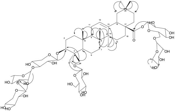
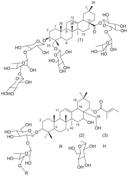
Compound 1 was obtained as whitish powder (10 mg), whose molecular formula was established to be C64H104O30 from the [M-H]− ion at m/z 1351.6274 (calcd. for C64H103O30 1351.6535), and the [M + Na]+ ion at m/z 1375.6396 (calcd. for C64H104O30Na 1375.6510), in the HR-ESIMS. The 1H NMR and 13C NMR spectra of 1 showed six tertiary methyl signals resonated at δ 0.89 (H-29), δ 0.90 (H-30), δ 0.99 (H-25), δ 1.13 (H-26), δ 1.15 (H-24) and δ 1.21 (H-27), one olefinic proton signal resonated at δ 5.42 (H-12, bs) with two typical olefinic carbon signals resonated at δ 123.6 and δ 144.8, and one carbonyl signal resonated at δ 177.3, revealed that its aglycone was hederagenin (Li et al., 1990). The 13C NMR and DEPT-NMR spectra showed the presence of eight methyl, fifteen methylene, thirty three methine and eight quaternary carbons. The 2D-NMR spectra showed six anomeric carbons resonated at δ 96.3, 101.9, 103.4, 105.3, 104 and 107.9, and their corresponding protons were deduced from HMQC spectrum, and were attached to anomeric protons resonated at δ 6.24 (d, J = 8 Hz), δ 6.33 (s), δ 5.84 (s), δ 5.09 (d, J = 6.5 Hz), δ 5.01 (d, J = 7.5 Hz) and δ 5.35 (d, J = 7.5 Hz), respectively. The complete assignments of each glycosidic proton system were achieved by analysis of COSY and TOCSY experiments. The units with anomeric protons at δ 5.01 (d, J = 7.5 Hz) and δ 6.24 (d, J = 8 Hz) corresponded to two hexoses and were identified as two β-d-glucoses, while the units with anomeric protons at δ 5.09 (d, J = 6.5 Hz) and δ 5.35 (d, J = 7.5 Hz) corresponded to two pentoses and were identified as two β-d-xyloses, and the units with the anomeric protons at δ 6.33 (s) and δ 5.84 (s) were identified as α-l-rhamnoses. The HMBC correlations (Fig. 2) showed a set of correlations which confirmed the attachment of sugars in three positions of the aglycone; the correlation of the proton resonated at δ 5.09 (H-1‵) with the carbon signal resonated at δ 81.7 (C-3) confirmed the attachment of xylose-1 to C-3 of hederagenin, while the correlation of the proton resonated at δ 5.01 (H-1‵‵‵‵) with the carbon signal resonated at δ 64.7 (C-23) confirmed the attachment of glucose-1 to C-23 of hederagenin, and the correlation of the proton resonated at δ 6.24 (H-1‵‵‵‵‵) with the carbon signal resonated at δ 177.3 (C-28) confirmed the attachment of glucose-2 to C-28 of hederagenin. The down-fielded carbon signals at δ 76.2 and at δ 83.3 for both of C-3‵ of xylose-1 and C-3‘‵ of rhamnose-1 moiety respectively, confirmed that the C-3‵ positions for both sugars are attached to another sugar moieties, as well as the HMBC correlations of protons resonated at δ 6.33 (H-1‵‵) and at δ 5.35 (H-1‵‵‵) with the carbon signal resonated at δ 83.3 (C-3‵‵) confirmed the attachment of xylose-2 to rhamnose-1 was by C-3‵ of rhamnose-1 (Borges et al., 2009; Wang et al., 2014). The up-fielded carbon signals at δ 73.2 and 74.5 for C-3‵ and C-4‵ of rhamnose-2, confirmed that the rhamnose-2 is terminal and not attached to another sugars (Wang et al., 2014). The up-fielded carbon signal at δ 61.9 for C-6‵ of glucose-1 confirmed that its C-6‵ is free, while the down-fielded carbon signal at δ 69.7 for C-6‵ of glucose-2 confirmed the attachment of glucose-2 to rhamnose-2 in the C-6‵ position of glucose-2 (Fu et al., 2006; Viana et al., 2004; Voutquenne et al., 2003; Wang et al., 2014). Acid hydrolysis of 1 gave the aglycon hederagenin along with xylose, glucose and rhamnose. Thus, the structure of 1 proved to be the new compound (3β-O-β-d-xylopyranosyl-(1 → 3)-α-l-rhamnopyranosyl-(1 → 3)-β-d-xylopyranosyl-23-O-β-d-glucopyranosyl-hederagenin-28-O-α-l-rhamnopyranosyl-(1 → 6)-β-d-glucopyranosyl ester) (Fig. 1).
Fig. 2.

HMBC Correlations of compound (1).
Fig. 1.
Chemical Structure of Compounds 1-3.
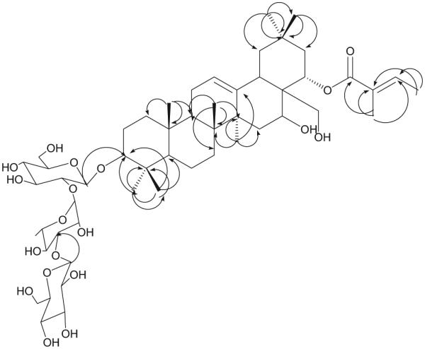
Compound 2 was obtained as whitish powder (12 mg), whose molecular formula was established to be C53H86O19 from the [M-Angeloyl-Glucose]− ion at m/z 763.4771 (calcd. for C42H67O12 763.4634), in the HR-ESIMS. The 1H NMR and 13C NMR spectra of 2 showed eight tertiary methyl signals resonated at δ 0.84 (H-25), δ 0.91 (H-26), δ 1.08 (H-29), δ 1.19 (H-23), δ 1.24 (H-24), δ 1.32 (H-30), δ 1.88 (H-27) and δ 2.01 (H-5 Angeloyl), two doublet methyl signal resonated at δ 2.13 (H-3 angeloyl, J = 5.6 Hz), and at δ 1.58 (H-6 rhamnose, J = 5.6 Hz), two olefinic proton signals at δ 5.95 (H-3 angeloyl, d, J = 5.6 Hz) and δ 5.44 (H-12, br s) with four typical olefinic carbon signals at δ 124.0, δ 130.0, δ 137.2 and δ 144.3, and one carbonyl signal at δ 168.7. The HMQC spectrum, showed a proton signal at δ 6.22 (H-22, m) assigned for a methine carbon resonated at δ 72.7 (C-22), and two methylene proton signals resonated at δ 2.06 and at δ 2.85 (H-21, m) assigned for a methylene carbon resonated at δ 42.2(C-21), furthermore this proton signal at δ 6.22 (H-22, m) showed correlation only with the two methylene proton signals resonated at δ 2.06 and at δ 2.85 in TOCSY revealed that the angeloyl moiety is attached to C-22, and thus the aglycone of 2 was identified as camelliagenin-A (Voutquenne et al., 1998). The 13C NMR and DEPT-NMR spectra showed the presence of ten methyl, eleven methylene, twenty three methine and nine quaternary carbons. The 2D-NMR spectra showed a three anomeric carbons resonated at δ 103.6, 104.9 and 105.3, and their corresponding protons were deduced from HMQC spectrum, and were attached to protons resonated at δ 6.14 (s), δ 5.21 (d, J = 5.6 Hz) and δ 4.79 (d, J = 5.2 Hz) respectively. The complete assignments of each glycosidic proton system were achieved by analysis of COSY and TOCSY experiments. The units with anomeric protons at δ 5.21 and δ 4.79 were corresponded to two hexoses and were identified as two β-d-glucoses, while the units with the anomeric proton at δ 6.14 (s) was identified as α-l-rhamnose. The HMBC correlations of 2 (Fig. 3) showed a set of correlations confirmed the attachment of glucose in C-3 of the aglycone. The HMBC correlation of the proton resonated at δ 4.79 (H-1‵‵) with the carbon signal resonated at δ 90.7 (C-3) confirmed the attachment of glucose-1 to C-3 of camelliagenin-A. The down-fielded carbon signal at δ 83.3 for C-3‵‵ of rhamnose moiety, confirmed its attachment in such position to another sugar moiety. The up-fielded carbon signal at δ 61.9 for C-6‵‵ and C-6‵‵‵‵ confirmed that both C-6 of glucose-1 and glucose-2 were free. Thus the structure of 2 proved to be the new compound (3β-O-β-d-glucopyranosyl-(1 → 3)-α-l-rhamnopyranosyl-(1 → 2)-β-d-glucopyranosyl-22α-O-angeloyl-camelliagenin-A) (Fig. 1).
Fig. 3.

HMBC Correlations of compound (2).
Compound 3 was obtained as whitish powder (6 mg). The analysis of the NMR data for 3 confirmed that 3 was very close to the structure of 2. The 1H NMR and 13C NMR spectra for 3 are very close to the spectra of 2 and showed signals for eight tertiary methyl signals, one doublet methyl signal, three olefinic proton signals with four typical olefinic carbon signals, and one carbonyl signal, revealed that the aglycone of 3 was the same as 2 and it was camelliagenin-A (Voutquenne et al., 1998). The 13C NMR and DEPT-NMR spectrum for 3 showed the presence of ten methyl, ten methylene, eighteen methine and nine quaternary carbons. The 2D-NMR spectra of 3 showed a two anomeric carbons resonated at δ 103.6 and 105.5, and their corresponding protons were deduced from HMQC spectrum, and were attached to protons resonated at δ 6.18 (s) and δ 4.79 (d, J = 5.2 Hz) respectively. The HMBC correlations of 3 showed a set of correlations confirmed the attachment of glucose in C-3 of the aglycone. The HMBC correlation of the proton resonated at δ 4.79 (H-1‵) with the carbon signal resonated at δ 90.7 (C-3) confirmed the attachment of glucose-1 to C-3 of camelliagenin-A. The absence of the carbon signal at δ 83.3 for C-3‘‵ of rhamnose moiety, confirmed that the rhamnose is terminal and not attached to another sugar. The up-fielded carbon signal at δ 61.9 for C-6‵ confirmed that C-6 of glucose was free. Thus the structure of 3 proved to be the new compound (3β-O-α-l-rhamnopyranosyl-(1 → 2)-β-d-glucopyranosyl-22α-O-angeloyl-camelliagenin-A) (Fig. 1).
Acid hydrolysis of 1-3: The acid hydrolysis of saponins 1-3 were done according to the method mentioned in (Wanas et al., 2010), About 2 mg of each compound was hydrolyzed with 1 N HCl (0.1 mL) at 88 °C for 2 h, the reaction mixture was cooled to room temperature, then partitioned with an equal amount of EtOAc (0.1 mL), and the water layer was evaporated to dryness in vacuo and analyzed for its sugar components. The sugar mixture was compared with a standard mixture of xylose, glucose and rhamnose using silica gel TLC and CHCl3-MeOH-H2O (6:4:0.5) as solvent.
Compounds 4-14 were identified by using 1H NMR, 13C NMR, DEPT-135, HMQC and HMBC as well as HRESIMS experiments, as: 5-O-methyl-luteolin 4 (Abou-Zaid et al., 2001), loliolide 5 (Kimura and Maki, 2002), kaempferol 7-O-α-l-rhamnoside 6 (Chua et al., 2008), kaempferol-3-O-α-l-rhamnoside (Afzelin) 7 (Cheng et al., 2013), methyl myo-inositol (Quebrachitol) 8 (HuiCong et al., 2013), β-sitosterol 9 (Lu et al., 2012), β-sitosterol-β-d-glucoside 10 (Wei et al., 2011), palmitic acid monoglyceride 11, ethyl gallate 12 (Yang et al., 1999), methyl gallate 13 (Lin et al., 2002) and gallic acid 14 (Qu et al., 2011). Compounds 4-6 were reported to be isolated for the first time from family Sapindaceae. Compounds 4–13 were evaluated for their antimalarial, antileishmanial, antifungal and antimicrobial activities. Compound 12 showed weak antibacterial activity against Escherichia coli with an IC50 value of 101 μM. Compounds 12 and 13 showed antimalarial activity against chloroquine-sensitive (D6) Plasmodium falciparum protozoan with IC50 values of 6.46 and 6.95 μM, and against chloroquine-resistant (W2) Plasmodium falciparum protozoan with IC50 values of 9.34 and 4.18 μM.
3. Experimental
3.1. General experimental procedures
NMR spectra were recorded on a Bruker Avance DRX- 500 instrument at 500 (1H) and 125 MHz (13C), and a Varian Mercury 400 MHz spectrometer at 400 (1H) and 100 MHz (13C). The HRESIMS spectra were measured using a Bruker Bioapex-FTMS with electrospray ionization (ESI). Column chromatographic separation was performed on silica gel 60 (0.04-0.063 mm), SPE Cartridges, (C-18), Supelc, and Sephadex LH-20 (0.25–0.1 mm, Aldrich). TLC was performed on precoated TLC plates with silica gel 60 F254 (0.2 mm, Merck). Preparative HPLC (Waters delta prep 4000) was performed using Waters Atlantis® RP-18 (5 μ, 250, 20 mm).
3.2. Plant material
The aerial parts of Koelreuteria paniculata Laxm were collected in May 2012 from Orman garden, Giza, Egypt. The plant was authenticated by Dr. Mohammad El-Gebaly, Consultant Taxonomist, Cairo University, Egypt. A voucher specimen (KP 2) has been deposited in the Pharmacognosy Department, Faculty of Pharmacy, Al-Azhar University, Cairo, Egypt.
3.3. Extraction and isolation
The air-dried powdered aerial parts of Koelreuteria paniculata (1.40 kg) were exhaustively extracted by maceration with 70% EtOH (6 L × 3), at room temperature. The combined ethanolic extracts were concentrated under reduced pressure to afford (138 g) residue. The residue was subjected to (VLC) on silica gel (1500 g, 45 cm, 15 cm) using n-hexane, n-hexane – EtOAc (50:50), EtOAc, EtOAc –MeOH (50:50) and MeOH, each 2.0 L to give five fractions (A-E). Fraction B (2.48 g) was subjected to CC (silica gel, 60 g, 60 cm, 4 cm, eluted with n-hexane-EtOAc mixtures in a manner of increasing polarities) to obtain nine subfractions (B1-B9). Subfr. B2 (280 mg) was subjected to further purifications on CC silica gel and sephadex, to obtain compound 9 (7 mg). Subfr. B4 (213 mg) was subjected to repeated CC on silica gel and sephadex, to obtain compound 11 (20 mg). Fraction C (1.22 g) was subjected to CC (silica gel, 30 g, 40 cm, 5 cm, eluted with n-hexane-EtOAc mixtures in a manner of increasing polarities) to obtain four subfractions (C1-C4). Subfr. C2 was subjected to further purification on CC silica gel and sephadex to obtain compounds 5 (3 mg) and 13 (7 mg). Subfr. C3 was purified on repeated CC silica gel and sephadex to afford compound 12 (88 mg). Fraction D (7.68 g) was subjected to CC silica gel (200 g, 60 cm, 8 cm) eluted with DCM-MeOH mixtures in a manner of increasing polarities) to obtain seven subfractions (D1-D7). Subfr. D1 (890 mg) was subjected to further CC silica gel columns to afford compound 10 (25 mg). Subfr. D3 was purified on repeated CC silica gel and sephadex to afford compound 7 (11 mg). Subfr. D6 was subjected to CC (sephadex LH-20, 45 cm, 1 cm) eluting with MeOH, to afford compound 4 (8 mg). Fraction E (22.3 g) was subjected to (VLC) on silica gel (600 g, 30 cm, 12 cm) using DCM, DCM-MeOH (95:5) DCM-MeOH (90:10), DCM-MeOH (80:20), DCM-MeOH (50:50) and MeOH, each 2.0 L to give six subfractions (E1-E6). Subfr. E3 was subjected to CC silica gel (100 g, 50 cm, 6 cm) eluted with DCM-MeOH mixtures in a manner of increasing polarities) to afford 8 subfractions (E-3-1-E-3-8). Subfr. E-3-3 was subjected to subsequent purification on preparative RP-C18 HPLC gradually eluted with mixture of MeOH and H2O, using Atlantis® RP-18(5 μg, 250, 20 mm) at a flow rate 17.0 mL/min and detection at 268 nm to yield compounds: 14 (6 mg) and 6 (5 mg). Subfr. E4 was subjected to SPE column RP C-8 and eluted with Water-MeOH mixtures in a manner of decreasing polarities) to afford compound 8 (30 mg). Subfr. E-5 was subjected to further purification on SPE (RP- C-18) and eluted with Water-MeOH mixtures in a manner of decreasing polarities) to afford compounds 1 (10 mg), 2 (12 mg) and 3 (6 mg).
3.4. Spectral data
3β-O-β-d-xylopyranosyl-(1 → 3)-α-l-rhamnopyranosyl-(1 → 3)-β-d-xylopyranosyl-23-O-β-d-glucopyranosyl-hederagenin-28-O-α-l-rhamnopyranosyl-(1 → 6)-β-d-glucopyranosyl ester 1: whitish powder; for 1H NMR and 13C NMR spectroscopic data Table 1; for HR-ESIMS m/z 1351.6274 [M-H]− (calcd. for C64H103O30 1351.6535), and m/z 1375.6396 [M+Na]+ (calcd. for C64H104O30Na 1375.6510), consistent with the molecular formula C64H104O30.
Table 1.
1H NMR and 13C NMR spectral data (400MHz for δH, 125 MHz for δC, δ in ppm, J values in Hz) for compounds (1–3) in Pyridine-d5.
| (1) | (2) | (3) | ||||
|---|---|---|---|---|---|---|
|
|
|
|
||||
| Position | δH | δC | δH | δC | δH | δC |
| 1 | 1.02m, 1.53m | 39.7 | 1.36, m | 39.4 | 1.36, m | 39.4 |
| 2 | 1.81m | 27.1 | 1.80, m | 27.1 | 1.80, m | 27.1 |
| 3 | 4.33 | 81.7 | 3.22, m | 90.7 | 3.19, m | 90.7 |
| 4 | – | 44.2 | – | 40.3 | – | 40.3 |
| 5 | 1.73, m | 48.1 | 0.74, m | 56.3 | 0.74, m | 56.3 |
| 6 | 18.8 | 1.39, m | 18.8 | 1.39, m | 18.8 | |
| 7 | 1. 27 m, 1.61 m | 33.1 | 2.13, m | 35.7 | 2.13, m | 35.7 |
| 8 | – | 40.5 | – | 41. 5 | – | 41. 5 |
| 9 | 48.7 | 47.5 | 47.5 | |||
| 10 | – | 37.5 | – | 37.4 | – | 37.4 |
| 11 | 1.89, m | 23.9 | 1.74, m | 24.4 | 1.74, m | 24.4 |
| 12 | 5.42, bs | 123.6 | 5.44, bs | 124 | 5.44, bs | 124 |
| 13 | – | 144.8 | 144.3 | 144.3 | ||
| 14 | – | 42.8 | – | 44.8 | – | 44.8 |
| 15 | 1.17, m | 28.9 | 33.7 | 33.7 | ||
| 16 | 1.89, m | 24.5 | 70.4 | 70.4 | ||
| 17 | – | 47.7 | – | 42.3 | – | 42.3 |
| 18 | 3.17, m | 42.3 | na | na | ||
| 19 | 1.18, m | 46.7 | 2.87, m | 48.1 | 2.87, m | 48.1 |
| 20 | – | 30.6 | – | 32.6 | – | 32.6 |
| 21 | 1.03, m | 34.6 | 2.06,m, 2.85,m | 42.2 | 2.85, m | 42.2 |
| 22 | 1.28, m | 33.3 | 6.22, m | 72.7 | 6.22, m | 72.7 |
| 23 | 3.94m, 4.33 m | 64.7 | 1.19, s | 17. 4 | 1.19, s | 17. 4 |
| 24 | 1.15, s | 14.8 | 1.24, s | 28.6 | 1.24, s | 28.6 |
| 25 | 0.99, s | 16.9 | 0.84, s | 16.3 | 0.84, s | 16.3 |
| 26 | 1.13, s | 18.3 | 0.91, s | 16.4 | 0.91, s | 16.4 |
| 27 | 1.21, s | 26.8 | 1.88, s | 28.2 | 1.88, s | 28.2 |
| 28 | – | 17 7. 3 | 3.59, 3.74, m | 64.3 | 3.59, 3.74, m | 64.3 |
| 29 | 0.89, s | 33.8 | 1.08, s | 34.1 | 1.08, s | 34.1 |
| 30 | 0.90, s | 24.4 | 1.32, s | 25.9 | 1.32, s | 25.9 |
| Angeloyl | ||||||
| 1 | – | 168.7 | – | 168.7 | ||
| 2 | – | 130 | – | 130 | ||
| 3 | 5.95, d | 137. 2 | 5.95, d | 137. 2 | ||
| 4 | 2.13, d, 5.6 | 17. 5 | 2.13, d, 5.6 | 17. 5 | ||
| 5 | 2.01, s | 21.7 | 2.01, s | 21.7 | ||
| 3-O-Sugar | ||||||
| Xylose-1 | Glucose-1 | Glucose-1 | ||||
| 1‵ | 5.09, d, 6.5 Hz | 105.3 | 4.79, d, 5.2 | 105.3 | 4.79, d, 5.2 | 105.3 |
| 2‵ | 4.03, m | 75.6 | 4.15, m | 76.4 | 4.15, m | 76.4 |
| 3‵ | 4.13, m | 76.2 | 4.23, m | 76.3 | 4.23, m | 76.3 |
| 4‵ | 4.12, m | 71.3 | 4.33, m | 72.6 | 4.33, m | 78.2 |
| 5‵ | 3.68, 4.45, m | 67.9 | 4.12,m | 77 | 4.12,m | 77 |
| 6‵ | 4.36, 4.68, m | 61.9 | 4.36, 4.68, m | 61.9 | ||
| Rhamnose-1 | Rhamnose | Rhamnose | ||||
| 1‵‵ | 6.33, s | 101. 9 | 6.14, s | 103.6 | 6.18, s | 103.8 |
| 2‵‵ | 4.88, bs | 71.7 | 4.88, bs | 72 | 4.88, bs | 72 |
| 3‵‵ | 4.81, m | 83.3 | 4.81, m | 83.3 | 4.72, m | 73.5 |
| 4‵‵ | 4.45, m | 73.2 | 4.45, m | 72.9 | 4.45, m | 72.9 |
| 5‵‵ | 4.71, m | 70.4 | 4.71, m | 70.2 | 4.71, m | 70.2 |
| 6‵‵ | 1.58, d, 5.6 | 19.2 | 1.58, d, 5.6 | 19.2 | 1.58, d, 5.6 | 19.2 |
| Xylose-2 | Glucose-2 | |||||
| 1‵‵‵ | 5.35, d, 7. 5 Hz | 10 7. 9 | 5.21, d, 5.6 Hz | 104.9 | ||
| 2‵‵‵ | 4.09, m | 73.3 | 3.95, m | 76.4 | ||
| 3‵‵‵ | 4.18, m | 75.9 | 4.14, m | 78.6 | ||
| 4‵‵‵ | 4.22, m | 71 | 4.44, m | 70.8 | ||
| 5‵‵‵ | 3.71, 4.49, m | 66.9 | 3.66, m | 77.9 | ||
| 6‵‵‵ | 4.08, 4.22, m | 61.9 | ||||
| 23-O-sugar | ||||||
| Glucose-1 | ||||||
| 1‵‵‵‵ | 5.01, d, 7. 5 Hz | 105.4 | ||||
| 2‵‵‵‵ | 4.15, m | 75.9 | ||||
| 3‵‵‵‵ | 4.23, m | 74.6 | ||||
| 4‵‵‵‵ | 4.33, m | 72.6 | ||||
| 5‵‵‵‵ | 4.12, m | 77.1 | ||||
| 6‵‵‵‵ | 4.36, 4.68, m | 61.9 | ||||
| 28-O-sugar | ||||||
| Glucose-2 | ||||||
| 1‵‵‵‵‵ | 6.24, d, 8Hz | 96.3 | ||||
| 2‵‵‵‵‵ | 3.95, m | 75.9 | ||||
| 3‵‵‵‵‵ | 4.14, m | 78.9 | ||||
| 4‵‵‵‵‵ | 4.44, m | 70.3 | ||||
| 5‵‵‵‵‵ | 3.66, m | 77.7 | ||||
| 6‵‵‵‵‵ | 4.08, 4.22, m | 69.7 | ||||
| Rhamnose-2 | ||||||
| 1‵‵‵‵‵‵ | 5.84, s | 103.4 | ||||
| 2‵‵‵‵‵‵ | 4.68, m | 71.7 | ||||
| 3‵‵‵‵‵‵ | 4.56, m | 73.2 | ||||
| 4‵‵‵‵‵‵ | 4.35, m | 74.5 | ||||
| 5‵‵‵‵‵‵ | 4.93, m | 71 | ||||
| 6‵‵‵‵‵‵ | 1.7, d, 5.6 | 19.1 | ||||
3β-O-β-d-glucopyranosyl-(1 → 3)-α-l-rhamnopyranosyl-(1 → 2)-β-d-glucopyranosyl-22α-O-angeloyl-camelliagenin-A 2: whitish powder; for 1H NMR and 13C NMR spectroscopic data Table 1; for HR-ESIMS m/z 763.4771 [M-Angeloyl-Glucose]− (calcd. for C42H67O12 763.46341).
3β-O-α-l-rhamnopyranosyl-(1 → 2)-β-d-glucopyranosyl-22α-O-angeloyl-camelliagenin-A 3: whitish powder; for 1H NMR and 13C NMR spectroscopic data Table 1.
3.5. Antimicrobial assay
Compounds 4-13 were tested for antimicrobial activity against Staphylococcus aureus ATCC 29213, methicillin-resistant S. aureus ATCC 33591 (MRS), Escherichia coli ATCC 35218, Pseudomonas aeruginosa ATCC 27853, Mycobacterium intracellulare ATCC 23068, Candida albicans ATCC 90028, Candida glabrata ATCC 90030, Candida krusei ATCC 6258, Cryptococcus neoformans ATCC 90113, and Aspergillus fumigatus ATCC 204305. Ciprofloxacin and Amphotericin B were used as positive controls for bacteria and fungi, respectively (Bharate et al., 2007; Radwan et al., 2007).
3.6. Antileishmanial assay
Compounds 4–13 were tested for antileishmanial activity in vitro against a culture of Leishmania donovani promastigotes; Pentamidine and Amphotericin B were used as a positive controls (Ma et al., 2004).
3.7. Antimalarial assay
Compounds 4-13 were tested for antimalarial activity in vitro against chloroquine sensitive (D6, Sierra Leone) and resistant (W2, Indo China) strains of Plasmodium falciparum by measuring plasmodial LDH activity as described earlier (Makler and Hinrichs, 1993). Chloroquine was used as positive control.
4. Conclusion
Compounds 12 and 13 showed antimalarial activity against chloroquine-sensitive (D6) Plasmodium falciparum protozoan with IC50 values of 6.46 and 6.95 μM, and against chloroquine-resistant (W2) Plasmodium falciparum protozoan with IC50 values of 9.34 and 4.18 μM. Compound 12 showed weak antibacterial activity against Escherichia coli with an IC50 value of 101 μM.
Supplementary Material
Acknowledgment
We are grateful to the Egyptian Government and National Center for Natural Products Research, University of Mississippi for financial support. We also thankful for Dr. Baharthi Avula for HRMS, Drs. Babu Tekwani and Surendra Jain for antileishmanial, Shabana Khan and John Trott for antimalarial assays, and Marsha Wright for antimicrobial testing. This investigation was conducted in part in a facility constructed with support from the Research facilities improvement Program C06 RR-14503-01, the NIH, NIAID, Division of AIDS, Grant No. AI 27094.
Footnotes
Appendix A. Supplementary data
Supplementary data associated with this article can be found, in the online version, at http://dx.doi.org/10.1016/j.phytol.2016.07.008.
References
- Abou-Zaid MM, Helson BV, Nozzolillo C, Arnason JT. Ethyl m-digallate from red maple, Acer rubrum L., as the major resistance factor to forest tent caterpillar, Malacosoma disstria Hbn. J. Chem. Ecol. 2001;27:2517–2527. doi: 10.1023/a:1013683600211. [DOI] [PubMed] [Google Scholar]
- Bharate SB, Khan SI, Yunus NA, Chauthe SK, Jacob MR, Tekwani BL, Khan IA, Singh IP. Antiprotozoal and antimicrobial activities of O-alkylated and formylated acylphloroglucinols. Bioorg. Med. Chem. 2007;15:87–96. doi: 10.1016/j.bmc.2006.10.006. [DOI] [PubMed] [Google Scholar]
- Borges RM, Tinoco LW, Souza Filho JDD, Barbi NDS, Silva AJRD. Two new oleanane saponins from Chiococca alba (L.) Hitch. J. Braz. Chem. Soc. 2009;20:1738–1741. [Google Scholar]
- Cheng, Hui-Ling, Zhang L-J, Liang, Yu-Han, Hsu, Ya-Wen, Lee, I-Jung, Liaw, Chia-Ching, Hwang, Syh-Yuan, Kuo, Yao-Haur Antiinflammatory and antioxidant flavonoids and phenols from Cardiospermum halicacabum. J. Tradit. Complement. Med. 2013:8. doi: 10.4103/2225-4110.106541. [DOI] [PMC free article] [PubMed] [Google Scholar]
- Chua M-T, Tung Y-T, Chang S-T. Antioxidant activities of ethanolic extracts from the twigs of Cinnamomum osmophloeum. Bioresour. Technol. 2008;99:1918–1925. doi: 10.1016/j.biortech.2007.03.020. [DOI] [PubMed] [Google Scholar]
- Fu W-W, Shimizu N, Dou D-Q, Takeda T, Fu R, Pei Y-H, Chen Y-J. Five new triterpenoid saponins from the roots of Platycodon grandiflorum. Chem. Pharm. Bull. 2006;54:557–560. doi: 10.1248/cpb.54.557. [DOI] [PubMed] [Google Scholar]
- Hostettmann K, Marston A. Cambridge University Press; Saponins: 2005. [Google Scholar]
- HuiCong W, ZieHen W, XuMing H, GuiBing H, HouBin C. Determination of quebrachitol Litchi chinensis and Dimocarpus longan in Sapindacea family. J. South China Agric. Univ. 2013;34:315–319. [Google Scholar]
- Kimura J, Maki N. New loliolide derivatives from the brown alga Undaria pinnatifida. J. Nat. Prod. 2002;65:57–58. doi: 10.1021/np0103057. [DOI] [PubMed] [Google Scholar]
- Li X-C, Wang D-Z, Wu S-G, Yang C-R. Triterpenoid saponins from Pulsatilla campanella. Phytochemistry. 1990;29:595–599. [Google Scholar]
- Lin W-H, Deng Z-W, Lei H-M, Fu H-Z, Li J. Polyphenolic compounds from the leaves of Koelreuteria paniculata Laxm. J. Asian Nat. Prod. Res. 2002;4:287–295. doi: 10.1080/1028602021000049087. [DOI] [PubMed] [Google Scholar]
- Lu REN, Lu Z, He BAO, Xiaojuan YOU, Di T, Fang DOU, Junxian W. Chemical constituents of coumarins from flowers of Koelreuteria paniculata. Northwest Pharm. J. 2012;1:5. [Google Scholar]
- Ma G, Khan SI, Jacob MR, Tekwani BL, Li Z, Pasco DS, Walker LA, Khan IA. Antimicrobial and antileishmanial activities of hypocrellins A and B. Antimicrob. Agents Chemother. 2004;48:4450–4452. doi: 10.1128/AAC.48.11.4450-4452.2004. [DOI] [PMC free article] [PubMed] [Google Scholar]
- Mahmoud I, Moharram FA, Marzouk MS, Soliman HS, El-Dib RA. Two new flavonol glycosides from leaves of Koelreuteria paniculata. Die Pharmazie. 2001;56:580–582. [PubMed] [Google Scholar]
- Makler MT, Hinrichs DJ. Measurement of the lactate dehydrogenase activity of Plasmodium falciparum as an assessment of parasitemia. Am. J. Trop. Med. Hyg. 1993;48:205–210. doi: 10.4269/ajtmh.1993.48.205. [DOI] [PubMed] [Google Scholar]
- Qu QH, Zhang L, Bao H, Zhang JH, You XJ, Wang JX. Chemical constituents of flavonoids from flowers of Koelreuteria paniculata. Zhong yao cai = Zhongyaocai = J. Chin. Med. Mater. 2011;34:1716–1719. [PubMed] [Google Scholar]
- Radwan MM, Manly SP, Ross SA. Two new sulfated sterols from the marine sponge Lendenfeldia dendyi. Nat. Prod. Commun. 2007;2:901–904. [Google Scholar]
- Seigler DS, Butterfield CS. The origin of cyanolipids in Koelreuteria paniculata. Phytochemistry. 1976;15:842–844. [Google Scholar]
- Sutiashvili MG. Koelreuteria saponin B from seeds of Koelreuteria paniculata. Chem. Nat. Compd. 2000;36:99. [Google Scholar]
- Viana FA, Braz-Filho R, Pouliquen Y, Andrade Net M, Santiago GMP, Rodrigues-Filho E. Triterpenoid saponins from stem bark of Pentaclethra macroloba. J. Braz. Chem. Soc. 2004;15:595–602. [Google Scholar]
- Voutquenne L, Lavaud C, Massiot G, Delaude C. Saponins from Harpullia cupanioides. Phytochemistry. 1998;49:2081–2085. doi: 10.1016/s0031-9422(98)00406-3. [DOI] [PubMed] [Google Scholar]
- Voutquenne L, Guinot P, Thoison O, Sevenet T, Lavaud C. Oleanolic glycosides from Pometia ridleyi. Phytochemistry. 2003;64:781–789. doi: 10.1016/s0031-9422(03)00380-7. [DOI] [PubMed] [Google Scholar]
- Wanas AS, Matsunami K, Otsuka H, Desoukey SY, Fouad MA, Kamel MS. Triterpene glycosides and glucosyl esters, and a triterpene from the leaves of Scheffiera actinophylla. Chem. Pharm. Bull. 2010;58:1596–1601. doi: 10.1248/cpb.58.1596. [DOI] [PubMed] [Google Scholar]
- Wang X, Wang M, Xu M, Wang Y, Tang H, Sun X. Cytotoxic oleanane-Type triterpenoid saponins from the rhizomes of Anemone rivularis var. flore-minore. Molecules. 2014;19:2121–2134. doi: 10.3390/molecules19022121. [DOI] [PMC free article] [PubMed] [Google Scholar]
- Wei J, Chen J, Cai S, Lu R, Lin S. Chemical constituents in whole herb of Cardiospermum halicacabum (I) Zhongcaoyao. 2011;42:1509–1511. [Google Scholar]
- Yang XF, Fu HZ, Lei HM, Lin WH, Ma GE. Chemical constituents from the leaves of Koelreuteria paniculata Laxm. Acta Pharm. Sin. 1999;34:462–465. [Google Scholar]
Associated Data
This section collects any data citations, data availability statements, or supplementary materials included in this article.


