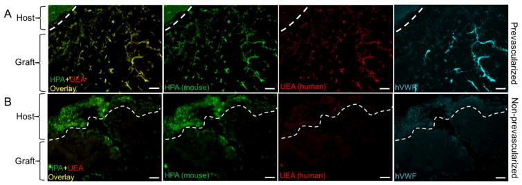
Fig. 7. Perfusion of mouse and human specific lectins after two-week subcutaneous implantation.
(A) In the prevascularized tissue, mouse-specific lectin (HPA) and human-specific lectin (UEA) are chimeric in the graft area, and the host tissue is only stained with mouse-specific lectin. Further staining of hVWF confirms the endothelial network formed by human-origin HUVECs. (B) In the nonprevascularized tissue, HPA stains the host tissue and minor regions of the graft area, no UEA or hVWF staining is observed. Scale bars: 100μm.

