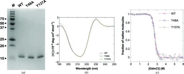
Figure 1.
Characterization of wild-type FimH lectin and mutants. (a) Affinity-purified wild-type FimH and the tyrosine-gate mutants Y48A and Y137A were subjected to SDS–PAGE under reducing conditions on a 16% acrylamide/bisacrylamide SDS–PAGE gel. Lane M, molecular-weight marker (labelled in kDa). (b) CD spectra of WT FimH lectin and the Y48A and Y137A mutants. All samples were measured at 10 µM concentration in 10 mM sodium phosphate buffer pH 7.4 and at 25°C using a thermostat-controlled 0.1 cm cell as described in §2. (c) GdmCl-dependent equilibrium unfolding profiles at 25°C and pH 7.4 were monitored by changes in fluorescence at 350 nm upon excitation at 280 nm. The transition midpoint values D 1/2 are 2.75 M (WT), 2.77 M (Y48A) and 2.73 M (Y137A) GdmCl.

