Abstract
Natural compounds capable of inducing apoptosis in cancer cells have always been of considerable interest as potential anti-cancer agents. Many such compounds are under screening and development with their potential evolution as a clinical drug benefiting many of the cancer patients. Guggulsterone (GS), a phytosterol isolated gum resin of the tree Commiphora mukul has been widely used in Indian traditional medicine as a remedy for various diseses. GS has been shown to possess cancer chemopreventive and therapeutic potential as established by in vitro and in vivo studies. GS has been shown to target constitutively activated survival pathways such as PI3-kinase/AKT, JAK/STAT, and NFκB signaling pathways that are involved in the regulation of growth and inflammatory responses via regulation of antiapoptotic and inflammatory genes. The current review focuses on the molecular targets of GS, cellular responses, and the animal model studies in various cancers. The mechanistic action of GS in different types of cancers also forms a part of this review. The perspective of translating this natural compound into a clinically approved drug with its pros and cons is also discussed.
Keywords: Natural compounds, Guggulsterone, Cancers, Chemoprevention, Molecular targets
Background
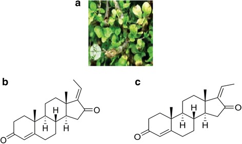
The important aspect in cancer chemoprevention is to identify drugs or compounds that kill the tumor cells with lower toxicity to healthy tissue. The other alternative approach could be to identify agents, which either slows or even halts the tumor progression. The growing evidence linked the deregulation of apoptosis in cancer cells and supports the hypothesis that targeting deregulated apoptotic signaling pathways could serve as a tool for cancer prevention [1, 2]. Interestingly, the chemotherapeutic drugs that are currently used exert their cytotoxic effect through the induction of apoptosis [3–5]. Therefore, the success of cancer therapy depends on the sensitivity of cancer cells to respond to the therapeutic agents that turn on the apoptotic process. The signaling cascade that leads to apoptosis can be induced by a vast variety of drugs with diverse chemical structures and different mechanisms of action, notably through the inhibition of survival and increased expression of pro-apoptotic genes. Natural compounds have been shown to have less cytotoxicity and efficient in blocking prosurvival/ growth signal transduction pathways. Interestingly, most of the natural compounds currently available as pharmaceutical products isolated from plant extracts have proven to exhibit anti-tumor properties in vivo and in vitro. One such traditional medicine, guggulsterone (GS), [4, 17(20)-pregnadiene- 3, 16-dione], a plant polyphenol extracted from the gum resin of the Commiphora mukul tree has been broadly used for centuries to treat multiple human diseases [6–8]. The active ingredients found in the extract from the gum resin of Mukul tree are the isomers E- and Z-GS (Fig. 1) and both these forms have been extensively used to treat multiple disorders. It has been shown that GS is an antagonist of bile acid farnesoid X receptor (FXR) [9–11] and inhibition of FXR expression by GS causes anticancer activity in many cancer cells [12–17]. There is also accumulating evidence about the role of GS in cholesterol homeostasis regulation by increasing the transcription of bile salt export pump [10, 11, 17]. GS has been shown to play an important role in nutritional metabolism as it has been found to inhibit cholesterol synthesis in the liver via antagonism of the FXR and the bile-acid receptor [18]. GS has been widely used for the treatment of hyperlipidemia in humans [5, 19]. A number of studies have demonstrated that GS efficiently decreases low density lipoprotein cholesterol and triglyceride levels in serum and increases high density lipoprotein cholesterol levels [20, 21]. Specifically, E and Z isoforms of GS have been identified as active ingredients for lipid-lowering [22]. GS has been shown to bind FXR and prevent the expression of FXR agonist-mediated genes [8, 23]. Furthermore, it has been demonstrated that the lipid lowering effect of GS in liver are due to inhibition of FXR as confirmed from FXR knockout mice studies [8].
Fig. 1.

a The Plant Commiphora mukul. The chemical structure of Guggulsterone isoforms, E-Guggulsterone (b) and Z-Guggulsterone (c)
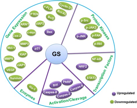
GS has been found to induce apoptotic cell death in many types of cancer [24–28] via activation of caspases, increased expression of genes of Bcl-2 family members and generation of reactive oxygen intermediates. A number of studies have shown that GS strongly inhibits the activation of various survival signaling pathways including, PI3-kinase/AKT, JAK/STAT and nuclear factor-kB (NF-kB) in various cancer cells [29–31] (Table 1). Constitutive activity of NF-kB plays a crucial role in growth and proliferation of malignant cells via regulating expression of several antiapoptotic genes. GS was found to efficiently suppress the expression of these antiapoptotic genes in many cancer cells (Fig. 2). In addition, GS has also been shown to suppress the ionizing radiation (IR)-mediated activation of NF-κB and augments the radiosensitivity of human cancer cell lines [32]. Further, GS is reported to reduce cell growth as well as prevents IR-induced DNA damage repair [32] and GS has been shown to induce apoptosis in a wide rangeof cancer cells [24, 25, 27, 28, 33–36]. The detailed molecular targets of GS and mechanisms regulating apoptosis in various cancers are discussed in this review.
Table 1.
Anticancer activity of GS in in vitro experimental model and underlying molecular targets
| Cancer Type | Model/System | Molecular Targets | References |
|---|---|---|---|
| Pancreatic cancer | Human pancreatic cancer cell lines | ↓FXR reduced ↓ NF-κB, ↓Cyclin D1, ↓Bcl-2, ↓XIAP↓MPP9, ↓STAT3, ↓FAK, ↓Src, p-AKT,c-June, ↑Caspase-3,↑Bax | [33, 38, 39] |
| Head and Neck cancer | Head and neck carcinoma cell line | ↓ PI3-kinase/AKT, ↑Bax, ↑Bad | [88] |
| Esophagael cancer | Esophageal adenocarcinoma cell lines | ↓caudal type homeobox 2,↓Cox2,↓NFkB, ↓FXR, ↓ RAR-β2, ↑caspase-8,caspase-9,caspase-3 | [47, 48] |
| Colon cancer | Colon cancer cell line | ↓cIAP-1, ↓cIAP-2, ↓Bcl-2, ↓STAT3, ↓VEGF, ↑truncated Bid, ↑Fas, ↑p-JNK, ↑p-c-Jun | [50, 51] |
| Breast cancer | Breast cancer cell lines | ↓cyclin D1, ↓C-myc, ↓survivin, ↓TCF-4, ↓IKK/NF-κB, ↓MAPK/AP-1, ↓MMP-9 ↓ | [34, 89] |
| Prostate cancer | Prostate cancer cell lines | ↑caspase-9, ↑caspase-8, ↑caspase-3, ↑Bax ↓Bcl-2 and ↓Bcl-xL |
[27] |
| Hepatocellular carcinoma | Hepatocellular carcinoma cell lines |
↓TGF-β1, ↓VEGF,↓ Bcl-2,↑Bax, ↓ NF-κB, ↓STAT3 |
[83, 85] |
| Hematological malignancies | Leukemic cell lines | ↓Bfl-1/A1, ↓XIAP, ↓cFLIP, ↓Bcl-2, ↓BclXL, ↓survivin ↑caspase 8, ↑bid cleavage, ↑cytochrome c release, ↑caspase 9, ↑ caspase 3, ↑ PARP cleavage. |
[25] |
Fig. 2.

Biochemical and molecular targets of Guggulsterone. Guggulsterone exerts anti-cancer effects through activation or suppression of protein kinases, transcription factors, anti-oxidant enzymes, cell cycle regulators, proapoptotic and antiapoptotic proteins. GS exerts anti-inflammatory effects through suppression of nuclear factor-kB (NF-kB), which plays a crucial role in the inflammatory processes by regulating the expression of diverse proinflammatory proteins, including cyclooxygenase-2 (COX-2). GS fortifies cellular defense against oxidative stress by inducing the de novo synthesis of the powerful antioxidant enzyme heme oxygenase-1 (HO-1). GS induces apoptosis by increasing the expression of proapoptotic proteins while decreasing the levels of antiapoptotic proteins (e.g., IAP1, XIAP, Bfl-1/A1, Bcl-2, cFLIP, Survivin, etc.). GS induces apoptosis by increasing the expression of proapoptotic proteins while decreasing the levels of antiapoptotic proteins (e.g., IAP1, XIAP, Bfl-1/A1, Bcl-2, cFLIP, Survivin, etc.). GS suppresses invasion and metastasis by targeting MMPs, FXR etc
Guggulsterone and cancer
Since several decades extensive research has revealed that many chronic illnesses are caused by the deregulation of multiple genes mainly involved in cell cycle control enabling the cells to divide uncontrollably leading to metastasis [1–4]. Most of the conventional drugs primarily target a single gene product or signaling pathway at a given time, thus having a limited scope for the treatment. In addition these medicines exhibit many toxic side effects. Due to these limitations, there is a growing trend towards alternative medicines such as traditional medicine derived from natural compounds which are safe and have broad spectrum activity [37]. GS is one such ancient medicine that targets multiple signaling molecules with a varied range of mechanisms with its proven antiproliferative and proapoptotic effects in vitro and in vivo studies (Tables 1 and 2). The following sections describe GS-mediated anticancer effects and its potential targets in various cancers.
Table 2.
Anticancer activity of GS in in vivo animal experimental models
| Cancer Type | Model/System | Antitumor effects | References |
|---|---|---|---|
| Pancreatic cancer | Pancreatic cancer cell line xenograft tumors | ↓ tumor size | [39] |
| Colon Cancer | HT-29 xenograft tumors | ↓ tumor size | [50] |
| Esophageal cancer | Esophageal adenocarcinoma cell lines | ↓ tumor size | [47] |
| Breast cancer | MCF7 xenograft tumors | ↓Bcl-2 and P-glycoprotein expression ↑Chemosensivity of doxorubicin in vivo |
[75] |
| Prostate cancer | DU145 prostate cancer cells implanted in mouse | ↓ tumor size, ↓ angiogenesis↓ VEGFR-2 | [28] |
Pancreatic cancer
Pancreatic tumors are highly aggressive, and there is an urgent need of therapeutic strategy for the better management of this cancer. The existing chemotherapeutic drugs cause high toxicity and drug resistance. Macha et al. [33] using in vitro model have shown that GS prevents cell proliferation, inhibits cell motility reduces cell invasion and induces apoptotic cell death in many pancreatic cancer cell lines. The anti-cancer activity of GS was correlated with the down-regulation of anti-apoptotic proteins, cell cycle progression proteins and up-regulation of proapoptotic proteins. Furthermore, the reduced motility and suppressive effects on invasion in pancreatic cancer cells by GS were associated with the disruption of cytoskeletal organization, inhibition of FAK and Src kinase signaling. GS treatment was also found to reduce mucin MUC4 gene expression by inhibition of JAK kinase mediated signaling [33]. A recent study using pancreatic cancer cell lines has reported a significant reduction in cell migration and invasion by GS-mediated FXR inhibition indicating the potential role of FXR overexpression in lymphatic metastasis of pancreatic cancer [38].
GS has been found to increase the sensitivity of conventional chemotherapeutic agents such as gemcitabine. Treatment of pancreatic cancer cells with GS augmented apoptotic cell death when combined with gemcitabine as compared to treatment either with gemcitabine or GS alone. In addition, the tumors from xenografted mice in vivo model showed a better antitumor response to GS combination treatment compared to gemcitabine or GS alone.
Studies from in vitro and in vivo (Tables 1 and 2) settings have shown that the combination treatment leads to increased growth inhibition as well as apoptosis through a cascade of events involving the down-regulation of NF-κB, inhibition of AKT activity, downregulation of antiapoptotic gene BcL-2, upregulation of proapoptotic gene Bax and, activation of c-Jun NH(2)-terminal kinase (JNK) [39].
Head and neck cancer
A number of studies provide substantial evidence that GS suppressed the growth/or induced apoptotic cell death of head and neck squamous cell carcinoma (HNSCCC) [40–42]. GS-mediated inhibition of HNSCC proliferation is caused by inactivation of NF-κB and STAT3 signaling cascades. GS treatment of HNSCC cells prevents NF-κB activation and leads to its degradation resulting in the inhibition of inflammatory and angiogenic responses as well as progression and metastasis [35, 36]. GS was also able to inhibit COX-2 and vascular endothelial growth factor (VEGF) which contributes to inflammation and angiogenesis [35]. The antiproliferative and pro-apoptotic action of GS in SCC4 cells has been due to downregulation of various antiapoptotic genes including Bcl-2, XIAP, Mcl1, survivin, cyclin D1 and c-myc. Furthermore GS-mediated downregulation of these genes resulted sequential activation of caspas-9,-3 and cleavage, of poly-ADP-ribose phosphate (PARP) [25, 43].
Recently it has been reported that combination of GS and bortezomib, a proteasome inhibitor synergize the inhibition of signaling molecules that are essential for the proliferation and survival of malignant cells [44, 45]. These reports further suggest that co-treatment of HNSCC cells with bortezomib and GS potentiated effects on cell death and inhibited clonogenic survival. These findings correlated with inhibition of NF-κB signaling pathway [44] and induction of the proapoptotic proteins Bik, Bim, and Noxa [45]. These results suggest that GS could significantly improve the therapeutic activity of bortezomib against HNSCC in cotreatment strategy. Similar studies with erlotinib, cetuximab, and cisplatin in HNSCC cell lines further supported the synergistic activity of GS towards enhanced efficacy to apoptosis, cell cycle arrest and invasion [46].
Esophageal adenocarcinoma
Chronic Gastroesophageal Reflux Disease (GERD) is the main risk factor for the development of Barrett's esophagus (BE) and its progression to esophageal adenocarcinoma (EAC). Studies have shown the significant overexpression of bile acid receptor FXR in Barrett's esophagus and treatment of Barrett's esophagus-derived cell line with GS was found to significantly reduce the expression of FXR.. Treatment of Barrett's esophagus-derived cell line with GS was linked with a significant increase in the percentage of apoptotic cells and of the caspase -3 activity signifying that FXR may contribute to the regulation of apoptosis [12]. A similar study further showed that the inhibition of FXR in esophageal adenocarcinoma tissues either with an expression of FXR shRNA or treatment with GS suppressed tumor cell viability and induced apoptosis in vitro and in vivo [16] suggesting GS as a potential antagonist to FXR overexpression and a cancer chemotherapeutic. GS has been shown to have additive effect in suppressing esophageal cancer cell growth in vitro and in nude mouse xenografts when combined with amiloride and this activity is due to the inhibition of gastric acid-inducing gene Na + /H + exchanger-1 (NHE-1) [47]. In another study, GS has been shown to suppress bile acid induced caudal type homeobox 2 (CDX2) and cyclooxygenase-2 (COX-2) expression, which are critical in the development of Barrett’s Esophagus and esophageal adenocarcinoma and this effect is due to inhibition of NF-kB activity [48].
Colon cancer
Although there is a major progress in the advancement of chemotherapeutic agents for the treatment of colon cancer, however, the relapse rates still remain to be elevated for the existing drugs. The current chemotherapy has been benefecial only to the patients with advanced colorectal cancerregarding their survival and quality of life [49]. The other problems that happen to be with the conventional chemotherapy are systemic side effects and increased chemoresistance. Because of these limitations, there is an urgent need for more efficient and safer therapeutic strategies for advanced or untreatable colorectal cancer. Interestingly, a considerable amount of attention towards the anti-tumor properties of various phytochemicals has developed in the last few years both in vivo and in vitro. In connection to this, GS was found to possess potential anti-tumor effects in several cancer cell types including colorectal cancer [24–28, 33]. Emerging studies have also demonstrated that GS and its derivatives significantly reduced dextran sulfate sodium (DSS)-induced acute murine colitis. Further, more this effect was found due to GS-mediated down-regulation of NF-kB signaling pathway. These findings suggest that GS- mediated targeting of signaling pathways may be an attractive strategy for the treatment of inflammatory bowel disease (IBD) [30]. GS has also been shown to induce apoptotic cell death via activation of caspase cascade, downregulation of inhibitor of apoptosis proteins Bcl-2, and activation of JNK kinase [50]. GS treatment of colon cancer cell lines has been shown to block angiogenesis and metastasis by inactivation of STAT3 activity and downregulation of VEGF expression [51].
Bile acid receptor FXR besides playing a role in lipid metabolism, has also been shown to play an important role in intestinal carcinogenesis. Reduced expression of FXR mRNA has been found in human colon polyps and even more pronounced in colon adenocarcinomas [52, 53]. FXR has been shown to suppress intestinal tumorigenesis in both the Apc Min+/− and chronic colitis mouse models of intestinal neoplasia by regulating Wnt signaling and apoptosis [54]. FXR-deficient mice have been shown to exhibit increased intestinal epithelial cell proliferation and tumor development [55]. Recently, Peng et al. [15] have demonstrated that treatment of colon cancer cell lines with FXR antagonist GS or FXR siRNA lead to phosphorylation of EGFR and ERK whereas treatment with GW4064 or FXR overexpression prevented cell proliferation by dephosphorylation of EGFR and ERK. In addition treatment of colon cancer cell lines with GS and GW4064 also caused dose-dependent changes in Src (Tyr416) phosphorylation.
Breast cancer
Advanced early screening as well as detection methods have been developed, and several are under development for various cancers, including breast cancer. However, breast cancer continues to be the most challenging owing to its high frequency among women worldwide [56, 57]. A number of studies have suggested that various mechanisms are responsible for the incidence and development of breast carcinogenesis. NF-κB pathway is highly activated in breast cancer [58, 59]. NF-κB is one of the key molecules shown to regulate the expression of various antiapoptotic genes that plays a crucial role in tumorigenesis [29, 60]. These genes are bcl-2, and bcl-xl, adhesion molecule encoding genes, chemokines, and inflammatory cytokines; and cell cycle regulators. Therefore, targeting NF-κB and its associated partners could be an important therapeutic strategy for the management of breast cancer. Previously it has been demonstrated that GS inhibits NF-κB activation via IκBα kinase suppression, along with the dephosphorylation and degradation of IκBα. Moreover, GS also interferes nuclear translocation of p65 and NF-κB- mediated reporter gene activity [29]. GS treatment of cancer cells has been found to abrogate the expression of NF-κB-mediated antiapoptotic genes, as well the genes involved in regulation of inflammation and tumor metastasis [29–31]. Activation of pro-survival signaling pathways play a crucial role in suppression of apoptotic cell death during radiosensitization of cancer cells [61]. Ionizing radiation (IR) has been found to activate NF-κB and associated pathways that are involved in growth and survival of cancer cells [62]. Interestingly, GS treatment abrogated the IR-mediated activation of NF-κB and augmented the radiosensitivity of human tumor cell lines. Expression of hormone receptors by breast cancer cells makes them sensitive to hormonal therapy. However, the use of ER antagonists is restricted due to unwanted side effects [58]. Therefore, the development of new, safe and affordable therapeutics against ER-breast cancer cells harboring ER as well is much needed. GS has been found to downregulate the expression of ERα in breast cancer cells implicating that it could be a viable therapeutic useful in the treatment of ER-positive tumors that are resistant to tamoxifen [32].
It has been shown that the isomer of GS, cis-GS prevented TPA-upregulated MMP expression via obstructing IKK/NF-kB signaling. On the other hand, trans-GS was found to inhibit MAPK/AP-1 signaling pathway in MCF7 breast cancer cell line. Furthermore, co-treatment of breast cancer cells with these isomers exhibited additive effects on the inhibition of cell invasion. Another key signaling in growth and development of tumors is the Wnt/β-Catenin and its associated pathways [34]. This pathway has been shown to play a significant role in the initiation, progression, and metastasis of breast cancers [63–65]. The Wnt signaling exerts its effects on TCF-mediated transcription via β-Catenin [63–66]. Therefore, intercepting the signaling between Wnt and β-Catenin may prove a better way in developing new cancer therapeutics. Research in this direction, using natural products have already been shown to be promising [67–69]. It has been demonstrated that the expression of c-Myc, cyclin D1, and survivin, downstream of Wnt/β-Catenin signaling, were inhibited by guggulipid (GL) and z-GS in breast cancer significantly. In addition, treatment with GL in human breast cancer cells results in downregulation of TCF-4 protein expression significantly.
DNA methylation, as well as histone modifications, are other attractive targets for therapeutics strategy for the management of cancer [70]. Deregulated methylation can result in silencing of various functional genes including tumor suppressors that often lead to cancer development and progression [70]. Inhibition of DNA methyltransferases has been shown to suppress tumor formation [71]. Interestingly, some epigenetic modifications that regulate normal cellular activity via dietary phytochemicals have proved to be reducing cancer susceptibility [72]. It has been demonstrated that GS treatment of breast cancer cells inhibits the expression of DNA (cytosine-5)-methyltransferase 1(DNMT1) and HDAC1 [73].
Current chemotherapy for breast cancer faces a major problem of drug resistance. A viable approach to avoid drugs causing drug resistance is by utilizing non-toxic compounds in combination with conventional chemotherapeutic agents. Xu et al. [74] have reported that multidrug resistance developed by the expression of P-glycoprotein in breast cancer cells (MCF/Dox) against doxorubicin can be improved by treatment with GS. Co-treatment of MCF/DOX cells with GS and doxorubicin results in a significant increase in chemosensitivity. A similar observation was found in xenografts generated from MCF-7/DOX cells [75]. BCRP/ABCG2, an ABC transporter is overexpressed in breast cancer cells and has been shown to be involved in multidrug resistance [76]. Combined treatment of GS with bexarotene a retinoid X receptor agonist resulted in cytotoxicity via downregulation of BCRP/ABCG2 in breast cancer cell line.
Prostate cancer
Prostate cancer progression is a slow multistep process which begins with localized and low-grade lesions to high-grade and metastatic carcinomas resulting in a significant number of deaths in men [77, 78]. The slow progression and late diagnosis of prostate cancer allow a substantial opportunity for intervention to prevent this malignancy [79].
There are a considerable number of preclinical studies showing the anticancer activity of GS in prostate cancers. Singh and colleagues [24], have demonstrated that the GS treatment of human prostate cancer cell line, PC-3 resulted in efficient cytotoxic effects without affecting the normal prostate epithelial cell line (PrEC). In addition, GS-mediated growth inhibition of PC-3 cells occurs due to apoptosis rather than the cell cycle progression arrest. Furthermore, GS-induced apoptotic cell death correlated with the enhanced expression of Bcl-2 family members such as Bax and Bak and sequential activation of caspase cascade [27]. Furthermore, Xu and Sing [28] reported that z-GS treatment of DU145 implanted cells in mice angiogenesis via suppression of VEGF–VEGF-R2–Akt signaling axis. These findings were in accordance with the later reports in which treatment of GS was shown to downregulate the expression of antiapoptotic gene products including XIAP, survivin, cFLIP, Bcl-2, Bcl-Xl, c-myc and COX-2 [24, 25]. The mechanism by which GS-induced apoptosis in prostate cancer cells is not known, however, GS-mediated generation of ROS, which leads to activation of JNK has been implicated as one of the mechanisms leading to cell death in these cancer cells [24]. Treatment of LNCaP and PC3 cells with GS-causes the activation of JNK and p38. Interestingly, GS treatment activates extracellular signal-regulated kinase 1/2 (ERK1/2) only in LNCaP cells. GS treatment of prostate cancer cells resulted in the generation of ROS but not in normal PrEC prostate cells. In addition, PrEC cells showed resistance to GS-mediated activation of JNK kinase. Furthermore, overexpression of catalase and superoxide dismutase in prostate cancer cells prevented GS-mediated apoptosis and JNK activation [25]. In another study from the same group, treatment of prostate cancer cells, LNCaP withGL, a crude extract from which GS has been isolated, showed a dose-dependent inhibition of cell viability [80]. GL-mediated inhibition of cell viability in prostate cancer cells correlated with apoptosis as supported by an increase in cytoplasmic histone-associated DNA fragmentation and cleavage of PARP. Further, GL-induced apoptosis has been found to be associated with the generation of ROS and JNK activation along with the upregulation of proapoptotic proteins Bax and Bak and downregulation of Bcl-2 expression. During GS-mediated apoptosis, activation of JNK preceded before upregulation of Bax activation [80]. It was further shown that z-GS, another form of GS causes inhibition of angiogenesis via inactivation of AKT, and suppression of angiogenic factors such as VEGF and G-CSF [28]. The growth inhibitory effect of GS in prostate cancer has also been proposed to be due to inactivation of ATP citrate lyase (ACL or ACLY) which has been shown to exhibit crosstalk with the AKT signaling [81]. ACL is an extra-mitochondrial enzyme that has been demonstrated to play a crucial role in cellular lipogenesis, and its dysregulated expression is reported various cancers such as colon, prostate, liver, lung cancers as well as in many immortalized cells. Aberrant expression of ACL was reported to be inversely associated with tumor stage as well as differentiation and serves as a negative prognostic marker [82]. Thus, targeting ACL with GS in prostate cancermight be of potential therapeutic intervention strategy.
Liver, lung and ovarian cancer
It is well-known fact that the growth inhibitory and proapoptotic effects of GS are mediated through various mechanisms and certain cancers share the common mechanism. In liver cancer, the mechanism of cell death has been through sensitizing hepatoma cells to tumor necrosis factor-related apoptosis inducing ligand (TRAIL) mediated apoptosis. TRAIL at higher doses has been shown to cause toxicity to the healthy cells in addition to cytotoxicity to the cancer cells. Therefore using other agent/drugs in combination with TRAIL could be a viable strategy to induce maximum cytotoxic effects at subtoxic doses of TRAIL. It has been shown that subtoxic doses of GS and TRAIL can generate efficient apoptotic death in hepatoma cells. This GS/TARIL combination has proved to be efficient in inducing the apoptosis by disrupting the disrupting mitochondrial membrane potential resulting in the release of cytochrome C to the cytosol and consequent activation of caspases. In addition GS-mediated ROS generation can lead to upregulation of the death receptor DR5 via eIF2α and C/EBP homologous protein (CHOP). TRAIL binding to DR5 can result in the efficient induction of TRAIL-mediated apoptosis in hepatoma cell [83, 84]. GS has been shown to induce apoptotic cell death in hepatocellular carcinoma cell lines by activating intrinsic mitochondrial pathway [85].
GS was found to have the antifibrotic activity as it mediates reduced activation and survival of hepatic stellate cells (HSCs), which serve as the primary source of the matrix proteins. Accumulation of extracellular matrix has been shown to be involved in liver fibrosis that can lead to cirrhosis of the liver. During cirrhosis, the blood flow through the liver becomes disrupted due to damage in an architectural organization of the liver. Once cirrhosis is developed, the risk of developing liver cancer is significantly increased. It was found that the GS inhibits the growth of immortalized LX-2 HSC cells via induction of apoptosis. GS-induced apoptosis in HSC was accompanied by activation of c-Jun N-terminal kinase and mitochondrial apoptotic signaling. GS-induced HSC growth inhibition was also found to involve AKT and adenosine monophosphate-activated protein kinase (AMPK) phosphorylation modifications resulting in the activation of proapoptotic proteins and downregulation of antiapoptotic proteins [83]. GS has also been shown to inhibit NF-κB activation in LX-2 cells where the constitutive activation of this pathway leads to increased growth of these cells [86]. Besides NF-κB activation, increased collagen α1 synthesis and α-smooth muscle actin expression plays a significant role in the growth of HSCs resulting in enhanced liver cirrhosis, and treatment with GS significantly decreased the extent of collagen deposition via inhibiting collagen α1 synthesis and α-smooth muscle actin expression. These findings strongly implicate GS as an antifibrotic agent inhibiting various survival pathways via induction of apoptotic cell death in HSC cells.
Limited studies have also shown that GS induces anticancer activities in lung and ovarian cancers. Treatment of lung and ovarian cancer cell lines with GS resulted in inhibition of cell proliferation and downregulation of cyclin D1 and cdc2 expression leading to inhibition of DNA synthesis. In addition GS treatment also increased the expression of cyclin-dependent kinase inhibitor p21 and p27. Moreover, GS-mediated apoptosis correlated with the activation of JNK, caspase-cascade, accompanied with inhibition of the expression of various anti-apoptotic genes [25].
Hematological malignancies
Hematological malignancies constitute approximately 6.5% of all cancer incidences worldwide [87]. The primary causes of these liquid cancers are due to defect at the level of bone marrow and lymphatic system [87]. These malignancies are classified into three main groups including leukemia, lymphoma, and multiple myeloma (MM). The anticancer activity of GS in these hematological malignancies is not studied in detail, and this review would enable to pursue further research in this field. Antileukemic effect of GS has been reported by Samudio and colleagues [26] where they examined the anticancer effects of three isomeric pregnadienedione steroids [i.e., cis-GS, trans-GS, and 16-dehydroprogesterone] in HL60 and U937 cells as well as in primary leukemic blasts in culture [26]. They demonstrated that the treatment of HL60 and U937 cells with these compounds prevented cell proliferation via mitochondrial-dependent but caspase-independent apoptosis. All three compounds were shown to induce the generation of ROS which can be one of their mechanisms of cell death. Furthermore, these compounds resulted in the dephosphorylation of constitutive extracellular signal-regulated kinase phosphorylation status in these leukemic cells. Interestingly only cis-GS caused a rapid depletion of glutathione levels as well as oxidation of the mitochondrial phospholipid cardiolipin [26]. The other study carried by Shishodia and colleagues [25] observed that the treatment of leukemia, myeloma and melanoma cell lines with GS resulted in decreased proliferation along with reduced levels of cyclin D1 and cdc2 which inhibited DNA synthesis. They found an increased levels of cyclin-dependent kinase inhibitor p21 and p27 as well as induction of apoptosis by activation of JNK, caspase-cascade, PARP cleavage and downregulation of anti-apoptotic products [25].
Conclusion
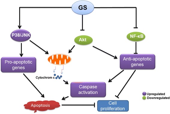
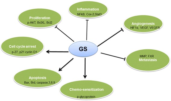
There is a growing evidence now that GS is capable of preventing tumor growth and proliferation through activation of pro-apoptotic and inhibition of anti-apoptotic signaling pathways (Fig. 3). GS has been shown to cause effects on the biological function of cells including cell proliferation, angiogenesis, inflammatory response and apoptotic cells death in cancers cells (Fig. 4). GS mediated anticancer effects are due to inhibition of kinase activity of AKT and its downstream targets such as GSK3, FOXO1 and mTOR signaling. GS has been shown to inhibit the activity of many transcription factors such as NF-κB and AP1 that can lead to down regulation of various gene products including c-myc, Bcl-2, COX-2, NOS, Cyclin D1, interleukins and MMP-9. Furthermore, GS affects many growth factor receptors and angiogenic factors such as VEGF, which play a pivotal role in tumor growth, metastasis and angiogenesis. Not only GS-induces apoptosis in cancer cells and inhibits cell proliferation but it is also useful in reducing the cytotoxicity associated with conventional chemotherapeutic agents by sensitizing or causing the additive apoptotic effects. Despite, GS has been extensively studied, the conclusive mechanisms responsible for its anticancer effects are still not fully understood. Outcomes of various preclinical studies suggest that anticancer action of GS and its isomers are due to combined effects on proliferation and invasiveness in cancer cells.
Fig. 3.

Schematic diagram illustrating the main biological targets of Guggulsterone. The apoptotic effects of guggulsterone are preceded by activation of JNK, suppression of Akt and NF-kB activity. Activation of JNK leads to induction of propapoptic proteins and release of cytochrome c from the mitochondria which in turn activates caspases, resulting in apoptosis. Down regulation of NF-kB activity leads to inhibition of anti apoptotic proteins which in turn activates caspases, resulting in apoptosis and inhibition of proliferation
Fig. 4.

Schematic representation of Guggulsterone mediated effects on various biological processes. i) Abrogating pro-inflammatory signaling by inhibiting activity/expression of IKK-NF-kB, STAT3, COX-2,iNOS, etc. ii) Inhibition of cancer cell proliferation through cell cycle arrest by modulating cyclins, CDKs, etc. iii) Induction of apoptosis of cancerous or transformed cells by modulating expression/activity of caspases, IAPs, Bcl-2 family proteins, etc. iv) Inhibition of angiogenesis by targeting HIF-1a, VEGF, VEGF-R, etc. v) Sensitization of tumor cells to apoptosis induced by chemotherapeutic drugs and reversal of multidrug resistance
Future perspective
The rate of incidence of cancer and resulted deaths are alarming around the world despite the accessibility of various therapeutic options for cancer patients. Most modern medicines currently available for treating cancer are synthetic, mono-targeted, very expensive, less efficient and often possess severe side effects. Therefore, there is a critical need to develop alternative drugs for the management of cancer.
Phytochemicals, a family of naturally occurring compounds including polyphenols, carotenoids and steroids have been demonstrated to have anticancer activities against a variety of cancers both in vitro and in vivo. Among these compounds, guggulsterone (GS), a steroid by nature recently has attracted the attention of cancer researchers and investigators for its anticancer potentials. GS has been shown to induce efficient apoptotic cell death in a variety of cancer cells. Interestingly, no apoptotic death was seen in healthy cells. A number of studies further showed significant cellular changes induced by GS via modulating distinct signaling molecules involved in carcinogen detoxification, cell proliferation, angiogenesis, metastasis, multi-drug resistance, etc. In addition, GS has been shown to sensitize the effects of chemotherapeutic drugs in in vitro system. These anticancer activities in preclinical settings are potentially beneficial in treating cancer. Further studies directed towards target identification and pathway analysis could pave the way for the addition of GS to the management of anticancer therapy. Despite the availability of extensive preclinical data on anticancer potentials of GS, there is a lack of studies accounting for its safety and bioavailability, which needs to be pursued. Safety of long-term use of GS needs to be evaluated in clinical settings, but appears to be devoid of acute, subacute, chronic toxicity in rats, dogs, and monkeys; no mutagenic or teratogenic effects have been reported. Ayurvedic system of medicine describes GS as safe and efficient medicine; however, it should be used cautiously in combination with prescribed drugs as it may modulate the activity of drug metabolizing enzymes. As soon as a consensus on its safety and bioavailability emerges, a planned Phase I clinical trials should be perused to validate its usefulness as anticancer agents and must be prioritized for different site-specific cancers. The outcome of these studies may lead to development of new and efficient therapeutic trategies for the management of cancer.
Acknowledgements
This study was supported by grant from the Medical Research Center (Grant # 16354/16), Hamad Medical Corporation, Doha, and State of Qatar.
The authors appreciate the work of the editors and anonymous reviewers.
Funding
Medical Research Center (Grant # 16354/16), Hamad Medical Corporation, Doha, and State of Qatar.
Availability of data and materials
Not applicable.
Authors’ contributions
AAB, KSB, SK, RK, RMM and SU conceived and designed the study. All authors read and approved the final manuscript.
Competing interest
The authors declare that they have no competing interests.
Consent for publication
Not applicable.
Ethics approval and consent to participate
Not applicable.
Abbreviations
- AMPK
Adenosine monophosphate-activated protein kinase
- DSS
Dextran sulfate sodium
- ERK1/2
Extracellular signal-regulated kinase 1/2
- FXR
Farnesoid X receptor
- GL
Guggul lipid
- GS
Guggulsterone
- HNSCCC
Squamous cell carcinoma
- HSCs
Hepatic stellate cells
- IBD
Inflammatory bowel disease
- IR
Ionizing radiation
- JNK
c-Jun NH(2)-terminal kinase
- NF-kB
Nuclear factor-kB
- PARP
Poly-ADP-ribose phosphate
- PrEC
Prostate epithelial cell line
- TRAIL
Tumor necrosis factor-related apoptosis inducing ligand
- VEGF
Vascular endothelial growth factor
Contributor Information
Ajaz A. Bhat, Email: Abhat1@hamad.qa
Kirti S. Prabhu, Email: KPrabhu@hamad.qa
Shilpa Kuttikrishnan, Email: SKuttikrishnan@hamad.qa.
Roopesh Krishnankutty, Email: RKishnankutty@hamad.qa.
Jayaprakash Babu, Email: s.uppada@unmc.edu.
Ramzi M. Mohammad, Email: RMohammad2@hamad.qa
Shahab Uddin, Phone: 974 40253220, Email: Skhan34@hamad.qa.
References
- 1.Evan GI, Vousden KH. Proliferation, cell cycle and apoptosis in cancer. Nature. 2001;411:342–348. doi: 10.1038/35077213. [DOI] [PubMed] [Google Scholar]
- 2.Hanahan D, Weinberg RA. Hallmarks of cancer: the next generation. Cell. 2011;144:646–674. doi: 10.1016/j.cell.2011.02.013. [DOI] [PubMed] [Google Scholar]
- 3.Ferreira CG, Epping M, Kruyt FA, Giaccone G. Apoptosis: target of cancer therapy. Clin Cancer Res. 2002;8:2024–2034. [PubMed] [Google Scholar]
- 4.Elmore S. Apoptosis: a review of programmed cell death. Toxicol Pathol. 2007;35:495–516. doi: 10.1080/01926230701320337. [DOI] [PMC free article] [PubMed] [Google Scholar]
- 5.Satyavati GV. Gum guggul (Commiphora mukul)--the success story of an ancient insight leading to a modern discovery. Indian J Med Res. 1988;87:327–335. [PubMed] [Google Scholar]
- 6.Satyavati GV, Dwarakanath C, Tripathi SN: Experimental studies on the hypocholesterolemic effect of Commiphora mukul Engl. (Guggul). 1969. Indian J Med Res. 2013; 137:14 p following p403. [PubMed]
- 7.Sinal CJ, Gonzalez FJ. Guggulsterone: an old approach to a new problem. Trends Endocrinol Metab. 2002;13:275–276. doi: 10.1016/S1043-2760(02)00640-9. [DOI] [PubMed] [Google Scholar]
- 8.Urizar NL, Liverman AB, Dodds DT, Silva FV, Ordentlich P, Yan Y, Gonzalez FJ, Heyman RA, Mangelsdorf DJ, Moore DD. A natural product that lowers cholesterol as an antagonist ligand for FXR. Science. 2002;296:1703–1706. doi: 10.1126/science.1072891. [DOI] [PubMed] [Google Scholar]
- 9.Wu J, Xia C, Meier J, Li S, Hu X, Lala DS. The hypolipidemic natural product guggulsterone acts as an antagonist of the bile acid receptor. Mol Endocrinol. 2002;16:1590–1597. doi: 10.1210/mend.16.7.0894. [DOI] [PubMed] [Google Scholar]
- 10.Owsley E, Chiang JY. Guggulsterone antagonizes farnesoid X receptor induction of bile salt export pump but activates pregnane X receptor to inhibit cholesterol 7alpha-hydroxylase gene. Biochem Biophys Res Commun. 2003;304:191–195. doi: 10.1016/S0006-291X(03)00551-5. [DOI] [PubMed] [Google Scholar]
- 11.Cui J, Huang L, Zhao A, Lew JL, Yu J, Sahoo S, Meinke PT, Royo I, Pelaez F, Wright SD. Guggulsterone is a farnesoid X receptor antagonist in coactivator association assays but acts to enhance transcription of bile salt export pump. J Biol Chem. 2003;278:10214–10220. doi: 10.1074/jbc.M209323200. [DOI] [PubMed] [Google Scholar]
- 12.De Gottardi A, Dumonceau JM, Bruttin F, Vonlaufen A, Morard I, Spahr L, Rubbia-Brandt L, Frossard JL, Dinjens WN, Rabinovitch PS, Hadengue A. Expression of the bile acid receptor FXR in Barrett's esophagus and enhancement of apoptosis by guggulsterone in vitro. Mol Cancer. 2006;5:48. doi: 10.1186/1476-4598-5-48. [DOI] [PMC free article] [PubMed] [Google Scholar]
- 13.Ahn KS, Sethi G, Sung B, Goel A, Ralhan R, Aggarwal BB. Guggulsterone, a farnesoid X receptor antagonist, inhibits constitutive and inducible STAT3 activation through induction of a protein tyrosine phosphatase SHP-1. Cancer Res. 2008;68:4406–4415. doi: 10.1158/0008-5472.CAN-07-6696. [DOI] [PubMed] [Google Scholar]
- 14.Kapoor S. Guggulsterone: a potent farnesoid X receptor antagonist and its rapidly evolving role as a systemic anticarcinogenic agent. Hepatology. 2008;48:2090–2091. doi: 10.1002/hep.22601. [DOI] [PubMed] [Google Scholar]
- 15.Peng Z, Raufman JP, Xie G. Src-mediated cross-talk between farnesoid X and epidermal growth factor receptors inhibits human intestinal cell proliferation and tumorigenesis. PLoS One. 2012;7:e48461. doi: 10.1371/journal.pone.0048461. [DOI] [PMC free article] [PubMed] [Google Scholar]
- 16.Guan B, Li H, Yang Z, Hoque A, Xu X. Inhibition of farnesoid X receptor controls esophageal cancer cell growth in vitro and in nude mouse xenografts. Cancer. 2013;119:1321–1329. doi: 10.1002/cncr.27910. [DOI] [PMC free article] [PubMed] [Google Scholar]
- 17.Deng R, Yang D, Radke A, Yang J, Yan B. The hypolipidemic agent guggulsterone regulates the expression of human bile salt export pump: dominance of transactivation over farsenoid X receptor-mediated antagonism. J Pharmacol Exp Ther. 2007;320:1153–1162. doi: 10.1124/jpet.106.113837. [DOI] [PMC free article] [PubMed] [Google Scholar]
- 18.Szapary PO, Wolfe ML, Bloedon LT, Cucchiara AJ, DerMarderosian AH, Cirigliano MD, Rader DJ. Guggulipid for the treatment of hypercholesterolemia: a randomized controlled trial. JAMA. 2003;290:765–772. doi: 10.1001/jama.290.6.765. [DOI] [PubMed] [Google Scholar]
- 19.Dev S. Ancient-modern concordance in Ayurvedic plants: some examples. Environ Health Perspect. 1999;107:783–789. doi: 10.1289/ehp.99107783. [DOI] [PMC free article] [PubMed] [Google Scholar]
- 20.Nityanand S, Srivastava JS, Asthana OP. Clinical trials with gugulipid. A new hypolipidaemic agent. J Assoc Physicians India. 1989;37:323–328. [PubMed] [Google Scholar]
- 21.Singh RB, Niaz MA, Ghosh S. Hypolipidemic and antioxidant effects of Commiphora mukul as an adjunct to dietary therapy in patients with hypercholesterolemia. Cardiovasc Drugs Ther. 1994;8:659–664. doi: 10.1007/BF00877420. [DOI] [PubMed] [Google Scholar]
- 22.Beg M, Singhal KC, Afzaal S. A study of effect of guggulsterone on hyperlipidemia of secondary glomerulopathy. Indian J Physiol Pharmacol. 1996;40:237–240. [PubMed] [Google Scholar]
- 23.Urizar NL, Moore DD. GUGULIPID: a natural cholesterol-lowering agent. Annu Rev Nutr. 2003;23:303–313. doi: 10.1146/annurev.nutr.23.011702.073102. [DOI] [PubMed] [Google Scholar]
- 24.Singh SV, Choi S, Zeng Y, Hahm ER, Xiao D. Guggulsterone-induced apoptosis in human prostate cancer cells is caused by reactive oxygen intermediate dependent activation of c-Jun NH2-terminal kinase. Cancer Res. 2007;67:7439–7449. doi: 10.1158/0008-5472.CAN-07-0120. [DOI] [PubMed] [Google Scholar]
- 25.Shishodia S, Sethi G, Ahn KS, Aggarwal BB. Guggulsterone inhibits tumor cell proliferation, induces S-phase arrest, and promotes apoptosis through activation of c-Jun N-terminal kinase, suppression of Akt pathway, and downregulation of antiapoptotic gene products. Biochem Pharmacol. 2007;74:118–130. doi: 10.1016/j.bcp.2007.03.026. [DOI] [PMC free article] [PubMed] [Google Scholar]
- 26.Samudio I, Konopleva M, Safe S, McQueen T, Andreeff M. Guggulsterones induce apoptosis and differentiation in acute myeloid leukemia: identification of isomer-specific antileukemic activities of the pregnadienedione structure. Mol Cancer Ther. 2005;4:1982–1992. doi: 10.1158/1535-7163.MCT-05-0247. [DOI] [PubMed] [Google Scholar]
- 27.Singh SV, Zeng Y, Xiao D, Vogel VG, Nelson JB, Dhir R, Tripathi YB. Caspase-dependent apoptosis induction by guggulsterone, a constituent of Ayurvedic medicinal plant Commiphora mukul, in PC-3 human prostate cancer cells is mediated by Bax and Bak. Mol Cancer Ther. 2005;4:1747–1754. doi: 10.1158/1535-7163.MCT-05-0223. [DOI] [PubMed] [Google Scholar]
- 28.Xiao D, Singh SV. z-Guggulsterone, a constituent of Ayurvedic medicinal plant Commiphora mukul, inhibits angiogenesis in vitro and in vivo. Mol Cancer Ther. 2008;7:171–180. doi: 10.1158/1535-7163.MCT-07-0491. [DOI] [PubMed] [Google Scholar]
- 29.Shishodia S, Aggarwal BB. Guggulsterone inhibits NF-kappaB and IkappaBalpha kinase activation, suppresses expression of anti-apoptotic gene products, and enhances apoptosis. J Biol Chem. 2004;279:47148–47158. doi: 10.1074/jbc.M408093200. [DOI] [PubMed] [Google Scholar]
- 30.Cheon JH, Kim JS, Kim JM, Kim N, Jung HC, Song IS. Plant sterol guggulsterone inhibits nuclear factor-kappaB signaling in intestinal epithelial cells by blocking IkappaB kinase and ameliorates acute murine colitis. Inflamm Bowel Dis. 2006;12:1152–1161. doi: 10.1097/01.mib.0000235830.94057.c6. [DOI] [PubMed] [Google Scholar]
- 31.Ichikawa H, Aggarwal BB. Guggulsterone inhibits osteoclastogenesis induced by receptor activator of nuclear factor-kappaB ligand and by tumor cells by suppressing nuclear factor-kappaB activation. Clin Cancer Res. 2006;12:662–668. doi: 10.1158/1078-0432.CCR-05-1749. [DOI] [PubMed] [Google Scholar]
- 32.Choudhuri R, Degraff W, Gamson J, Mitchell JB, Cook JA. Guggulsterone-mediated enhancement of radiosensitivity in human tumor cell lines. Front Oncol. 2011;1:19. doi: 10.3389/fonc.2011.00019. [DOI] [PMC free article] [PubMed] [Google Scholar]
- 33.Macha MA, Rachagani S, Gupta S, Pai P, Ponnusamy MP, Batra SK, Jain M. Guggulsterone decreases proliferation and metastatic behavior of pancreatic cancer cells by modulating JAK/STAT and Src/FAK signaling. Cancer Lett. 2013;341:166–177. doi: 10.1016/j.canlet.2013.07.037. [DOI] [PMC free article] [PubMed] [Google Scholar]
- 34.Jiang G, Xiao X, Zeng Y, Nagabhushanam K, Majeed M, Xiao D. Targeting beta-catenin signaling to induce apoptosis in human breast cancer cells by z-guggulsterone and Gugulipid extract of Ayurvedic medicine plant Commiphora mukul. BMC Complement Altern Med. 2013;13:203. doi: 10.1186/1472-6882-13-203. [DOI] [PMC free article] [PubMed] [Google Scholar]
- 35.Macha MA, Matta A, Chauhan SS, Siu KW, Ralhan R. Guggulsterone (GS) inhibits smokeless tobacco and nicotine-induced NF-kappaB and STAT3 pathways in head and neck cancer cells. Carcinogenesis. 2011;32:368–380. doi: 10.1093/carcin/bgq278. [DOI] [PubMed] [Google Scholar]
- 36.Leeman-Neill RJ, Wheeler SE, Singh SV, Thomas SM, Seethala RR, Neill DB, Panahandeh MC, Hahm ER, Joyce SC, Sen M, et al. Guggulsterone enhances head and neck cancer therapies via inhibition of signal transducer and activator of transcription-3. Carcinogenesis. 2009;30:1848–1856. doi: 10.1093/carcin/bgp211. [DOI] [PMC free article] [PubMed] [Google Scholar]
- 37.Aggarwal BB, Shishodia S. Molecular targets of dietary agents for prevention and therapy of cancer. Biochem Pharmacol. 2006;71:1397–1421. doi: 10.1016/j.bcp.2006.02.009. [DOI] [PubMed] [Google Scholar]
- 38.Lee JY, Lee KT, Lee JK, Lee KH, Jang KT, Heo JS, Choi SH, Kim Y, Rhee JC. Farnesoid X receptor, overexpressed in pancreatic cancer with lymph node metastasis promotes cell migration and invasion. Br J Cancer. 2011;104:1027–1037. doi: 10.1038/bjc.2011.37. [DOI] [PMC free article] [PubMed] [Google Scholar]
- 39.Ahn DW, Seo JK, Lee SH, Hwang JH, Lee JK, Ryu JK, Kim YT, Yoon YB. Enhanced antitumor effect of combination therapy with gemcitabine and guggulsterone in pancreatic cancer. Pancreas. 2012;41:1048–1057. doi: 10.1097/MPA.0b013e318249d62e. [DOI] [PubMed] [Google Scholar]
- 40.Grandis JR, Drenning SD, Zeng Q, Watkins SC, Melhem MF, Endo S, Johnson DE, Huang L, He Y, Kim JD. Constitutive activation of Stat3 signaling abrogates apoptosis in squamous cell carcinogenesis in vivo. Proc Natl Acad Sci U S A. 2000;97:4227–4232. doi: 10.1073/pnas.97.8.4227. [DOI] [PMC free article] [PubMed] [Google Scholar]
- 41.Rubin Grandis J, Zeng Q, Drenning SD. Epidermal growth factor receptor--mediated stat3 signaling blocks apoptosis in head and neck cancer. Laryngoscope. 2000;110:868–874. doi: 10.1097/00005537-200005000-00016. [DOI] [PubMed] [Google Scholar]
- 42.Grandis JR, Drenning SD, Chakraborty A, Zhou MY, Zeng Q, Pitt AS, Tweardy DJ. Requirement of Stat3 but not Stat1 activation for epidermal growth factor receptor- mediated cell growth In vitro. J Clin Invest. 1998;102:1385–1392. doi: 10.1172/JCI3785. [DOI] [PMC free article] [PubMed] [Google Scholar]
- 43.Macha MA, Matta A, Chauhan S, Siu KM, Ralhan R. 14-3-3 zeta is a molecular target in guggulsterone induced apoptosis in head and neck cancer cells. BMC Cancer. 2010;10:655. doi: 10.1186/1471-2407-10-655. [DOI] [PMC free article] [PubMed] [Google Scholar]
- 44.Van Waes C, Chang AA, Lebowitz PF, Druzgal CH, Chen Z, Elsayed YA, Sunwoo JB, Rudy SF, Morris JC, Mitchell JB, et al. Inhibition of nuclear factor-kappaB and target genes during combined therapy with proteasome inhibitor bortezomib and reirradiation in patients with recurrent head-and-neck squamous cell carcinoma. Int J Radiat Oncol Biol Phys. 2005;63:1400–1412. doi: 10.1016/j.ijrobp.2005.05.007. [DOI] [PubMed] [Google Scholar]
- 45.Li C, Li R, Grandis JR, Johnson DE. Bortezomib induces apoptosis via Bim and Bik up-regulation and synergizes with cisplatin in the killing of head and neck squamous cell carcinoma cells. Mol Cancer Ther. 2008;7:1647–1655. doi: 10.1158/1535-7163.MCT-07-2444. [DOI] [PMC free article] [PubMed] [Google Scholar]
- 46.Leeman-Neill RJ, Seethala RR, Singh SV, Freilino ML, Bednash JS, Thomas SM, Panahandeh MC, Gooding WE, Joyce SC, Lingen MW, et al. Inhibition of EGFR-STAT3 signaling with erlotinib prevents carcinogenesis in a chemically-induced mouse model of oral squamous cell carcinoma. Cancer Prev Res (Phila) 2011;4:230–237. doi: 10.1158/1940-6207.CAPR-10-0249. [DOI] [PMC free article] [PubMed] [Google Scholar]
- 47.Guan B, Hoque A, Xu X. Amiloride and guggulsterone suppression of esophageal cancer cell growth in vitro and in nude mouse xenografts. Front Biol (Beijing) 2014;9:75–81. doi: 10.1007/s11515-014-1289-z. [DOI] [PMC free article] [PubMed] [Google Scholar]
- 48.Yamada T, Osawa S, Ikuma M, Kajimura M, Sugimoto M, Furuta T, Iwaizumi M, Sugimoto K. Guggulsterone, a plant-derived inhibitor of NF-TB, suppresses CDX2 and COX-2 expression and reduces the viability of esophageal adenocarcinoma cells. Digestion. 2014;90:208–217. doi: 10.1159/000365750. [DOI] [PubMed] [Google Scholar]
- 49.Kelly H, Goldberg RM. Systemic therapy for metastatic colorectal cancer: current options, current evidence. J Clin Oncol. 2005;23:4553–4560. doi: 10.1200/JCO.2005.17.749. [DOI] [PubMed] [Google Scholar]
- 50.An MJ, Cheon JH, Kim SW, Kim ES, Kim TI, Kim WH. Guggulsterone induces apoptosis in colon cancer cells and inhibits tumor growth in murine colorectal cancer xenografts. Cancer Lett. 2009;279:93–100. doi: 10.1016/j.canlet.2009.01.026. [DOI] [PubMed] [Google Scholar]
- 51.Kim ES, Hong SY, Lee HK, Kim SW, An MJ, Kim TI, Lee KR, Kim WH, Cheon JH. Guggulsterone inhibits angiogenesis by blocking STAT3 and VEGF expression in colon cancer cells. Oncol Rep. 2008;20:1321–1327. [PubMed] [Google Scholar]
- 52.De Gottardi A, Touri F, Maurer CA, Perez A, Maurhofer O, Ventre G, Bentzen CL, Niesor EJ, Dufour JF. The bile acid nuclear receptor FXR and the bile acid binding protein IBABP are differently expressed in colon cancer. Dig Dis Sci. 2004;49:982–989. doi: 10.1023/B:DDAS.0000034558.78747.98. [DOI] [PubMed] [Google Scholar]
- 53.Lax S, Schauer G, Prein K, Kapitan M, Silbert D, Berghold A, Berger A, Trauner M. Expression of the nuclear bile acid receptor/farnesoid X receptor is reduced in human colon carcinoma compared to nonneoplastic mucosa independent from site and may be associated with adverse prognosis. Int J Cancer. 2012;130:2232–2239. doi: 10.1002/ijc.26293. [DOI] [PubMed] [Google Scholar]
- 54.Modica S, Murzilli S, Salvatore L, Schmidt DR, Moschetta A. Nuclear bile acid receptor FXR protects against intestinal tumorigenesis. Cancer Res. 2008;68:9589–9594. doi: 10.1158/0008-5472.CAN-08-1791. [DOI] [PubMed] [Google Scholar]
- 55.Maran RR, Thomas A, Roth M, Sheng Z, Esterly N, Pinson D, Gao X, Zhang Y, Ganapathy V, Gonzalez FJ, Guo GL. Farnesoid X receptor deficiency in mice leads to increased intestinal epithelial cell proliferation and tumor development. J Pharmacol Exp Ther. 2009;328:469–477. doi: 10.1124/jpet.108.145409. [DOI] [PMC free article] [PubMed] [Google Scholar]
- 56.Althuis MD, Dozier JM, Anderson WF, Devesa SS, Brinton LA. Global trends in breast cancer incidence and mortality 1973-1997. Int J Epidemiol. 2005;34:405–412. doi: 10.1093/ije/dyh414. [DOI] [PubMed] [Google Scholar]
- 57.Siegel R, DeSantis C, Virgo K, Stein K, Mariotto A, Smith T, Cooper D, Gansler T, Lerro C, Fedewa S, et al. Cancer treatment and survivorship statistics, 2012. CA Cancer J Clin. 2012;62:220–241. doi: 10.3322/caac.21149. [DOI] [PubMed] [Google Scholar]
- 58.Gilmore TD. Introduction: The Rel/NF-kappaB signal transduction pathway. Semin Cancer Biol. 1997;8:61–62. doi: 10.1006/scbi.1997.0056. [DOI] [PubMed] [Google Scholar]
- 59.Rayet B, Gelinas C. Aberrant rel/nfkb genes and activity in human cancer. Oncogene. 1999;18:6938–6947. doi: 10.1038/sj.onc.1203221. [DOI] [PubMed] [Google Scholar]
- 60.Singh RP, Dhanalakshmi S, Agarwal C, Agarwal R. Silibinin strongly inhibits growth and survival of human endothelial cells via cell cycle arrest and downregulation of survivin, Akt and NF-kappaB: implications for angioprevention and antiangiogenic therapy. Oncogene. 2005;24:1188–1202. doi: 10.1038/sj.onc.1208276. [DOI] [PubMed] [Google Scholar]
- 61.Chautard E, Loubeau G, Tchirkov A, Chassagne J, Vermot-Desroches C, Morel L, Verrelle P. Akt signaling pathway: a target for radiosensitizing human malignant glioma. Neuro Oncol. 2010;12:434–443. doi: 10.1093/neuonc/nop059. [DOI] [PMC free article] [PubMed] [Google Scholar]
- 62.Li N, Karin M. Ionizing radiation and short wavelength UV activate NF-kappaB through two distinct mechanisms. Proc Natl Acad Sci U S A. 1998;95:13012–13017. doi: 10.1073/pnas.95.22.13012. [DOI] [PMC free article] [PubMed] [Google Scholar]
- 63.Khalil S, Tan GA, Giri DD, Zhou XK, Howe LR. Activation status of Wnt/ss-catenin signaling in normal and neoplastic breast tissues: relationship to HER2/neu expression in human and mouse. PLoS One. 2012;7:e33421. doi: 10.1371/journal.pone.0033421. [DOI] [PMC free article] [PubMed] [Google Scholar]
- 64.Ayyanan A, Civenni G, Ciarloni L, Morel C, Mueller N, Lefort K, Mandinova A, Raffoul W, Fiche M, Dotto GP, Brisken C. Increased Wnt signaling triggers oncogenic conversion of human breast epithelial cells by a Notch-dependent mechanism. Proc Natl Acad Sci U S A. 2006;103:3799–3804. doi: 10.1073/pnas.0600065103. [DOI] [PMC free article] [PubMed] [Google Scholar]
- 65.Zhang J, Li Y, Liu Q, Lu W, Bu G. Wnt signaling activation and mammary gland hyperplasia in MMTV-LRP6 transgenic mice: implication for breast cancer tumorigenesis. Oncogene. 2010;29:539–549. doi: 10.1038/onc.2009.339. [DOI] [PMC free article] [PubMed] [Google Scholar]
- 66.Clevers H. Wnt/beta-catenin signaling in development and disease. Cell. 2006;127:469–480. doi: 10.1016/j.cell.2006.10.018. [DOI] [PubMed] [Google Scholar]
- 67.Tarapore RS, Siddiqui IA, Mukhtar H. Modulation of Wnt/beta-catenin signaling pathway by bioactive food components. Carcinogenesis. 2012;33:483–491. doi: 10.1093/carcin/bgr305. [DOI] [PMC free article] [PubMed] [Google Scholar]
- 68.Chen HJ, Hsu LS, Shia YT, Lin MW, Lin CM. The beta-catenin/TCF complex as a novel target of resveratrol in the Wnt/beta-catenin signaling pathway. Biochem Pharmacol. 2012;84:1143–1153. doi: 10.1016/j.bcp.2012.08.011. [DOI] [PubMed] [Google Scholar]
- 69.Sundram V, Chauhan SC, Ebeling M, Jaggi M. Curcumin attenuates beta-catenin signaling in prostate cancer cells through activation of protein kinase D1. PLoS One. 2012;7:e35368. doi: 10.1371/journal.pone.0035368. [DOI] [PMC free article] [PubMed] [Google Scholar]
- 70.Ptak C, Petronis A. Epigenetics and complex disease: from etiology to new therapeutics. Annu Rev Pharmacol Toxicol. 2008;48:257–276. doi: 10.1146/annurev.pharmtox.48.113006.094731. [DOI] [PubMed] [Google Scholar]
- 71.Lyko F, Brown R. DNA methyltransferase inhibitors and the development of epigenetic cancer therapies. J Natl Cancer Inst. 2005;97:1498–1506. doi: 10.1093/jnci/dji311. [DOI] [PubMed] [Google Scholar]
- 72.Li Y, Tollefsbol TO. Impact on DNA methylation in cancer prevention and therapy by bioactive dietary components. Curr Med Chem. 2010;17:2141–2151. doi: 10.2174/092986710791299966. [DOI] [PMC free article] [PubMed] [Google Scholar]
- 73.Mirza S, Sharma G, Parshad R, Gupta SD, Pandya P, Ralhan R. Expression of DNA methyltransferases in breast cancer patients and to analyze the effect of natural compounds on DNA methyltransferases and associated proteins. J Breast Cancer. 2013;16:23–31. doi: 10.4048/jbc.2013.16.1.23. [DOI] [PMC free article] [PubMed] [Google Scholar]
- 74.Xu HB, Li L, Liu GQ. Reversal of multidrug resistance by guggulsterone in drug-resistant MCF-7 cell lines. Chemotherapy. 2011;57:62–70. doi: 10.1159/000321484. [DOI] [PubMed] [Google Scholar]
- 75.Xu HB, Shen ZL, Fu J, Xu LZ. Reversal of doxorubicin resistance by guggulsterone of Commiphora mukul in vivo. Phytomedicine. 2014;21:1221–1229. doi: 10.1016/j.phymed.2014.06.003. [DOI] [PubMed] [Google Scholar]
- 76.Kong JN, He Q, Wang G, Dasgupta S, Dinkins MB, Zhu G, Kim A, Spassieva S, Bieberich E. Guggulsterone and bexarotene induce secretion of exosome-associated breast cancer resistance protein and reduce doxorubicin resistance in MDA-MB-231 cells. Int J Cancer. 2015;137:1610–20. [DOI] [PMC free article] [PubMed]
- 77.Whittemore AS, Kolonel LN, Wu AH, John EM, Gallagher RP, Howe GR, Burch JD, Hankin J, Dreon DM, West DW, et al. Prostate cancer in relation to diet, physical activity, and body size in blacks, whites, and Asians in the United States and Canada. J Natl Cancer Inst. 1995;87:652–661. doi: 10.1093/jnci/87.9.652. [DOI] [PubMed] [Google Scholar]
- 78.Jemal A, Siegel R, Ward E, Hao Y, Xu J, Thun MJ. Cancer statistics, 2009. CA Cancer J Clin. 2009;59:225–249. doi: 10.3322/caac.20006. [DOI] [PubMed] [Google Scholar]
- 79.Gilligan T, Kantoff PW. Chemotherapy for prostate cancer. Urology. 2002;60:94–100. doi: 10.1016/S0090-4295(02)01583-2. [DOI] [PubMed] [Google Scholar]
- 80.Xiao D, Zeng Y, Prakash L, Badmaev V, Majeed M, Singh SV. Reactive oxygen species-dependent apoptosis by gugulipid extract of Ayurvedic medicine plant Commiphora mukul in human prostate cancer cells is regulated by c-Jun N-terminal kinase. Mol Pharmacol. 2011;79:499–507. doi: 10.1124/mol.110.068551. [DOI] [PMC free article] [PubMed] [Google Scholar]
- 81.Shishodia S, Azu N, Rosenzweig JA, Jackson DA. Guggulsterone for Chemoprevention of Cancer: Curr Pharm Des. 2016;22:294–306. [DOI] [PubMed]
- 82.Zu XY, Zhang QH, Liu JH, Cao RX, Zhong J, Yi GH, Quan ZH, Pizzorno G. ATP citrate lyase inhibitors as novel cancer therapeutic agents. Recent Pat Anticancer Drug Discov. 2012;7:154–167. doi: 10.2174/157489212799972954. [DOI] [PubMed] [Google Scholar]
- 83.Moon DO, Park SY, Choi YH, Ahn JS, Kim GY. Guggulsterone sensitizes hepatoma cells to TRAIL-induced apoptosis through the induction of CHOP-dependent DR5: involvement of ROS-dependent ER-stress. Biochem Pharmacol. 2011;82:1641–1650. doi: 10.1016/j.bcp.2011.08.019. [DOI] [PubMed] [Google Scholar]
- 84.Hussain AR, Uddin S, Bu R, Khan OS, Ahmed SO, Ahmed M, Al-Kuraya KS. Resveratrol suppresses constitutive activation of AKT via generation of ROS and induces apoptosis in diffuse large B cell lymphoma cell lines. PLoS One. 2011;6:e24703. doi: 10.1371/journal.pone.0024703. [DOI] [PMC free article] [PubMed] [Google Scholar]
- 85.Shi JJ, Jia XL, Li M, Yang N, Li YP, Zhang X, Gao N, Dang SS. Guggulsterone induces apoptosis of human hepatocellular carcinoma cells through intrinsic mitochondrial pathway. World J Gastroenterol. 2015;21:13277–13287. doi: 10.3748/wjg.v21.i47.13277. [DOI] [PMC free article] [PubMed] [Google Scholar]
- 86.Kim BH, Yoon JH, Yang JI, Myung SJ, Lee JH, Jung EU, Yu SJ, Kim YJ, Lee HS, Kim CY. Guggulsterone attenuates activation and survival of hepatic stellate cell by inhibiting nuclear factor kappa B activation and inducing apoptosis. J Gastroenterol Hepatol. 2013;28:1859–1868. doi: 10.1111/jgh.12314. [DOI] [PubMed] [Google Scholar]
- 87.Lichtman MA. Battling the hematological malignancies: the 200 years' war. Oncologist. 2008;13:126–138. doi: 10.1634/theoncologist.2007-0228. [DOI] [PubMed] [Google Scholar]
- 88.Macha MA, Matta A, Chauhan SS, Siu KW, Ralhan R. Guggulsterone targets smokeless tobacco induced PI3K/Akt pathway in head and neck cancer cells. PLoS One. 2011;6:e14728. [DOI] [PMC free article] [PubMed]
- 89.Noh EM, Chung EY, Youn HJ, Jung SH, Hur H, Lee YR, Kim JS. Cis-guggulsterone inhibits the IKK/NF-kappaB pathway, whereas trans-guggulsterone inhibits MAPK/AP-1 in MCF7 breast cancer cells: guggulsterone regulates MMP9 expression in an isomer-specific manner. Int J Mol Med. 2013;31:393–99. [DOI] [PubMed]
Associated Data
This section collects any data citations, data availability statements, or supplementary materials included in this article.
Data Availability Statement
Not applicable.


