The authors present a successfully treated case of Mycobacterium goodii cardiac device pocket infection, complicated by inherent resistance and drug reactions. This case highlights the complexity of treating these infections. This article outlines treatment approaches and caveats, including duration of therapy and when device reimplantation is appropriate.
Key Words: cardiac, device, infection, mycobacterium, nontuberculous, nosocomial, sulfamethoxazole/trimethoprim
Abstract
Mycobacterium goodii, a rapidly growing nontuberculous mycobacterium, is an emerging pathogen in nosocomial infections. Its inherent resistance patterns make it a challenging organism to treat, and delays in identification can lead to poor outcomes. We present a case of cardiac device pocket infection with M. goodii, complicated by both antibiotic resistance and drug reactions that highlight the challenges faced by clinicians trying to eradicate these infections. We also present a brief review of the English literature surrounding this disease, including a table of all reported cases of M. goodii infections and their outcomes to act as guide for clinicians formulating treatment plans for these infections. A clear understanding of diagnostic methods and treatment caveats is essential to curing infections caused by these organisms.
Mycobacterium goodii, a rapidly growing nontuberculous mycobacterium (NTM), is a Gram-positive, acid, and alcohol fast bacillus. It is related to Mycobacterium smegmatis; however, in 1999, Brown et al1 reclassified it as its own species based on gene sequencing and antimicrobial resistance data. Since then, it has been identified in several infections, most of which are nosocomial. We present the third cardiac device pocket infection with M. goodii.
CASE REPORT
A 77-year-old man with a medical history of heart failure with reduced ejection fraction and chronic kidney disease was admitted for elective placement of an automated implantable cardiac defibrillator (AICD). He underwent placement of the AICD in the cardiac catheterization laboratory without complication. He was discharged the same day.
The patient returned 4 weeks later with pus draining from the incision site. He reported erythema and tenderness had initially developed approximately 2 weeks after the procedure. He denied fevers or chills and had no other systemic manifestations of infection.
In the clinic, a 2 × 2-cm fluctuant mass was noted over the superior edge of the healed surgical incision. The patient otherwise had normal vital signs and an unremarkable physical examination, including lack of thromboembolic phenomena. His laboratory results were notable for a white blood cell count of 10.6 × 10² with a normal differential and a creatinine level of 2.03 mg/dL (his baseline). Two sets of blood cultures (4 bottles) were drawn and were negative for any bacterial growth. Pus was expressed from the AICD pocket and sent for culture. The patient was started on cephalexin 500 mg by mouth twice daily.
Later that week, the patient underwent complete AICD removal with pocket debridement and washout. Tissue from the pocket, a wound swab, and AICD leads were all sent for routine bacterial cultures. The patient was treated empirically with vancomycin 15 mg/kg intravenous (IV) every 12 hours and piperacillin/tazobactam 3.375 g IV every 12 hours (both renally adjusted). A wound vacuum was placed over the pacemaker pocket with excellent clinical response. The patient underwent a transesophageal echocardiogram, which did not show any evidence of endocarditis.
Four days into incubation, all tissue cultures grew Gram-positive rods. The patient's antibiotics were narrowed to vancomycin IV monotherapy, and the cultures were sent to the New Mexico State Laboratory for further identification. The organism was identified as a rapidly growing NTM. Cultures were then sent to Advanced Diagnostic Laboratories, National Jewish Health in Denver, Colo, for identification. The isolate was grown on a Lowenstein-Jensen slant and identified as M. goodii by RNA polymerase Beta subunit (rpoB) gene sequencing.
The patient's antibiotic coverage was changed from vancomycin to sulfamethoxazole/trimethoprim one double strength tablet by mouth twice daily and doxycycline 100 mg by mouth twice daily. Unfortunately, 4 days after treatment with this regimen, he developed worsening renal dysfunction with a creatinine level increase to 4 mg/dL and a serum urea nitrogen level of 40 mg/dL. Sulfamethoxazole/trimethoprim was stopped, and the patient was continued on doxycycline with the addition of both ethambutol 900 mg by mouth daily and ciprofloxacin 500 mg by mouth daily (renally adjusted). Two weeks later, ciprofloxacin was discontinued due to concerns for QTC prolongation in the setting of recent AICD removal. At this point, antimicrobial susceptibilities were available, and the patient's isolate was found to be sensitive to ampicillin-sulbactam. The patient was switched to a regimen of amoxicillin-clavulanate 875 mg by mouth twice daily and doxycycline 100 mg by mouth twice daily to complete a 4-month course.
There was a question about whether to treat the patient for endocarditis versus a pocket infection because the AICD leads grew M. goodii. On discussion with the laboratory and the operating surgeons, the entire lead was cultured, including the section of the leads attached to the battery within the infected pocket, making a pure ventricular tip lead infection impossible to determine. Given that the patient had no major or minor Duke criteria, was never septic, and had both a negative transesophageal echocardiogram and negative blood cultures, he was treated for an isolated pocket infection. His wounds are currently completely healed, and his inflammatory markers are down trending.
We present the third case of M. goodii cardiac device pocket infection reported in the English literature.
DISCUSSION
Infections, particularly nosocomial, with NTM are on the rise.2 These bacteria, though indolent, are resistant to many forms of decontamination and sterilization, with postsurgical outbreak reports in South America and the United States.3–5 Mycobacterium goodii presents a unique challenge due to its particular resistance pattern. This case highlights the diagnostic and treatment challenges of this particular NTM organism. We will summarize the current literature about diagnosing and treating M. goodii.
Mycobacterium goodii is a rapidly growing NTM that was differentiated from M. smegmatis in 1999 by Brown et al. These small Gram-positive rods grow in 2 to 4 days on most media (blood agar, chocolate agar, trypticase soy agar, Middlebrook 7H10 or 7H11 agar, and Lowenstein-Jensen agar).1
Treatment of M. goodii can be complex. All M. goodii infections associated with surgical intervention or implants in the literature required adequate surgical debridement and removal of contaminated material for clinical cure, with the exception of 2 cases.6,7 Treatment subsequently requires prolonged appropriate antibiotic therapy.2 Because of delays in diagnosis, empiric therapy is often begun for rapidly growing NTM, usually clarithromycin and rifampin. Unfortunately, M. goodii is inherently resistant to these medications due to 2 factors: overexpression of the Wag31 gene and the presence of the erm gene.8 Overexpression of the Wag31 gene results in increased thickness of the peptidoglycan layer leading to decreased permeability of lipophilic drugs like rifampin.9 The erm gene causes irregularities at the ribosomal binding site for macrolides, causing clarithromycin resistance.8,10
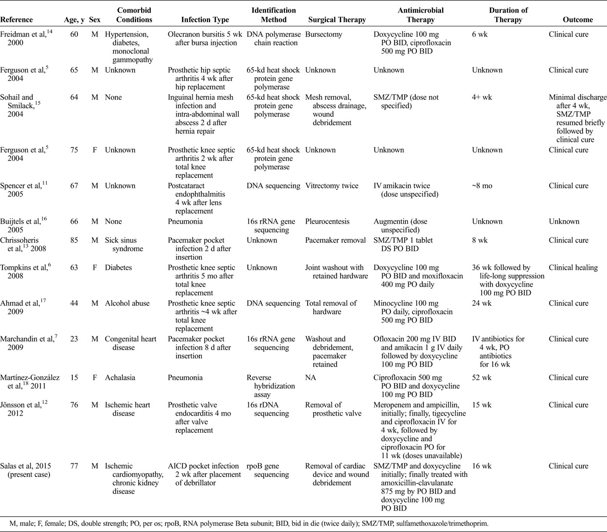
Treatment should be guided by antimicrobial susceptibility testing. The most commonly used drugs to treat this organism are sulfamethoxazole/trimethoprim and ethambutol, followed by doxycycline and ciprofloxacin, depending on susceptibilities.1,2 For more serious infections, amikacin and meropenem have been reported.11,12 When tolerated, sulfamethoxazole/trimethoprim has the most evidence for treatment success; however, allergies and renal toxicity, as seen in our case, limit its use (Table 1). Combination therapy is often used, but monotherapy, particularly with sulfamethoxazole/trimethoprim, has been reported (Table 1). Table 1 lists all of the cases of M. goodii reported in the English literature as well as the medications and durations of therapy that have been used. Please note that Brown et al investigated 28 isolates that were later classified as M. goodii using 16s RNA sequencing and DNA-DNA hybridization. The infections listed were predominantly posttraumatic wounds and bone infections, nosocomial infections, and pulmonary disease, including 3 cases with lipoid pneumonia. These clinical isolates are not included in Table 1 due to lack of clinical data surrounding the infections.
TABLE 1.
Clinical Infections With Mycobacterium goodii

Treatment of M. goodii usually is prolonged and depends on the clinical syndrome10 ranging from 4 weeks to 12 months (Table 1). In the case of cardiac device infections, there is no clear evidence as to when a new device can be implanted. In one case report, successful reimplantation occurred within 2 weeks of removal of the infected device.13 In our patient, reimplantation was delayed until after completion of therapy (16 weeks).
In conclusion, M. goodii is a rare but challenging pathogen, most often associated with nosocomial infection. No specific host risk factors have been reported. Identification of the organism requires gene sequencing to differentiate it from other rapidly growing NTM, which is essential given its unique resistance pattern. Treatment includes adequate surgical debridement, removal of contaminated exogenous material, and the use of 1 to 2 antimicrobial agents, usually including sulfamethoxazole/trimethoprim, guided by susceptibility testing. Duration of therapy is determined by the clinical syndrome.10 Physicians need to be aware of the challenges to treating NTM infections as the incidence is increasing, and failed recognition can lead to poor outcomes.
ACKNOWLEDGMENTS
The authors thank Staci Lee, MD, Division of Infectious Diseases, University of New Mexico, for her work on revising and editing the text.
Footnotes
Editorial assistance in the preparation of this manuscript was provided by Dr Staci Lee who has no financial interests or conflicts of interest to declare. No funding or sponsorship was received for this study or publication of this article. All named authors meet the International Committee of Medical Journal Editors criteria for authorship for this manuscript, take responsibility for the integrity of the work as a whole, and have given final approval to the version to be published. Natalie Mariam Salas and Nicole Klein declare they have no conflicts of interest. This article does not contain any new studies with human or animal subjects performed by any of the authors.
REFERENCES
- 1.Brown BA, Springer B, Steingrube VA, et al. Mycobacterium wolinskyi sp. nov. and Mycobacterium goodii sp. nov., two new rapidly growing species related to Mycobacterium smegmatis and associated with human wound infections: a cooperative study from the International Working Group on Mycobacterial Taxonomy. Int J Syst Bacteriol. 1999;49(pt 4):1493–1511. [DOI] [PubMed] [Google Scholar]
- 2.Griffith DE, Aksamit T, Brown-Elliott BA, et al. An official ATS/IDSA statement: diagnosis, treatment, and prevention of nontuberculous mycobacterial diseases. Am J Respir Crit Care Med. 2007;175(4):367–416. [DOI] [PubMed] [Google Scholar]
- 3.Wallace RJ, Brown BA, Griffith DE. Nosocomial outbreaks/pseudo-outbreaks caused by nontuberculous mycobacteria. Annu Rev Microbiol. 1998;52:453–490. [DOI] [PubMed] [Google Scholar]
- 4.Maurer F, Castelberg C, von Braun A, et al. Postsurgical wound infections due to rapidly growing mycobacteria in Swiss medical tourists following cosmetic surgery in Latin America between 2012 and 2014. Euro Surveill. 2014;19(37):20905. [DOI] [PubMed] [Google Scholar]
- 5.Ferguson DD, Gershman K, Jensen B, et al. Mycobacterium goodii infections associated with surgical implants at Colorado hospital. Emerg Infect Dis. 2004;10(10):1868–1871. [DOI] [PMC free article] [PubMed] [Google Scholar]
- 6.Tompkins JC, Harrison MS, Witzig RS. Mycobacterium goodii infection of a total knee prosthesis. Infect Med. 2008;25(11):522–525. [Google Scholar]
- 7.Marchandin H, Battistella P, Calvet B, et al. Pacemaker surgical site infection caused by Mycobacterium goodii. J Med Microbiol. 2009;58(pt 4):517–520. [DOI] [PubMed] [Google Scholar]
- 8.Nash KA, Andini N, Zhang Y, et al. Intrinsic macrolide resistance in rapidly growing mycobacteria. Antimicrob Agents Chemother. 2006;50(10):3476–3478. [DOI] [PMC free article] [PubMed] [Google Scholar]
- 9.Xu W, Zhang L, Mai JT, et al. The Wag31 protein interacts with AccA3 and coordinates cell wall lipid permeability and lipophilic drug resistance in Mycobacterium smegmatis. Biochem Biophys Res Commun. 2014;448(3):255–260. [DOI] [PubMed] [Google Scholar]
- 10.Brown-Elliott BA, Nash KA, Wallace RJ. Antimicrobial susceptibility testing, drug resistance mechanisms, and therapy of infections with nontuberculous mycobacteria. Clin Microbiol Rev. 2012;25(3):545–582. [DOI] [PMC free article] [PubMed] [Google Scholar]
- 11.Spencer TS, Teske MP, Bernstein PS. Postcataract endophthalmitis caused by Mycobacterium goodii. J Cataract Refract Surg. 2005;31(6):1252–1253. [DOI] [PubMed] [Google Scholar]
- 12.Jönsson G, Rydberg J, Sturegård E. A case of Mycobacterium goodii prosthetic valve endocarditis in a non-immunocompromised patient: use of 16S rDNA analysis for rapid diagnosis. BMC Infect Dis. 2012;12:301. [DOI] [PMC free article] [PubMed] [Google Scholar]
- 13.Chrissoheris MP, Kadakia H, Marieb M, et al. Pacemaker pocket infection due to Mycobacterium goodii: case report and review of the literature. Conn Med. 2008;72(2):75–77. [PubMed] [Google Scholar]
- 14.Friedman ND, Sexton DJ. Bursitis due to Mycobacterium goodii, a recently described, rapidly growing mycobacterium. J Clin Microbiol. 2001;39(1):404–405. [DOI] [PMC free article] [PubMed] [Google Scholar]
- 15.Sohail MR, Smilack JD. Hernia repair mesh-associated Mycobacterium goodii infection. J Clin Microbiol. 2004;42(6):2858–2860. [DOI] [PMC free article] [PubMed] [Google Scholar]
- 16.Buijtels PC, Petit PL, Verbrugh HA, et al. Isolation of nontuberculous mycobacteria in Zambia: eight case reports. J Clin Microbiol. 2005;43(12):6020–6026. [DOI] [PMC free article] [PubMed] [Google Scholar]
- 17.Ahmad S, Khakoo RA. Left knee prosthesis-related Mycobacterium goodii infection. Int J Infect Dis. 2010;14(12):e1115–e1116. [DOI] [PubMed] [Google Scholar]
- 18.Martínez-González D, Franco J, Navarro-Ortega D, et al. Achalasia and Mycobacterium goodii pulmonary infection. Pediatr Infect Dis J. 2011;30(5):447–448. [DOI] [PubMed] [Google Scholar]


