Abstract
Protease-activated receptor (PAR)4 is a low affinity thrombin receptor with less understood function relative to PAR1. PAR4 is involved in platelet activation and hemostasis, but its specific actions on myocyte growth and cardiac function remain unknown. This study examined the role of PAR4 deficiency on cardioprotection after myocardial ischemia-reperfusion (IR) injury in mice. When challenged by in vivo or ex vivo IR, PAR4 knockout (KO) mice exhibited increased tolerance to injury, which was manifest as reduced infarct size and a more robust functional recovery compared to wild-type mice. PAR4 KO mice also showed reduced cardiomyocyte apoptosis and putative signaling shifts in survival pathways in response to IR. Inhibition of PAR4 expression in isolated cardiomyocytes by shRNA offered protection against thrombin and PAR4-agonist peptide-induced apoptosis, while overexpression of wild-type PAR4 significantly enhanced the susceptibility of cardiomyocytes to apoptosis, even under low thrombin concentrations. Further studies implicate Src- and epidermal growth factor receptor-dependent activation of JNK on the proapoptotic effect of PAR4 in cardiomyocytes. These findings reveal a pivotal role for PAR4 as a regulator of cardiomyocyte survival and point to PAR4 inhibition as a therapeutic target offering cardioprotection after acute IR injury.
Keywords: thrombin receptor, myocardial ischemia reperfusion, inflammation, apoptosis, cardioprotection
1. INTRODUCTION
Restoration of blood flow after acute myocardial infarction limits infarct size and reduces mortality. However, reestablishing blood flow is often followed by a second set of stresses, a phenomenon referred to as ischemia-reperfusion (IR) injury, which can result in additional myocardial damage and account for up to half of total infarct size [1]. The factors contributing to IR injury are complex and include microvascular obstruction, inflammation, release of oxygen radicals, myocardial stunning, and activation of mitochondrial apoptosis and necrosis [1,2]. Extensive research has explored the mechanisms responsible for the activation of inflammatory-derived cytokines/chemokines and reactive oxygen species and their roles in the infarcted heart. However, there is a paucity of information regarding the role of inflammatory serine proteases on cardiac injury post-IR [3]. These proteases not only cleave extracellular substrates, but also mediate cell signal transduction, in part, through protease-activated receptors (PARs) [4,5].
PARs are a family of seven transmembrane spanning domain G protein-coupled receptors that are activated by cleavage of the receptor N-terminal domain thereby exposing a new, previously cryptic sequence [6,7]. The exposed sequence remains tethered to the receptor and further acts as a receptor-activating tethered ligand. Four members of PARs have been characterized, PAR1, PAR3 and PAR4 are activated by thrombin, whereas PAR2 is activated by other proteases such as trypsin and tryptase, but not by thrombin [6,7]. PAR1 is the prototypical receptor to which most of the cellular and platelet actions of thrombin are attributed, and its role in fibroblast proliferation and cardiac remodeling has been well established [7–10]. In contrast, knowledge of PAR4 expression and function in the heart is still quite limited. PAR4 is generally recognized as a low affinity thrombin receptor [6,7], although it can also be activated by other proteases including trypsin [11], tissue kallikrein [12], and the neutrophilic granule protease cathepsin G (Cat.G) [13]. Synthetic peptides based on the receptor-activating sequence of the tethered ligand (e.g., GYPGKF for murine PAR4) are also capable of activating PAR4 by direct binding to the receptor [6,7]. PAR4-deficient mice show a normal phenotype, but hemostasis is impaired due to defective platelet thrombin signaling [14,15]. On the other hand, when administered locally at high doses, PAR4 agonists produce inflammation [16,17]. Although these results suggest that the predominant role of PAR4 is in platelet activation [7,9], these receptors are also expressed in cardiomyocytes[18] with their contribution to myocyte growth and function remaining largely unknown.
We previously showed that PAR4 stimulation is slower in onset but more prolonged in cardiac myocytes relative to PAR1 [18]. Moreover, pharmacological inhibition of PAR4 using the pepducin antagonist of PAR4, P4pal 10, or the trans-cinnamyol-YPGKF-amide peptide have suggested that antagonism of this receptor is protective in response to 2 hours of myocardial ischemia reperfusion injury [19]. However, the interpretation of data using antagonist peptides is potentially confounded by limitations of the bioavailability of these agents in vivo as well as the observation that both behave as agonists in some in vitro model systems [16]. The specific functions of PAR4 on cardiac function are still not well understood and no loss-of-function cardiac phenotypes of PAR4 gene have yet been described. In the present study, we show that PAR4-deficient mice display normal cardiac function but present reduced inflammation, less myocyte death and improved cardiac function in response to acute IR injury. Reciprocally, activation of PAR4 leads to myocyte apoptosis through activation of JNK signaling pathway. Our results suggest that inhibition of PAR4 could be a potential therapeutic target to protect the heart from acute IR injury.
2. METHODS
2.1. Myocardial IR injury procedure
All mice were maintained in accordance with protocols approved by the Animal Care and Use Committee of Temple University. PAR4 knockout (KO) mice were generated and genotyped as described previously [15]. Twelve week-old male wild-type (WT) and PAR4 KO mice were anesthetized with a mixture of ketamine (100 mg/kg) and xylazine (10 mg/kg) to perform a left thoracotomy under mechanical ventilation. Body temperature was maintained by a heated surgical platform and was monitored throughout surgery using a rectal sensor. A 6–0 suture with a slipknot was tied around the left anterior descending (LAD) coronary artery to produce ischemia. Consistent elevation of the ST segment was observed in lead II tracings following occlusion of the LAD coronary vessel. Regional ischemia was confirmed by visual inspection under a dissecting microscope (Nikon) by discoloration of the occluded distal myocardium. The ligation was released after 30 minutes of ischemia and the heart was allowed to reperfuse as confirmed by visual inspection. The chest wall was closed with 8–0 silk and then the animal was removed from the ventilator and kept warm in the cage maintained at 37°C overnight. A sham procedure constituted the surgical incision without LAD ligation. Hearts were harvested after 24 h of reperfusion.
2.2. Data Analysis
Data are expressed as the mean ± SEM. We used a 2-tailed, unpaired Student's t test for all pair-wise comparisons (GraphPad Prism version 5). P values of less than 0.05 were considered significant. All in vitro experiments were performed at least three times from three different cultures and the data values were scaled to controls. A value of P<0.05 was considered statistically significant.
An Expanded Methods section is provided in the Supplemental material.
3. RESULTS
3.1. PAR4 expression is upregulated after acute myocardial IR
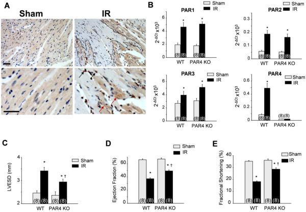
PAR4 expression was investigated in WT hearts after IR injury. In the absence of IR, PAR4 immunostaining was weak and was homogenously distributed throughout the myocardium (Fig. 1A). At 24 h after IR injury, PAR4 staining was strongly evidenced in inflammatory cells which occupied most of the damaged area, and in surviving cardiac muscle fibers at the border infarct zone which exhibited aggregated cytoplasmic labeling. However, PAR4 immunolabeling was not detected in damaged myocytes of the infarct zone. No staining was detected when PAR4 immunostaining was performed in PAR4 KO heart sections or when the primary antibody was omitted (Supplemental Figs. 1A and 1B). Quantification of PAR4 mRNA levels in WT heart samples by real-time quantitative PCR showed 5.2-fold increase in IR hearts (infarct border zone) than in sham control (Fig. 1B). We also measured PAR4 expression in area opposite of the infarct and found a small increase in PAR4 expression relative to shams (+1.8-fold increase in IR hearts compared to shams). Expression levels of PAR1, PAR2 and PAR3 mRNA were also increased by 2-, 3.2-, and 1.5-fold in hearts after IR compared to WT sham and this increase was similar between PAR4 KO and WT hearts subjected to IR, suggesting that PAR4 loss did not significantly affect the expression of other PARs.
Fig. 1. PAR4 ablation protects against acute IR.
(A–E) The left anterior descending artery was ligated for 30 minutes to induce ischemia and the heart was subsequently reperfused for 24 h (IR). (A) Representative immunostaining of paraffin-embedded heart sections of wild-type (WT) sham or after IR injury stained for PAR4 and counterstained with hematoxylin. PAR4 staining was detected in infiltrating inflammatory cells (black arrows) and in surviving cardiomyocytes (red arrows). Scale bar: 40 μm. (B) PARs mRNA levels in hearts of WT and PAR4 KO as assessed by real-time qPCR. Data were normalized to GAPDH and were expressed relative to levels in WT sham hearts. (C–E) Echocardiography measurement of left ventricular (LV)-end systolic dimension (C), LV ejection fraction (D), and LV fractional shortening (E) in WT and PAR4 KO animals. Values are presented as mean ± SEM, *P<0.05 vs. WT shams, †P<0.05 vs. WT IR.
3.2. Genetic ablation of PAR4 is cardioprotective after acute myocardial IR
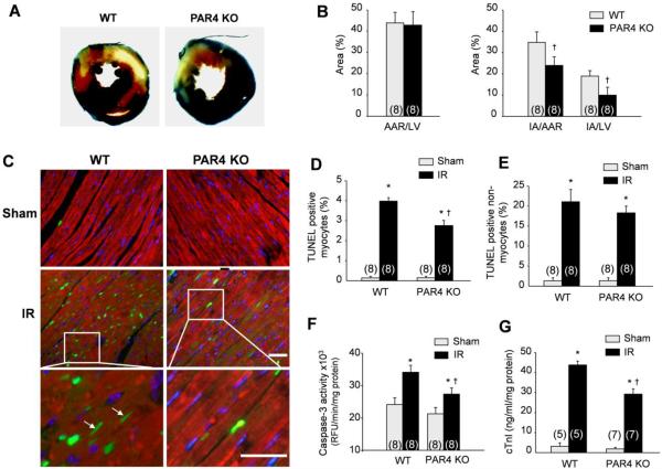
To explore the functions of PAR4 expression in vivo, PAR4 KO and their WT controls were subjected to IR injury. PAR4 deletion did not affect baseline heart weight-to-body weight ratio, heart rate, or left ventricular (LV) dilatation and function (Figs. 1C–1E and Supplemental Table 1). Interestingly, PAR4 KO mice showed reduced LV end-systolic dimensions (Fig. 1C) and a significant improvement in cardiac function in hearts subjected to IR injury as assessed by increased LV ejection fraction (Fig. 1D) and fractional shortening percentage (Fig. 1E) compared to WT mice. The percentage of infarcted area normalized to area at risk (AAR) or to LV was reduced in PAR4 KO hearts compared with WT mice (Figs. 2A and 2B). The AAR normalized to LV area was similar between PAR4 KO and WT mice after IR.
Fig. 2. PAR4 ablation reduces infarct size and cell apoptosis after acute IR.
Ischemia reperfusion (IR) was induced for 24 h. (A) Representative cross-sections were stained with triphenyl tetrazolium chloride and Evans blue to determine the extent of injury. (B) Quantification of infarct area (IA) vs. area at risk (AAR) after IR injury in the indicated groups. (C) LV tissue sections were assessed for apoptosis using TUNEL assay (green), tropomyosin (red), and DAPI (4',6-diamidino-2-phenylindole) (blue) staining. Arrows show TUNEL-positive myocytes. Scale bar, 40 μm. (D–E) The number of TUNEL-positive myocytes (D) and non-myocytes (E) in the reperfused area was expressed as a percentage of total nuclei detected by DAPI staining, n=8. (F) Quantification of caspase-3 activity in the LV using caspase-3 specific fluorogenic substrate, n=8. (G) Serum levels of cardiac troponin I (cTnI) after IR injury in WT (n=5) and PAR4 KO (n=7) mice. Values are presented as mean ±SEM, *P<0.05 vs. WT shams, †P<0.05 vs. WT IR.
The extent of myocardial infarct and impairment in cardiac performance depends on the levels of cardiomyocyte loss post-IR. We found that IR in WT mice increased Terminal deoxynucleotidyl transferase dUTP nick end labeling (TUNEL)-positive myocytes and non-myocytes (Figs. 2C–2E). In contrast, PAR4 KO hearts had reduced numbers of TUNEL-positive myocytes after 24 h of reperfusion. Caspase-3 activity was also reduced in PAR4 KO infarcted hearts compared to WT, thus confirming decreased death by apoptosis in PAR4 KO mice subjected to IR (Fig. 2F). Circulating plasma levels of the cardiac-specific isoform of troponin-I (cTnI) were evaluated as an additional marker of myocardial injury (Fig. 2G). Serum samples from PAR4 KO mice subjected to IR injury were found to contain less levels of cTnI than WT mice (43.2 ±2.2 in WT vs. 28.1 ±3.5 ng/ml per mg protein in PAR4 KO), thus confirming the cytoprotective effect of PAR4 deletion.
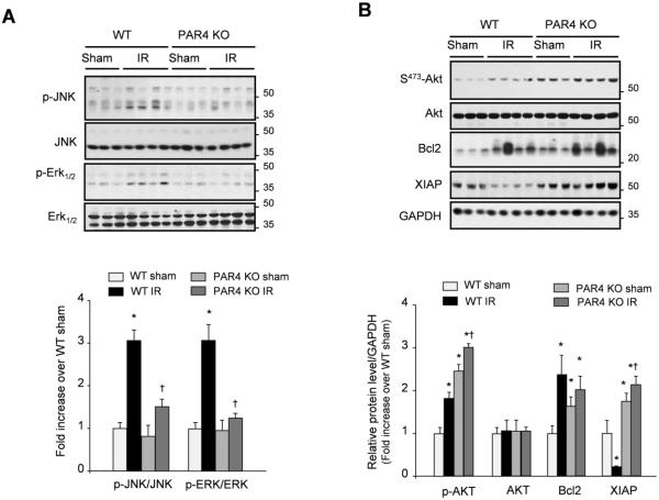
We next explored the molecular mechanisms downstream from PAR4 and has been shown to play a role in cardioprotection. Induction of IR injury in WT mice significantly increased the phosphorylation of MAPKs, JNK and Erk1/2, compared to shams (Fig. 3A), with only minor changes in p38 MAPK phosphorylation between the 2 groups (data not shown). PAR4 deletion attenuated the phosphorylation of JNK and Erk1/2 after IR injury compared to WT mice. PAR4 deficiency increased basal levels of phospho-AKT, Bcl2 and XIAP in shams compared to WT and the accumulation of phospho-AKT and XIAP was further enhanced in the infarcted heart after IR injury, indicating an increase in survival signaling molecules (Fig. 3B). Collectively, these data show that PAR4 deletion was associated with overall increase in cardioprotective signaling pathways.
Fig. 3. PAR4 deficiency is associated with enhanced anti-apoptotic signaling after IR.
(A–B) Immunoblot analysis for PAR4-downstream signaling indicates an increase in anti-apoptotic signaling response in the infarcted region of the PAR4 KO mice vs. the WT post-IR. Top: Representative immunoblots. Bottom: Quantification of experiments represented as fold change compared to WT animals (n=6 for each groups). GAPDH was included as a loading control. *P<0.05 vs. WT shams, †P<0.05 vs. WT IR.
3.3. PAR4 deletion reduces inflammation after acute IR injury
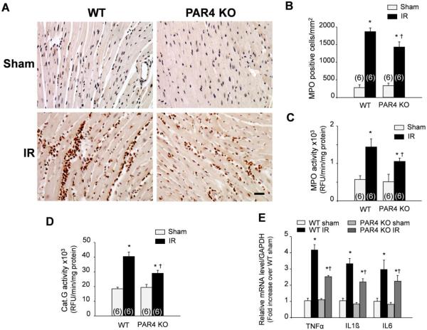
IR injury initiates a complex cycle of hypoxia and cell death that is perpetuated in part by inflammation [1,2]. Given the role of PAR4 activation in the modulation of different aspects of inflammation, including events associated with leukocyte recruitment [16,17], we next examined whether PAR4 deletion reduced inflammation after IR injury. Histological examination of PAR4 KO and WT hearts after 24 h of reperfusion revealed no differences in the numbers of infiltrating leukocytes in shams (Figs. 4A and 4B). Enzymatic assays for myeloperoxidase (MPO) and neutrophil-derived protease cathepsin G (Cat.G) activity also showed no differences between WT and PAR4 KO at baseline (Figs. 4C and 4D). However, the number of leukocytes and activity levels of MPO and Cat.G were markedly reduced in PAR4 KO infarcts compared to WT after IR. To further assess the extent of inflammation in PAR4 KO infarct hearts, we measured by RT-qPCR the expression of pro-inflammatory cytokines that mediate early infiltration of leukocytes in the infarcted myocardium including tumor necrosis factor (TNF)α, interleukin (IL)6 and IL1ß [20,21]. WT mice showed a greater increase in TNFα, IL6 and IL1ß mRNA expression in the infarct after IR injury, and this increase was attenuated in PAR4 KO infarcts (Fig. 4E). Collectively, these data show that PAR4 stimulation mediates early inflammatory responses following IR injury.
Fig. 4. Deletion of PAR4 reduces leukocyte infiltration and inflammation.
(A) Representative immunolabeling of paraffin-embedded heart sections from sham or mice subjected to ischemia reperfusion (IR) were stained for myeloperoxidase (MPO) and counterstained with hematoxylin. Scale bar, 40 μm. (B) Quantification of MPO-positive cells in PAR4 KO and WT infarcts. (C–D) Levels of MPO (C) and cathepsin G (Cat.G) (D) activity in the infarcted LV as determined by enzyme activity assay. Results are expressed as relative fluorescence unit (RFU)/min/mg protein. (E) Real-time qPCR analysis of TNFα, IL1ß and IL6 in PAR4 KO and WT infarcts. The data were normalized to GAPDH and were expressed as mean ± SEM (n=6 for each groups). *P<0.05 vs. WT shams, †P<0.05 vs. WT IR.
3.4. PAR4 KO mice show increased tolerance against IR injury in the isolated perfused heart
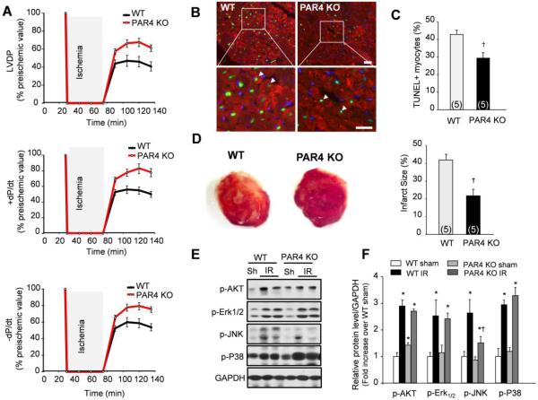
To exclude a role for endogenous systemic neurohormonal and inflammatory mechanisms in the cardioprotection seen in PAR4 KO mice, further studies were performed on isolated perfused hearts. Following a 45-minute period of global ischemia, LV-developed pressure (LVDP) recovered over 60 minutes of reperfusion to 61% of pre-ischemic values in the PAR4 KO compared with only 40% in the WT hearts (Fig. 5A). Post-ischemic maximum rate of contraction (+dP/dt) and maximum rate of relaxation (−dP/dt) also showed more rapid and significant recovery in PAR4 KO hearts than in WT. To directly assess myocardial cell damage, we examined myocyte apoptosis in infarcted hearts after a 60 minutes reperfusion period. The number of TUNEL-positive myocytes was ~35% lower in PAR4 KO hearts (Figs. 5B and 5C). Caspase-3 activity was also diminished in PAR4 KO infarcted hearts compared to WT, indicating resistance to cell apoptosis (Supplemental Fig. S2). Myocardial infarct size following reperfusion was likewise markedly reduced, from 42% ± 3% in WT, to 22% ± 4% in PAR4 KO mice (Fig. 5D). Analysis of AKT and MAPKs phosphorylation shows a marked reduction in JNK phosphorylation in PAR4 KO infarcted hearts compared to WT (Fig. 5E). In contrast, phosphorylation levels of AKT, Erk1/2 and p38 MAPK were similar between PAR4 KO and WT infarcted hearts. These data together demonstrate the role of PAR4 deficiency in ex vivo protection against IR injury.
Fig. 5. PAR4 deletion protects against IR injury in the isolated perfused heart.
Isolated hearts from WT and PAR4 KO mice were subjected to global ischemia for 45 minutes and reperfusion for 60 minutes (IR). (A) Time course recovery of contractility following ischemia measured by LVDP, +dP/dt, and −dP/dt. *P < 0.05 versus WT (n = 5). (B) LV tissue sections were assessed for apoptosis using TUNEL assay (green), tropomyosin (red), and DAPI (4',6-diamidino-2-phenylindole) (blue) staining. Arrows show TUNEL-positive myocytes. Scale bar, 40 μm. (C) The number of TUNEL-positive myocytes in the infarcted area was expressed as a percentage of total nuclei detected by DAPI staining. (D) Representative triphenyl tetrazolium chloride (TTC)-stained heart sections (left) and quantitative analysis of infarct size (right). (E) Representative blots showing AKT, JNK, Erk1/2 and p38 MAPK phosphorylation level. GAPDH was used as loading control, respectively. (F) Quantification of experiments as fold change compared to WT shams. Data are shown as mean ± SEM (n = 5 in each group). *P < 0.05 vs. WT. †P<0.05 vs. WT IR.
3.5. PAR4 stimulation leads to cardiomyocyte apoptosis
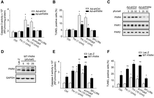
To directly demonstrate that the cardioprotective effects of PAR4 deletion after IR may result from endogenous expression of PAR4 in cardiomyocytes, we investigated the role of PAR4 in cardiomyocyte death. Physiological concentrations of thrombin that stimulate PAR1 have been shown to induce myocyte hypertrophy [22]. However, the effects of pathological concentrations of thrombin, that are able to activate both PAR1 and PAR4 [9,23], on myocyte growth are not yet defined. Treatment of neonatal rat cardiomyocytes (NRCMs) with PAR4-agonist peptide (AP) (AYPGKF) or high concentrations of thrombin (10 U/ml) for 24 h induced an increase in caspase-3 activity and in the number of TUNEL positive myocytes compared to untreated cells, indicating cell death by apoptosis (Figs. 6A and 6B). In contrast, treatment with PAR1/PAR2-AP (SFLLRN) or low concentrations of thrombin (1 U/ml) had no detectable effect on both markers of myocyte apoptosis.
Fig. 6. PAR4 stimulation mediates cardiomyocyte apoptosis.
(A–B) Neonatal rat cardiac myocytes were transduced with adenoviruses expressing shRNA PAR4 (Ad-shCbl, 10 pfu/cell) or shRNA control (Ad-shCtrl, 10 pfu/cell) for 48 h and then untreated or treated with 1 or 10 U/ml thrombin (Thr), 500 μM PAR4-AP (AYPGKF) or 300 μM PAR1/PAR2-AP (SFLLRN) for 24 h. Caspase-3 activity (A) and the percent of TUNEL-positive nuclei (B) were assayed in triplicate. (C–D) Lysates from myocytes infected with Ad-shCtrl, Ad-shPAR4 (C), Lac.Z (10 pfu/cell) or WT-PAR4 (D) adenoviruses for 48 h were assessed for immunoblot analysis. (E–F) Myocytes infected with Lac.Z or WT-PAR4 adenoviruses were treated with the indicated agonists for 24 h and assayed for caspase-3 activity (E) or TUNEL assay (F). Results are representative of 3 independent experiments. Data are mean ± SEM; *P<0.05 vs. control; †P<0.05 vs. treated myocytes.
To confirm the role of PAR4 stimulation on thrombin-induced myocyte apoptosis, we conducted loss-of-function experiments in which we transduced NRCMs with adenoviruses carrying shRNA PAR4 (Ad-shPAR4) or shRNA control (Ad-shCtrl). Transduction of Ad-shPAR4 reduced PAR4 expression by ~85% compared to cells infected with Ad-shCtrl (Fig. 6C) and had no detectable effect on markers of apoptosis in untreated myocytes. In contrast, expression of Ad-shPAR4 significantly decreased caspase-3 activity and TUNEL positive myocytes induced by treatment with high concentrations of thrombin or PAR4-AP (Figs. 6A and 6B).
To further explore the influence of PAR4 on myocyte apoptosis, we conducted gain-of-function experiments in which NRCMs were infected with adenoviruses carrying wild type PAR4 (WT-PAR4). Myocytes infected with WT-PAR4 viruses at 5, 10 and 25 pfu/cell showed an increased level of PAR4 expression by 3, 5, and 6 fold, respectively over ß-galactosidase (Lac.Z) infected controls (Fig. 6D). Expression of WT-PAR4 increased myocyte apoptosis compared to control cells infected with Lac.Z (Figs. 6E and 6F). Additional treatment with high concentrations of thrombin or PAR4-AP, but not with PAR1/PAR2-AP, further enhanced the increase in caspase-3 activity and the percentage of TUNEL positive cells in WT-PAR4-infected cells compared to Lac.Z-treated cells. It is noteworthy that low concentrations of thrombin, that do not stimulate apoptosis in Lac.Z infected myocytes, induced an increase in caspase-3 activity and TUNEL positive myocytes in WT-PAR4 infected cells. Collectively, these results show that stimulation of PAR4 leads to myocyte apoptosis and could be deleterious in the setting of IR injury.
3.6. PAR4 induces myocyte apoptosis through JNK pathway
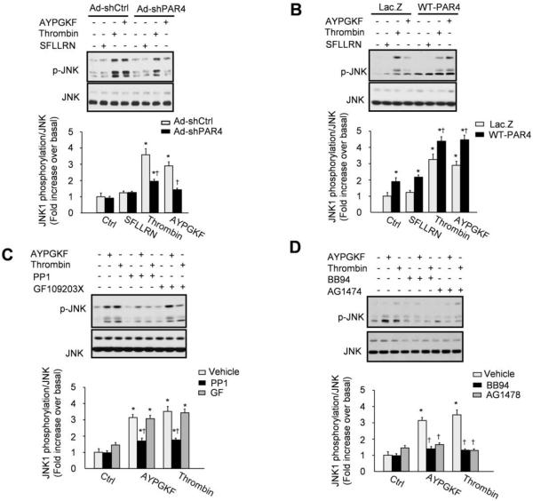
We next investigated the mechanisms by which PAR4 stimulation induces myocyte apoptosis in vitro. Since MAPKs mediate myocyte apoptosis during ischemia and/or reperfusion [24], we investigated which MAPK mediates PAR4-induced myocyte apoptosis. Treatment of NRCMs with high concentrations of thrombin induced a marked increase in JNK phosphorylation, which occurred early and was sustained for over 24 h (Figs. 7A and 7B and Supplemental Fig. S3). Chronic treatment with the PAR4-AP, but not with the PAR1/PAR2-AP, induced JNK phosphorylation, suggesting that PAR4 stimulation is mediating the effect of thrombin on JNK phosphorylation. To verify this hypothesis, we conducted gain- and loss-of-PAR4 function experiments. Expression of Ad-shPAR4 significantly attenuated JNK phosphorylation induced by PAR4-AP or thrombin compared to Ad-shCtrl infected myocytes (Fig. 7A). In contrast, expression of WT-PAR4 enhanced JNK phosphorylation in untreated, PAR1/PAR2-AP, PAR4-AP-, or thrombin-treated myocytes (Fig. 7B). Stimulation of Erk1/2 and p38-MAPK was slightly increased in response to PAR4-AP or thrombin treatment for 24 h and inhibition of PAR4 expression showed little effect on their phosphorylation state. In contrast, expression of WT-PAR4 enhanced Erk1/2 and p38-MAPK phosphorylation in response to thrombin or PAR4-AP (Supplemental Fig. S4).
Fig. 7. Delayed JNK activation after PAR4 stimulation involves Src and EGFR kinase activities.
(A–B) NRCMs were infected with Ad-shCtrl, Ad-shPAR4 (A), Lac.Z or WT-PAR4 (B) adenoviruses and were either untreated or treated with 10 U/ml thrombin, PAR1/PAR2- or PAR4-AP for 24 h. Cell lysates were processed for immunoblot analysis. (C, D) NRCMs were pretreated with vehicle, PP1 (5 μM), GF109203X (5 μM) (C), BB94 (10 μM), or AG1478 (5 μM) (D) for 30 minutes prior to treatment with 10 U/ml thrombin or PAR4-AP for 24 h. Cells were assessed for immunoblot analysis. Top: representative immunoblot with each lane from a single gel exposed for the same duration. Bottom: Quantification of experiments expressed as mean ± SEM from three separate cultures. *P<0.05 vs. control; †P<0.05 vs. treated myocytes.
Previous studies from our group have shown that thrombin-dependent activation of Src is mediated only by PAR4 in cardiomyocytes (and not PAR1) [18]. To determine whether Src lies upstream from JNK in the PAR4 signaling pathway, further studies were performed with PP1 (a specific inhibitor of Src family tyrosine kinases). Figure 7C shows that PP1 blocks JNK activation by PAR4-AP and thrombin. In contrast treatment with GF109203X (an inhibitor of phorbol ester-sensitive protein kinase C (PKC) isoforms that ablate the phospholipase C-PKC pathway) have no detectable effect on JNK phosphorylation induced by PAR4-AP and thrombin.
Given the evidence that signaling by cardiomyocyte G protein-coupled receptors and PAR4 also can involve epidermal growth factor receptor (EGFR) transactivation [18,25], PAR4-AP and thrombin responses also were examined in cultures pretreated with BB94 (a general MMP inhibitor) or AG1478 (a well-established inhibitor of the kinase activity of the EGFR). Figure 7D shows that BB94 or AG1478 treatment inhibits JNK activation induced by PAR4-AP and thrombin in cardiomyocytes. Collectively, these results implicate Src and EGFR kinases, but not PKC, activities in the pathway for PAR4-dependent activation of JNK in cardiomyocytes.
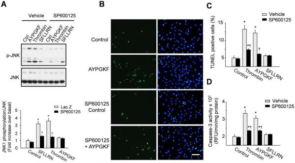
We next investigated the role of JNK pathway in PAR4-induced myocyte apoptosis. Treatment of cells with the specific inhibitor of JNK, SP600125, markedly attenuated JNK phosphorylation induced by PAR4-AP or thrombin treatment (Fig. 8A). SP600125 treatment also significantly attenuated the increase in TUNEL positive myocytes and caspase-3 activity induced in response to PAR4-AP or thrombin (Figs. 8B–8D). In contrast, treatment with inhibitors of Erk1/2 or p38 MAPK pathways by PD98059 or SB203580, respectively, had no effect on PAR4-AP- or thrombin-induced myocyte apoptosis (Supplemental Figs. S5A and S5B). Treatment with PP1 or AG1478 were also effective in blocking caspase-3 activity induced in response to thrombin and PAR4-AP treatment (Supplemental Fig. S6). Collectively, these studies resolve a JNK-dependent signaling pathway emanating from sustained stimulation of PAR4 that involves a Src-dependent pathway and EGFR transactivation and leads to cardiomyocyte apoptosis (schematized in Supplemental Fig. S7).
Fig. 8. PAR4 induces myocyte apoptosis through JNK pathway.
NRCMs were pretreated with vehicle or 5 μM SP600125 for 30 minutes prior to treatment with 10 U/ml thrombin, PAR1/PAR2- or PAR4-AP for 24 h. (A) Cells were assessed for immunoblot analysis. Top: representative immunoblot with each lane from a single gel exposed for the same duration. Bottom: Quantification of experiments expressed as mean ± SEM from three separate cultures. (B) Representative images of total nuclei (blue) and apoptotic nuclei (Green) from untreated and AYPGKF treated myocytes in presence or absence of SP600125. Scale bar: 100 μm. (C) Quantification of percent TUNEL-positive nuclei. Samples were assayed in triplicate and results are representative of 3 independent experiments. (D) Lysates from NRCMs were assayed for caspase-3 activity assay. Data are mean ± SEM; *P<0.05 vs. control; †P<0.05 vs. treated myocytes.
4. DISCUSSION
The results of this study reveal PAR4 as an important receptor mediating early IR stress responses in the heart. PAR4 expression is increased in heart after IR and its inhibition decreased inflammation and cardiomyocyte apoptosis while improving LV function after acute IR injury. PAR4 stimulation induces delayed JNK activation which subsequently mediates cardiomyocyte apoptosis. By inhibiting effectors of JNK signaling pathways, PAR4 deficiency can diminish the degree of cardiomyocyte death during IR. These data highlight the potential therapeutic role of PAR4 in cardioprotection in response to acute myocardial IR injury.
Our study provides evidence that this low affinity thrombin receptor is involved in the early responses induced by IR injury. At baseline, PAR4-deficient mice appear histologically and functionally normal, consistent with reports that PAR4 deletion in mice have minimal consequences under normal conditions [15]. In contrast, PAR4 deficient mice had smaller infarct sizes, reduced cardiomyocyte apoptosis, and showed preserved cardiac contractile function compared with WT controls after acute IR injury, indicating that endogenous PAR4 stimulation mediates myocardial tissue damage in vivo. When isolated PAR4 KO mouse hearts were challenged by IR, we observed profound tolerance to ex vivo IR injury. Protection was manifest as 52% reduction in infarct size, decreased myocyte death, and markedly improved post-ischemic contractile performance. Parallel loss-of-function experiments of PAR4 by siRNA demonstrated markedly decreased cardiomyocyte apoptosis when cardiomyocytes did not express PAR4. These data provide substantial support for the conclusion that endogenous PAR4 is activated in cardiomyocytes during IR, where it functions to reduce protection of myocytes against IR injury. However, our data do not rule out a role of PAR4 deletion on inflammatory responses in cardioprotection post-IR. PAR4 expression was observed in inflammatory cells infiltrating the myocardium after acute IR and hearts from PAR4 deficient mice show reduced levels of neutrophil accumulation, suggesting that PAR4 stimulation induces a pro-inflammatory state after acute IR. These data are consistent with previous findings showing the role of PAR4 stimulation on thrombin-induced platelet activation, which may affect the activation of several inflammatory cells [26], and in leukocyte rolling and adherence to endothelium in vivo [17,27]. Future studies are needed to determine the role of PAR4 expression in neutrophils on cardioprotection post-IR.
Our data provide strong evidence that PAR4 stimulation leads to myocyte apoptosis in vitro. Chronic treatment with thrombin or PAR4-AP increased caspase-3 activity and DNA fragmentation, while loss-of-function experiments of PAR4 by siRNA show reduced myocyte apoptosis in response to thrombin treatment. These data, in agreement with previous results obtained in vascular smooth muscle cells and in neurons [28,29], demonstrate a pro-apoptotic effect of thrombin on cardiomyocytes, requiring both higher concentrations of thrombin than those required to promote myocyte hypertrophy [22] and a more prolonged exposure (at least 24 h). However, in these studies the pro-apoptotic effect of thrombin was either independent of PAR1 or PAR4 stimulation (in vascular smooth muscle cells) [28] or dependent of PAR1 stimulation (in neurons) [29], suggesting that the pro-apoptotic effect of thrombin is cell type specific and could be related to levels of PAR4 expression. This hypothesis is supported by the fact that myocytes expressing WT-PAR4 showed enhanced apoptosis susceptibility even in response to low concentrations of thrombin that induce hypertrophy and not apoptosis [22]. WTPAR4 overexpression also increased myocyte apoptosis in absence of ligands, suggesting that overexpression of WT-PAR4 significantly increases the apparent constitutive activity of this GPCR. This constitutive activity of PAR4 is widely accepted and is a common property of many WT GPCRs [30]. Studies have shown that over-expression of a GPCR with even low constitutive activity increases the absolute number of GPCRs in the R* state at steady-state and renders the GPCR capable of fully activating the limited pools of G-proteins and effectors [30]. While the reasons of the constitutive activity of WT-PAR4 need to be investigated, differences in GPCR expression level, GPCR desensitization, G-protein complement and GPCR/G-protein stoichiometry can all contribute to such increase in PAR4 activity.
An important finding of the present study is the identification of the molecular mechanisms by which PAR4 exerts its protective effect. PAR4 deletion was associated with enhanced accumulation of cardioprotective signaling molecules such as phospho-AKT, Bcl2 and XIAP. This increase was not related to changes in PAR1, PAR2 or PAR3 expression levels in PAR4 deficient hearts, but rather involved endogenous stimulation of PAR4. Loss-of-function experiments using PAR4 siRNA corroborated this hypothesis and show that PAR4 knockdown enhances AKT activation and prevented myocytes apoptosis in response to thrombin. These data extend previous studies showing the effect of PI3 kinase/AKT pathway activation on the cardioprotective effects offered by PAR4 antagonist peptide in ex vivo model of IR [19]. Moreover, treatment with thrombin or chronic stimulation of Gαq have been shown to induce AKT signaling downregulation in several cells including cardiomyocytes [31,32]. Intriguingly, our study provides novel evidence of the role of PAR4 on JNK pathway activation. PAR4 knockdown reduced the activation of JNK induced after in vivo and ex vivo IR injury, while chronic stimulation of PAR4 in cardiomyocytes led to delayed JNK activation which was important in determining its effect on myocyte apoptosis. These findings are consistent with previous studies showing the effect of JNK inhibition or deletion on reducing myocyte apoptosis in response to hypoxia-reoxygenation [33]. It is also in agreement with cardioprotective effects observed in JNK KO mice after IR injury in vivo [34]. Additional studies implicate Src and EGFR kinase activities in the delayed activation of JNK by PAR4, providing evidence that transactivation of EGFR family members can contribute to JNK activation and PAR4 responses. These results extend our previous findings in cardiomyocytes where PAR4 stimulation induced EGFR and ErbB2 phosphorylation and led to slow activation of Src/p38 MAPK pathway, with only a minor associated increase in phospholipase C and Erk1/2 activation [18]. However, while the requirement of EGFR transactivation for PAR4-induced myocyte apoptosis seems paradoxical, studies have documented proapoptotic properties of EGFR activation in several cells including cardiomyocytes [35]. The proapoptotic effect of EGFR may be related to the signaling diversity associated with the EGFR Ligands, EGFR localization and/or EGFR interactions with tyrosine kinases and phosphatases that dictate the potency and duration of the signal activated by EGFRs. Thus, the delayed EGFR-dependent activation of JNK induced by PAR4 could be a prerequisite for its proapoptotic effect in cardiomyocytes.
The role of thrombin receptors PAR1 and PAR4 has been mainly characterized in thrombosis linking the coagulation cascade to the activation of platelets [6,7]. Pharmacological inhibition or genetic deletion of PAR1 improved cardiac function after myocardial IR injury,[10,36] although, only the former has been shown to reduce infarct size [36]. The study reported herein is the first to our knowledge to address PAR4 regulation of cardiomyocyte apoptosis both in vitro and in vivo. PAR4 has been shown to be activated by high concentrations of thrombin[37], a condition found in the setting of hemorrhagic infarction where the endothelial barrier is broken and cardiomyocytes come into direct contact with blood-borne substances. PAR4 has also been identified as a potential substrate for neutrophil-derived proteases that are released at sites of cardiac injury and/or inflammation [13]. Hence, the cardiac actions of PAR4 may become important in the context of cardiac remodeling during myocardial IR injury or in conditions that render PAR1 inactive. The unique capacity of PAR4 inhibition to block cardiomyocyte apoptosis while reducing platelet activation and subsequent blood coagulation represents a previously unrecognized constellation of features to prevent cardiac dysfunction post-IR. Correctly timed inhibition of PAR4 could be a potential approach to blunt cellular losses and to ameliorate the progression of degenerative changes accompanying acute IR damage.
In conclusion, the results of this study suggest that PAR4 deletion protects the heart against acute IR injury by reducing neutrophil infiltration and cardiomyocyte death. These findings provide the first evidence for an important role of PAR4 in direct modulation of cardiomyocyte apoptosis. Given that overexpression of thrombin and other serine proteases underlies cardiac injury post-IR and other forms of heart disease, it is likely that PAR4 deletion plays a cardioprotective role in a variety of acute stress settings. Thus, early inhibition of PAR4 signaling to attenuate JNK pathway and cardiomyocyte death may provide therapeutic benefit to acute IR injury.
Supplementary Material
Highlights.
PAR4 deficiency is cardioprotective after acute myocardial ischemia reperfusion injury.
PAR4 deletion reduces inflammation after acute ischemia reperfusion injury.
PAR4 mediates thrombin-induced myocyte apoptosis in vitro.
PAR4 induces myocyte apoptosis through activation of the JNK pathway.
ACKNOWLEDGEMENTS
This work was supported by the National Institute of Health (HL088626 and HL076799, AS) and American Heart Association (15SDG22650004, MAK; 14PRE20380518, BH).
Abbreviations
- AAR
Area at risk
- AP
Agonist peptide
- Cat.G
Cathepsin G
- cTnI
Cardiac Troponin I
- EGFR
Epidermal growth factor receptor
- IA
Infarct area
- IR
Ischemia-reperfusion
- KO
Knockout
- LAD
Left anterior descending
- LV
Left ventricular
- MPO
Myeloperoxidase
- NRCMs
Neonatal rat cardiac myocytes
- PAR
Protease-activated receptor
- TTC
Triphenyltretrazolium chloride
- TUNEL
Terminal deoxynucleotidyl transferase dUTP nick end labeling
- WT
Wild-type
Footnotes
Publisher's Disclaimer: This is a PDF file of an unedited manuscript that has been accepted for publication. As a service to our customers we are providing this early version of the manuscript. The manuscript will undergo copyediting, typesetting, and review of the resulting proof before it is published in its final citable form. Please note that during the production process errors may be discovered which could affect the content, and all legal disclaimers that apply to the journal pertain.
DISCLOSURES None
REFERENCES
- [1].Yellon DM, Hausenloy DJ. Myocardial reperfusion injury. N Engl J Med. 2007;357:1121–35. doi: 10.1056/NEJMra071667. [DOI] [PubMed] [Google Scholar]
- [2].Piper HM, Garcna-Dorado D, Ovize M. A fresh look at reperfusion injury. Cardiovas Res. 1998;38:291–300. doi: 10.1016/s0008-6363(98)00033-9. [DOI] [PubMed] [Google Scholar]
- [3].Heutinck K, ten Berge I, Hack C, Hamann J, Rowshani A. Serine proteases of the human immune system in health and disease. Mol Immunol. 2010;47:1943–55. doi: 10.1016/j.molimm.2010.04.020. [DOI] [PubMed] [Google Scholar]
- [4].Steinberg SF. The Cardiovascular actions of protease-activated receptors. Mol Pharmacol. 2005;67:2–11. doi: 10.1124/mol.104.003103. [DOI] [PubMed] [Google Scholar]
- [5].Adams MN, Ramachandran R, Yau M-K, Suen JY, Fairlie DP, Hollenberg MD, et al. Structure, function and pathophysiology of protease activated receptors. Pharmacol Therap. 2011;130:248–82. doi: 10.1016/j.pharmthera.2011.01.003. [DOI] [PubMed] [Google Scholar]
- [6].Coughlin SR. How the protease thrombin talks to cells. Proc Natl Acad Sci USA. 1999;96:11023–7. doi: 10.1073/pnas.96.20.11023. [DOI] [PMC free article] [PubMed] [Google Scholar]
- [7].Coughlin SR. Thrombin signalling and protease-activated receptors. Nature. 2000;407:258–64. doi: 10.1038/35025229. [DOI] [PubMed] [Google Scholar]
- [8].Sabri A, Short J, Guo J, Steinberg SF. Protease-activated receptor-1-mediated DNA synthesis in cardiac fibroblast is via epidermal growth factor receptor transactivation: Distinct PAR-1 signaling pathways in cardiac fibroblasts and cardiomyocytes. Circ Res. 2002;91:532–9. doi: 10.1161/01.res.0000035242.96310.45. [DOI] [PubMed] [Google Scholar]
- [9].Kahn ML, Zheng Y-W, Huang W, Bigornia V, Zeng D, Moff S, et al. A dual thrombin receptor system for platelet activation. Nature. 1998;394:690–4. doi: 10.1038/29325. [DOI] [PubMed] [Google Scholar]
- [10].Pawlinski R, Tencati M, Hampton CR, Shishido T, Bullard TA, Casey LM, et al. Protease-activated receptor-1 contributes to cardiac remodeling and hypertrophy. Circulation. 2007;116:2298–306. doi: 10.1161/CIRCULATIONAHA.107.692764. [DOI] [PMC free article] [PubMed] [Google Scholar]
- [11].Cottrell GS, Amadesi S, Grady EF, Bunnett NW. Trypsin IV, a novel agonist of protease-activated receptors 2 and 4. J Biol Chem. 2004;279:13532–9. doi: 10.1074/jbc.M312090200. [DOI] [PubMed] [Google Scholar]
- [12].Oikonomopoulou K, Hansen KK, Saifeddine M, Tea I, Blaber M, Blaber SI, et al. Proteinase-activated receptors, targets for kallikrein signaling. J Biol Chem. 2006;281:32095–112. doi: 10.1074/jbc.M513138200. [DOI] [PubMed] [Google Scholar]
- [13].Sambrano GR, Huang W, Faruqi T, Mahrus S, Craik C, Coughlin SR. Cathepsin G activates protease-activated receptor-4 in human platelets. J Biol Chem. 2000;275:6819–23. doi: 10.1074/jbc.275.10.6819. [DOI] [PubMed] [Google Scholar]
- [14].Hamilton JR, Cornelissen I, Coughlin SR. Impaired hemostasis and protection against thrombosis in protease-activated receptor 4-deficient mice is due to lack of thrombin signaling in platelets. J Thromb Haemost. 2004;2:1429–35. doi: 10.1111/j.1538-7836.2004.00783.x. [DOI] [PubMed] [Google Scholar]
- [15].Sambrano GR, Weiss EJ, Zheng Y-W, Huang W, Coughlin SR. Role of thrombin signalling in platelets in haemostasis and thrombosis. Nature. 2001;413:74–8. doi: 10.1038/35092573. [DOI] [PubMed] [Google Scholar]
- [16].Hollenberg MD, Saifeddine M, Sandhu S, Houle S, Vergnolle N. Proteinase-activated receptor-4: evaluation of tethered ligand-derived peptides as probes for receptor function and as inflammatory agonists in vivo. Br J Pharmacol. 2004;143:443–54. doi: 10.1038/sj.bjp.0705946. [DOI] [PMC free article] [PubMed] [Google Scholar]
- [17].Vergnolle N, Derian CK, D'Andrea MR, Steinhoff M, Andrade-Gordon P. Characterization of thrombin-induced leukocyte rolling and adherence: A potential proinflammatory role for proteinase-activated receptor-4. J Immunol. 2002;169:1467–73. doi: 10.4049/jimmunol.169.3.1467. [DOI] [PubMed] [Google Scholar]
- [18].Sabri A, Guo J, Elouardighi H, Darrow AL, Andrade-Gordon P, Steinberg SF. Mechanisms of protease-activated receptor-4 actions in cardiomyocytes. Role of Src tyrosine kinase. J Biol Chem. 2003;278:11714–20. doi: 10.1074/jbc.M213091200. [DOI] [PubMed] [Google Scholar]
- [19].Strande JL, Hsu A, Su J, Fu X, Gross GJ, Baker JE. Inhibiting protease-activated receptor 4 limits myocardial ischemia/reperfusion injury in rat hearts by unmasking adenosine signaling. J Pharmacol ExpTher. 2008;324:1045–54. doi: 10.1124/jpet.107.133595. [DOI] [PMC free article] [PubMed] [Google Scholar]
- [20].Kukielka GL, Smith CW, Manning AM, Youker KA, Michael LH, Entman ML. Induction of interleukin-6 synthesis in the myocardium: Potential role in postreperfusion inflammatory injury. Circulation. 1995;92:1866–75. doi: 10.1161/01.cir.92.7.1866. [DOI] [PubMed] [Google Scholar]
- [21].Hwang M-W, Matsumori A, Furukawa Y, Ono K, Okada M, Iwasaki A, et al. Neutralization of interleukin-1β in the acute phase of myocardial infarction promotes the progression of left ventricular remodeling. J Am Coll Cardiol. 2001;38:1546–53. doi: 10.1016/s0735-1097(01)01591-1. [DOI] [PubMed] [Google Scholar]
- [22].Sabri A, Muske G, Zhang H, Pak E, Darrow A, Andrade-Gordon P, et al. Signaling properties and functions of two distinct cardiomyocyte protease-activated receptors. Circ Res. 2000;86:1054–61. doi: 10.1161/01.res.86.10.1054. [DOI] [PubMed] [Google Scholar]
- [23].Xu WF, Andersen H, Whitmore TE, Presnell SR, Yee DP, Ching A, et al. Cloning and characterization of human protease-activated receptor-4. Proc Nat Acad Sci USA. 1998;95:6642–6. doi: 10.1073/pnas.95.12.6642. [DOI] [PMC free article] [PubMed] [Google Scholar]
- [24].Baines CP, Molkentin JD. Stress signaling pathways that modulate cardiac myocyte apoptosis. J Mol Cell Cardiol. 2005;38:47–62. doi: 10.1016/j.yjmcc.2004.11.004. [DOI] [PubMed] [Google Scholar]
- [25].Asakura M, Kitakaze M, Takashima S, Liao Y, Ishikura F, Yoshinaka T, et al. Cardiac hypertrophy is inhibited by antagonism of ADAM12 processing of HB-EGF: metalloproteinase inhibitors as a new therapy. Nat Med. 2002;8:35–40. doi: 10.1038/nm0102-35. [DOI] [PubMed] [Google Scholar]
- [26].Zarbock A, Polanowska-Grabowska RK, Ley K. Platelet-neutrophil-interactions: Linking hemostasis and inflammation. Blood Rev. 2007;21:99–111. doi: 10.1016/j.blre.2006.06.001. [DOI] [PubMed] [Google Scholar]
- [27].Asokananthan N, Graham PT, Fink J, Knight DA, Bakker AJ, McWilliam AS, et al. Activation of protease-activated receptor (PAR)-1, PAR-2, and PAR-4 stimulates IL-6, IL-8, and prostaglandin E2 release from human respiratory epithelial cells. J Immunol. 2002;168:3577–85. doi: 10.4049/jimmunol.168.7.3577. [DOI] [PubMed] [Google Scholar]
- [28].Rossignol P, Bouton M-C, Jandrot-Perrus M, Bryckaert M, Jacob M-P, Bezeaud A, et al. A paradoxical pro-apoptotic effect of thrombin on smooth muscle cells. Exp Cell Res. 2004;299:279–85. doi: 10.1016/j.yexcr.2004.05.034. [DOI] [PubMed] [Google Scholar]
- [29].Wu X, Zhang W, Li J-Y, Chai B-X, Peng J, Wang H, et al. Induction of apoptosis by thrombin in the cultured neurons of dorsal motor nucleus of the vagus. Neurogastroenter Motil. 2011;23:279–e124. doi: 10.1111/j.1365-2982.2010.01641.x. [DOI] [PMC free article] [PubMed] [Google Scholar]
- [30].Seifert R, Wenzel-Seifert K. Constitutive activity of G-protein-coupled receptors: cause of disease and common property of wild-type receptors. Naunyn-Schmiedeberg's Arch Pharmacol. 2002;366:381–416. doi: 10.1007/s00210-002-0588-0. [DOI] [PubMed] [Google Scholar]
- [31].Sabri A, Wilson BA, Steinberg SF. Dual actions of the Galpha(q) agonist Pasteurella multocida toxin to promote cardiomyocyte hypertrophy and enhance apoptosis susceptibility. Circ Res. 2002;90:850–7. doi: 10.1161/01.RES.0000016165.23795.1F. [DOI] [PMC free article] [PubMed] [Google Scholar]
- [32].Eto M, Kozai T, Cosentino F, Joch H, Lüscher TF. Statin prevents tissue factor expression in human endothelial cells: Role of Rho/Rho-kinase and Akt pathways. Circulation. 2002;105:1756–9. doi: 10.1161/01.cir.0000015465.73933.3b. [DOI] [PubMed] [Google Scholar]
- [33].Shao Z, Bhattacharya K, Hsich E, Park L, Walters B, Germann U, et al. c-Jun N-terminal kinases mediate reactivation of Akt and cardiomyocyte survival after hypoxic injury in vitro and in vivo. Circ Res. 2006;98:111–8. doi: 10.1161/01.RES.0000197781.20524.b9. [DOI] [PubMed] [Google Scholar]
- [34].Kaiser RA, Liang Q, Bueno O, Huang Y, Lackey T, Klevitsky R, et al. Genetic inhibition or activation of JNK1/2 protects the myocardium from ischemia-reperfusion-induced cell death in vivo. J Biol Chem. 2005;280:32602–8. doi: 10.1074/jbc.M500684200. [DOI] [PubMed] [Google Scholar]
- [35].Rafiq K, Hanscom M, Valerie K, Steinberg S, Sabri A. Novel mode for neutrophil protease cathepsin G-mediated signaling: Membrane shedding of epidermal grwoth factor is required for cardiomyocyte anoikis. Circ Res. 2008;102:32–41. doi: 10.1161/CIRCRESAHA.107.150573. [DOI] [PubMed] [Google Scholar]
- [36].Strande JL, Hsu A, Su J, Fu X, Gross GJ, Baker JE. SCH 79797, a selective PAR1 antagonist, limits myocardial ischemia/reperfusion injury in rat hearts. Basic Res Cardiol. 2007;102:350–8. doi: 10.1007/s00395-007-0653-4. [DOI] [PMC free article] [PubMed] [Google Scholar]
- [37].Shapiro MJ, Weiss EJ, Faruqi TR, Coughlin SR. Protease-activated receptors 1 and 4 are shut off with distinct kinetics after activation by thrombin. J Biol Chem. 2000;275:25216–21. doi: 10.1074/jbc.M004589200. [DOI] [PubMed] [Google Scholar]
Associated Data
This section collects any data citations, data availability statements, or supplementary materials included in this article.


