Abstract
[Purpose] The purpose of this study was to evaluate the validity of estimating step time and length asymmetries, using an accelerometer against force plate measurements in individuals with hemiparetic stroke. [Subjects and Methods] Twenty-four individuals who previously had experienced a stroke were asked to walk without using a cane or manual assistance on a 16-m walkway. Step time and length were measured using force plates, which is the gold standard for assessing gait asymmetry. In addition to ground reaction forces, trunk acceleration was simultaneously measured using an accelerometer. To estimate step time asymmetry using accelerometer data, the time intervals between forward acceleration peaks for each leg were calculated. To estimate step length asymmetry using accelerometer data, the integration of the positive vertical accelerations following initial contact of each leg was calculated. Asymmetry was considered the affected side value divided by the unaffected side value. [Results] Significant correlations were found between the accelerometer and the force plates for step time and length asymmetries (rho=0.83 and rho=0.64, respectively). [Conclusion] An accelerometer might be useful for assessing step time and length asymmetries in individuals with hemiparetic stroke, although improvements are needed for estimating the accuracy of step length asymmetry.
Key words: Gait analysis, Trunk acceleration, Rehabilitation
INTRODUCTION
Asymmetry is a gait characteristic exhibited by individuals following hemiparetic stroke1). Temporal and spatial gait asymmetries were identified in 55.5% and 33.3% of individuals with chronic stroke, respectively2). Previous studies have indicated that temporal asymmetry is significantly correlated with motor impairment, walking speed, and energy expenditure by gait2,3,4,5). Conversely, spatial asymmetry is correlated with spasticity and balance control4, 6). Assessment of gait asymmetry is essential to understand motor control7,8,9), identify the risk of falling6), and predict therapeutic effects during stroke rehabilitation9).
To assess gait asymmetry, a force plate and a pressure-sensitive mat have often been used to detect spatiotemporal parameters2,3,4,5,6). However, these instruments have not been widely utilized clinically for gait analysis because of limitations in the available testing space, expense of the device, and difficulty in recording a sufficient number of gait cycles10). One alternative strategy is the use of a wireless accelerometer. Some of the potential benefits of using a wearable accelerometer to assess spatiotemporal gait parameters include the low cost, small dimensions, and absence of limitations of the testing environment10, 11), as compared to using a force plate, a pressure-sensitive mat, and a three-dimensional motion analysis system.
Zijlstra et al.12) reported that the forward trunk acceleration peak during walking coincides with the instant of initial contact. Therefore, it was hypothesized that step time asymmetry can be estimated as the ratio of the time intervals between forward trunk acceleration peaks for each side. In addition, as trunk vertical displacement during walking becomes larger with increasing step length13), this study was also based on the hypothesis that step length asymmetry is reflected in the amplitude of trunk acceleration. However, the asymmetry of spatiotemporal gait parameters estimated using acceleration data has not been reported in individuals with stroke. The aim of this study was to determine the concurrent validity of step time and length asymmetries assessed using an accelerometer compared with the asymmetry measured using force plates in individuals following hemiparetic stroke.
SUBJECTS AND METHODS
Twenty-four individuals with post-stroke hemiparesis were recruited from a rehabilitation hospital between September 2012 and August 2013 (mean age of 65.8 ± 12.0 years, height of 164.1 ± 8.7 cm, and weight of 58.1 ± 11.6 kg) (Table 1). Eight individuals required the use of an ankle-foot orthosis to walk. The inclusion criteria for the study were as follows: (1) individuals aged 40–80 years; (2) initial stroke within 180 days prior to the experiment; and (3) an ability to walk independently at least 16 m without the use of a cane or manual assistance. The study exclusion criteria were as follows: (1) limited range of motion resulting from osteoarthropathy or pain in the lower extremities that affects walking; (2) unstable medical conditions, such as unstable angina or uncontrolled hypertension; (3) Mini-Mental State Examination14) score of 21 or lower that may affect the ability to follow directions; or (4) medical history with complications, such as a co-morbid neurological disorder. The study was approved by the ethics committee of Tokyo Bay Rehabilitation Hospital and Shinshu University. All participants provided written informed consent prior to enrollment. The study was conducted according to the Declarations of Helsinki.
Table 1. Participant characteristics.
| Variables | Values |
|---|---|
| Age (years) | 65.8 ± 12.0 |
| Height (cm) | 164.1 ± 8.7 |
| Weight (kg) | 58.1 ± 11.6 |
| Gender, Male/Female | 18/6 |
| Type of stroke, Ischemic/Hemorrhagic | 15/9 |
| Side affected by stroke, Right/Left | 8/16 |
| Time since stroke (days) | 96.5 ± 45.3 |
| Brunnstrom stage for the lower extremity, III/IV/V/VI | 3/5/12/4 |
Values are the mean ± standard deviation or the number of participants (N=24).
Participants performed five trials of walking at their preferred speed over a leveled 16-m walkway. Participants were allowed to walk at their preferred speed for each trial. To calibrate the acceleration data, participants were instructed to maintain a standing position for at least three seconds before and after walking in each trial. A wireless tri-axial accelerometer (WAA-006; Wireless Technologies Inc., Japan) was used to measure trunk accelerations. The accelerometer was mounted on the spinous process of the third lumbar vertebrae, using an elastic belt12). The accelerometer weighed 20 g and its dimensions were 39 mm wide × 44 mm tall × 12 mm deep. The sampling frequency of the accelerometer was set at 60 Hz. Acceleration signals were continuously measured from the static standing position before walking to the static standing position after walking. The ground reaction force was measured using four force plates (MG-1120, ANIMA Corp. Japan) located in the middle of the walkway. Ground reaction forces during walking were simultaneously sampled with the trunk acceleration signals at 60 Hz. The recorded signals were transferred to a personal computer and then low-pass filtered at 10 Hz prior to further analysis.
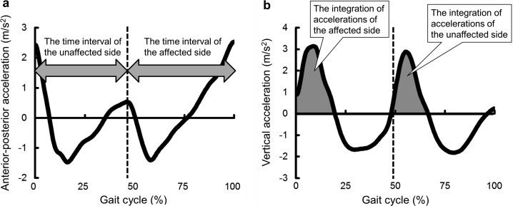
To remove linear trends and biases of tri-axial acceleration data, the data was adjusted to 0 m/s2 as the mean acceleration signal in the static standing position before and after walking in each trial. Trunk acceleration signals were averaged during the steady-state of five gait cycles, which include acceleration data during walking on the force plates. The beginning of the gait cycle was defined as the forward acceleration peak of the affected side12). To identify the forward acceleration peak of the affected side, the mediolateral trunk position was calculated using a double integration of the mediolateral trunk acceleration signal12). If the mediolateral trunk position changed toward the affected side following the forward acceleration peak, this forward acceleration peak was defined as the affected initial contact12). To average signals, gait cycles were time-normalized on a scale of 0–100%. To estimate step time asymmetry, anterior-posterior acceleration data were analyzed (Fig. 1a). In addition, to estimate step length asymmetry, the integration (m/s2) of the consecutive positive vertical accelerations that occurred after initial contact were calculated (Fig. 1b).
Fig. 1.
Data analysis procedures. The solid lines represent the mean acceleration patterns during five gait cycles
(a) Anterior-posterior acceleration during a gait cycle. The Y-axis represents the anterior-posterior acceleration. Positive values represent forward acceleration. The X-axis represents the time sequence expressed as a percentage of the gait cycle. The vertical dotted line indicates the initial contact of the unaffected side. The gray arrow indicates the time interval between the peaks of forward acceleration. (b) Vertical acceleration during a gait cycle. The Y-axis represents vertical acceleration. The X-axis represents the time sequence expressed as a percentage of the gait cycle. The vertical dotted line indicates the initial contact of the unaffected side, as defined by the forward acceleration peak. The gray area indicates the integration of consecutive vertical positive acceleration measures occurring immediately after initial contact.
To measure the reference values of the step time and length asymmetries, the center of pressure of initial contact during at least four consecutive steps on the force plates were calculated. Initial contact was defined as the time point when the vertical ground reaction force exceeded 5 N15). Affected step time (s) was defined as the time interval from unaffected initial contact to affected initial contact. Affected step length (m) was defined as the forward distance from center of the pressure position in the unaffected side to that in the affected side. Asymmetries were calculated by dividing the measurements of the affected side by those of the unaffected side16). Walking speed (m/s) was calculated by dividing the mean stride length by the mean stride time on the force plates.
The mean values of five trials were used for statistical analyses. Since all data, except walking speed, did not show normal distribution, according to the Shapiro-wilk test, non-parametric statistics were used for further analyses. To determine the concurrent validity of gait asymmetry estimation using an accelerometer, we evaluated the correlation between the values measured by an accelerometer and those measured by the force plates using the Spearman rank correlation coefficient. In addition, the Wilcoxon signed rank test was used to compare the accelerometer and the force plates for step time and length asymmetries. Since the amplitude of the trunk acceleration during walking becomes smaller with decreasing walking speed12), walking speed may affect the estimation of spatiotemporal asymmetries using the accelerometer. Therefore, we determined the value of these correlations between estimation errors in step time, length asymmetries, and walking speed, using the Spearman rank correlation coefficient. These estimation errors were calculated by determining the percentage of the absolute differences in measurement values between devices to measurement values derived from the force plates. Statistical analyses were performed using the Statistical Package for the Social Sciences version 20.0 (IBM Corp., Armonk, NY, USA) at a significance level of p<0.05.
RESULTS
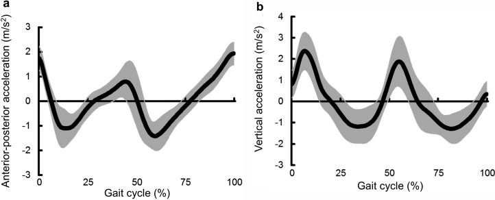
Participant walking speed was calculated to be 0.82 ± 0.25 (range 0.19–1.25) m/s. Anterior-posterior and vertical accelerations exhibited similar patterns among participants (Fig. 2). Step time asymmetry estimations using the accelerometer were significantly higher than those measured using the force plates (median: 1.22, interquartile range: 1.10–1.40 versus median: 1.12, interquartile range: 1.04–1.31, respectively; p<0.01). There was a significant correlation between devices for step time asymmetry (rho=0.83, p<0.01). Step length asymmetry was not significantly different between the accelerometer and the force plates (median: 1.17, interquartile range: 0.94–1.41 versus median: 1.06, interquartile range: 1.02–1.19, respectively; p=0.14). Step length asymmetry estimated by the accelerometer was significantly correlated with that measured from the force plates (rho=0.64, p<0.01).
Fig. 2.
Mean acceleration patterns of all participants. The solid line and the gray area represent the mean value of all participants and one standard deviation around this mean value, respectively. (a) Anterior-posterior acceleration during a gait cycle. The Y-axis represents anterior-posterior acceleration. Positive values represent forward acceleration. The X-axis represents the time sequence expressed as a percentage of the gait cycle. The 0% and 100% of gait cycle values indicate the affected initial contacts. (b) Vertical acceleration during a gait cycle. The Y-axis represents vertical acceleration. The X-axis represents the time sequence expressed as a percentage of the gait cycle.
The median estimation error of step time asymmetry was 7.7 (interquartile range: 4.9–11.2) % and that of step length asymmetry was 14.1 (interquartile range: 5.9–24.1) %. Walking speed was not significantly correlated with the estimation error of step time asymmetry (rho=−0.15, p=0.48) and that of step length asymmetry (rho=−0.21, p=0.32).
DISCUSSION
This is the first study that examines the concurrent validity of the accelerometer-based assessment of step time and length asymmetries in individuals with stroke. Previous studies have reported that there was sufficient validity in identifying step time by measuring the time interval between peaks of forward trunk acceleration in young12) and older adults17) as well as in individuals with lower limb prostheses18). In this study, there was a strong positive correlation between asymmetry calculated from the time intervals between peaks of forward trunk acceleration and step time asymmetry measured by the force plates. Therefore, these findings suggest that step time asymmetry assessment in hemiparetic gait by using an accelerometer has concurrent validity against measurements by force plates.
Previous studies have validated that step length could be estimated using a double integration of vertical acceleration, based upon leg length and changes in trunk vertical position during walking12, 17, 18). In the present study, we proposed an easier method for estimating step length asymmetry using only the integration of positive trunk vertical accelerations of each side during the stance phase. The integration of positive vertical accelerations was determined using the amplitude of vertical acceleration and the time during which positive vertical acceleration occurred, which may include the time of the loading response19). Roerdink et al.20) suggested dividing step length into two spatial components (i.e., trunk progression during step and forward foot placement relative to the trunk at initial contact). The amplitude of vertical acceleration upon initial contact may become larger with increasing trunk progression, since movements of the lower trunk during stepping are predicted well by an inverted pendulum model12, 13). In addition, the time of loading response may become longer with increasing forward foot placement relative to the trunk. These findings may support our results that there was a significant correlation between step length asymmetry estimated from vertical acceleration and that measured using the force plates. However, the accuracy of estimation of step length asymmetry should be improved using additional data, such as leg length12), prior to applying these findings to clinical practice.
To provide gait asymmetry assessment using an accelerometer for clinical applications, clinicians should consider the walking speed of individuals following hemiparetic stroke12). However, in the present study, we found that walking speed did not significantly correlate with estimation errors for step time and length asymmetries, even though individuals displayed a wide range of walking speeds from 0.19 to 1.25 m/s. Therefore, the assessment of gait asymmetry using an accelerometer may be applicable irrespective of walking speed.
Step time asymmetry was systematically overestimated by an accelerometer in this study. Although Houdijik et al.18) also reported similar results in individuals with lower limb prostheses, the causes of estimation errors have not been reported. In addition, we observed a wide range of estimation errors, particularly for step length asymmetry. The estimation errors may be affected by several factors, such as differences in the analyzed number of steps between devices, the incline of the accelerometer sensor axes21), and variability in spatiotemporal gait parameters22). In future studies, identifying the causes of estimation errors and establishing a method for improving estimation accuracy might enhance the clinical utility of the accelerometer-based assessment of gait asymmetry in individuals with hemiparetic stroke. We excluded participants who were unable to walk without walking devices in this study. In future studies, it is necessary to assess whether the walking device affects the assessment of step time and length asymmetries in individuals with hemiparetic stroke.
In conclusion, the step time asymmetry estimated from anterior-posterior acceleration data correlated with that measured using force plates. In addition, step length asymmetry estimated from trunk vertical acceleration significantly correlated to that measured using force plates. In this study, we have demonstrated, for the first time, that an accelerometer has the potential to estimate step time and length asymmetries in individuals with hemiparetic stroke.
Conflict of interest
The authors have declared that no competing interests exist.
Acknowledgments
This work was funded by JSPS KAKENHI Grant Number JP16J07949 to Kazuaki Oyake, and a grant from the Funds for a Grant-in Aid for Young Scientists (B) (15K16370) to Tomofumi Yamaguchi.
REFERENCES
- 1.Olney SJ, Richards C: Hemiparetic gait following stroke. Part I: characteristics. Gait Posture, 1996, 4: 136–148. [Google Scholar]
- 2.Patterson KK, Parafianowicz I, Danells CJ, et al. : Gait asymmetry in community-ambulating stroke survivors. Arch Phys Med Rehabil, 2008, 89: 304–310. [DOI] [PubMed] [Google Scholar]
- 3.Hsu AL, Tang PF, Jan MH: Analysis of impairments influencing gait velocity and asymmetry of hemiplegic patients after mild to moderate stroke. Arch Phys Med Rehabil, 2003, 84: 1185–1193. [DOI] [PubMed] [Google Scholar]
- 4.Lin PY, Yang YR, Cheng SJ, et al. : The relation between ankle impairments and gait velocity and symmetry in people with stroke. Arch Phys Med Rehabil, 2006, 87: 562–568. [DOI] [PubMed] [Google Scholar]
- 5.Awad LN, Palmer JA, Pohlig RT, et al. : Walking speed and step length asymmetry modify the energy cost of walking after stroke. Neurorehabil Neural Repair, 2015, 29: 416–423. [DOI] [PMC free article] [PubMed] [Google Scholar]
- 6.Lewek MD, Bradley CE, Wutzke CJ, et al. : The relationship between spatiotemporal gait asymmetry and balance in individuals with chronic stroke. J Appl Biomech, 2014, 30: 31–36. [DOI] [PubMed] [Google Scholar]
- 7.Malone LA, Bastian AJ: Thinking about walking: effects of conscious correction versus distraction on locomotor adaptation. J Neurophysiol, 2010, 103: 1954–1962. [DOI] [PMC free article] [PubMed] [Google Scholar]
- 8.Malone LA, Bastian AJ, Torres-Oviedo G: How does the motor system correct for errors in time and space during locomotor adaptation? J Neurophysiol, 2012, 108: 672–683. [DOI] [PMC free article] [PubMed] [Google Scholar]
- 9.Malone LA, Bastian AJ: Spatial and temporal asymmetries in gait predict split-belt adaptation behavior in stroke. Neurorehabil Neural Repair, 2014, 28: 230–240. [DOI] [PMC free article] [PubMed] [Google Scholar]
- 10.Iosa M, Morone G, Fusco A, et al. : Seven capital devices for the future of stroke rehabilitation. Stroke Res Treat, 2012, 2012: 187965. [DOI] [PMC free article] [PubMed] [Google Scholar]
- 11.Kavanagh JJ, Menz HB: Accelerometry: a technique for quantifying movement patterns during walking. Gait Posture, 2008, 28: 1–15. [DOI] [PubMed] [Google Scholar]
- 12.Zijlstra W, Hof AL: Assessment of spatio-temporal gait parameters from trunk accelerations during human walking. Gait Posture, 2003, 18: 1–10. [DOI] [PubMed] [Google Scholar]
- 13.Zijlstra W, Hof AL: Displacement of the pelvis during human walking: experimental data and model predictions. Gait Posture, 1997, 6: 249–262. [Google Scholar]
- 14.Folstein MF, Folstein SE, McHugh PR: “Mini-mental state”. A practical method for grading the cognitive state of patients for the clinician. J Psychiatr Res, 1975, 12: 189–198. [DOI] [PubMed] [Google Scholar]
- 15.Kim CM, Eng JJ: Symmetry in vertical ground reaction force is accompanied by symmetry in temporal but not distance variables of gait in persons with stroke. Gait Posture, 2003, 18: 23–28. [DOI] [PubMed] [Google Scholar]
- 16.Patterson KK, Gage WH, Brooks D, et al. : Evaluation of gait symmetry after stroke: a comparison of current methods and recommendations for standardization. Gait Posture, 2010, 31: 241–246. [DOI] [PubMed] [Google Scholar]
- 17.Hartmann A, Luzi S, Murer K, et al. : Concurrent validity of a trunk tri-axial accelerometer system for gait analysis in older adults. Gait Posture, 2009, 29: 444–448. [DOI] [PubMed] [Google Scholar]
- 18.Houdijk H, Appelman FM, Van Velzen JM, et al. : Validity of DynaPort GaitMonitor for assessment of spatiotemporal parameters in amputee gait. J Rehabil Res Dev, 2008, 45: 1335–1342. [PubMed] [Google Scholar]
- 19.Auvinet B, Berrut G, Touzard C, et al. : Reference data for normal subjects obtained with an accelerometric device. Gait Posture, 2002, 16: 124–134. [DOI] [PubMed] [Google Scholar]
- 20.Roerdink M, Beek PJ: Understanding inconsistent step-length asymmetries across hemiplegic stroke patients: impairments and compensatory gait. Neurorehabil Neural Repair, 2011, 25: 253–258. [DOI] [PubMed] [Google Scholar]
- 21.Meichtry A, Romkes J, Gobelet C, et al. : Criterion validity of 3D trunk accelerations to assess external work and power in able-bodied gait. Gait Posture, 2007, 25: 25–32. [DOI] [PubMed] [Google Scholar]
- 22.Balasubramanian CK, Neptune RR, Kautz SA: Variability in spatiotemporal step characteristics and its relationship to walking performance post-stroke. Gait Posture, 2009, 29: 408–414. [DOI] [PMC free article] [PubMed] [Google Scholar]


