Abstract
Background
The technique used to administer a selective nerve root block (SNRB) varies depending on individual expertise. Both the anteroposterior (AP) subpedicular approach and oblique Scotty dog subpedicular approach are widely practiced. However, the literature does not provide a clear consensus regarding which approach is more suitable. Hence, we decided to analyse the procedural parameters and clinical outcomes following SNRBs using these two approaches.
Methods
Patients diagnosed with a single lumbar herniated intervertebral disc (HIVD) refractory to conservative management but not willing for immediate surgery were selected for a prospective nonrandomized comparative study. An SNRB was administered as a therapeutic alternative using the AP subpedicular approach in one group (n = 25; mean age, 45 ± 5.4 years) and the oblique Scotty dog subpedicular approach in the other group (n = 22; mean age, 43.8 ± 4.7 years). Results were compared in terms of the duration of the procedure, the number of C-arm exposures, accuracy, pain relief, functional outcome and the duration of relief.
Results
Our results suggest that the oblique Scotty dog subpedicular approach took a significantly longer duration (p = 0.02) and a greater number of C-arm exposures (p = 0.001). But, its accuracy of needle placement was 95.5% compared to only 72% using the AP subpedicular approach (p = 0.03). There was no significant difference in terms of clinical outcomes between these approaches.
Conclusions
The AP subpedicular approach was simple and facile, but the oblique Scotty dog subpedicular approach was more accurate. However, a brief window period of pain relief was achieved irrespective of the approaching technique used.
Keywords: Intervertebral disc displacement, Nerve root compression, Radiculopathy
Sciatic pain due to nerve root compromise is a commonly encountered problem.1) It is characterised by radiating pain corresponding to the course of an affected nerve. The etiology of nerve root compromise varies and so does its management protocol.2,3) Selective nerve root blocks (SNRBs) are widely used as a diagnostic tool to localise such affected nerves.4) Even though the therapeutic efficacy of an SNRB is inconclusive, better outcomes can be obtained in selective patients.5) This procedure has evolved and been in practice for a considerable period; hence, there are certain variations in technique depending on individual preference.6)
Targeting the affected nerve root at the place where it exits is the key step of the procedure.7) This requires placement of the needle in an appropriate position. Currently, the commonly used approach involves identification of the “Scotty dog” in an oblique view C-arm image and placement of the needle tip just below the neck of the Scotty dog.8,9) However, considering the ease of the procedure, placing the needle tip at the so called “safe zone” just below and lateral to the pedicle in an anteroposterior (AP) view is also in practice.7,10) We intend to compare the procedural parameters and clinical outcomes following SNRBs using these approaches to analyse their efficacy.
METHODS
A prospective nonrandomized comparative study was formulated by selecting patients with symptoms of unilateral lumbar radiculopathy for a minimum duration of 3 months. The patients were diagnosed with a single lumbar herniated intervertebral disc (HIVD) based on magnetic resonance imaging (MRI). Despite being refractory to conservative management, they were not willing for immediate surgery and opted for a SNRB as a therapeutic alternative. Selection was further restricted to patients in whom a single lumbar nerve root had to be targeted. Hence, selected patients had either L4–L5 or L5–S1 HIVD, affecting L4 or L5 roots that would exit below L4 and L5 pedicles, respectively. We excluded those patients with L5–S1 disc prolapse affecting the S1 root that exits in the first sacral promontory because this requires a modified approach.
All selected patients had a positive straight leg raising (SLR) test on the affected side and none of them had any neurological deficit. Preprocedural numeric rating scale (NRS) pain score was assessed. Functional status was analysed using Roland-Morris disability questionnaire (RMDQ) score. MRI showed the discs appearing to be protruded or extruded causing significant nerve root compromise, which was graded using the criteria of Pfirrmann et al.11) This system incorporates all types of disc prolapse including protrusion, extrusion and sequestration, located centric or paracentric, and divides them into four grades depending on the nerve root compromise: normal (grade 0), contact (grade 1), deviation (grade 2), and compression (grade 3). However, our sample did not include patients with sequestrated discs, as most of these patients had severe symptoms with a definite indication for surgery.
We divided the patients into two groups; however, allocation of patients was nonrandomised as the initially enrolled patients were assigned to group 1 and the later enrolled patients to group 2. The AP subpedicular approach was used in group 1 and oblique Scotty dog subpedicular approach was used in group 2 to achieve appropriate needle placement; hence, the groups were named as AP group and oblique group, respectively. All procedures were done by a single performer with radiographic assistance using the same equipment.
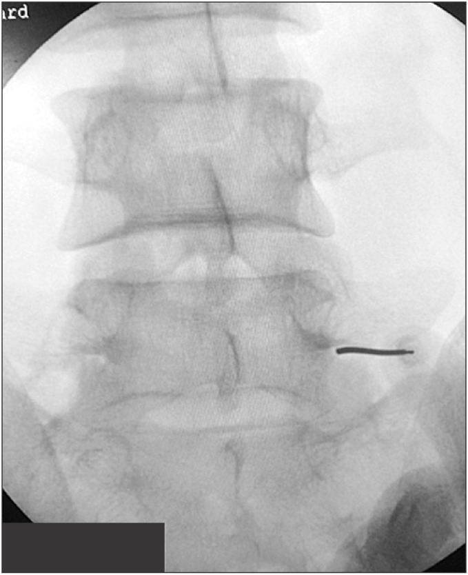
For the AP subpedicular approach, the patient was made to lie down in prone position on a radiolucent table and a C-arm was positioned appropriately for an AP view. Following local anaesthetic infiltration of the skin, an 18-gauge needle was directed to a point below and lateral to the pedicle of the affected side referring to the AP view; this corresponds to the so called “safe zone” (Fig. 1). This zone is an inverted right angled triangle with its base formed by the pedicle, lateral vertebral border on one side and the exiting root forming the hypotenuse.7) Depth of the needle was confirmed with a lateral view image where the needle tip had just entered the foraminal space.
Fig. 1. Needle placement using the anteroposterior subpedicular approach.

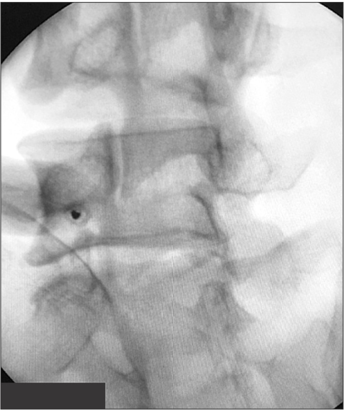
The oblique Scotty dog subpedicular approach requires similar patient positioning; however, the C-arm orientation is different. The C-arm must be precisely positioned for an oblique view where the classical Scotty dog can be visualized. Following local anaesthetic infiltration of the skin, an 18-gauge needle was introduced and directed to a point just below the neck of the Scotty dog (Fig. 2). Throughout the procedure, the needle must be maintained in an “end on” position along with the direction of C-arm so that it is visualized as a single point in the image. This also requires meticulous handling of the needle. As in the previous technique, the depth of the needle was confirmed with a lateral view image.
Fig. 2. Needle placement using the oblique Scotty dog subpedicular approach. The needle is visualized almost as a single point just below the neck of the Scotty dog.

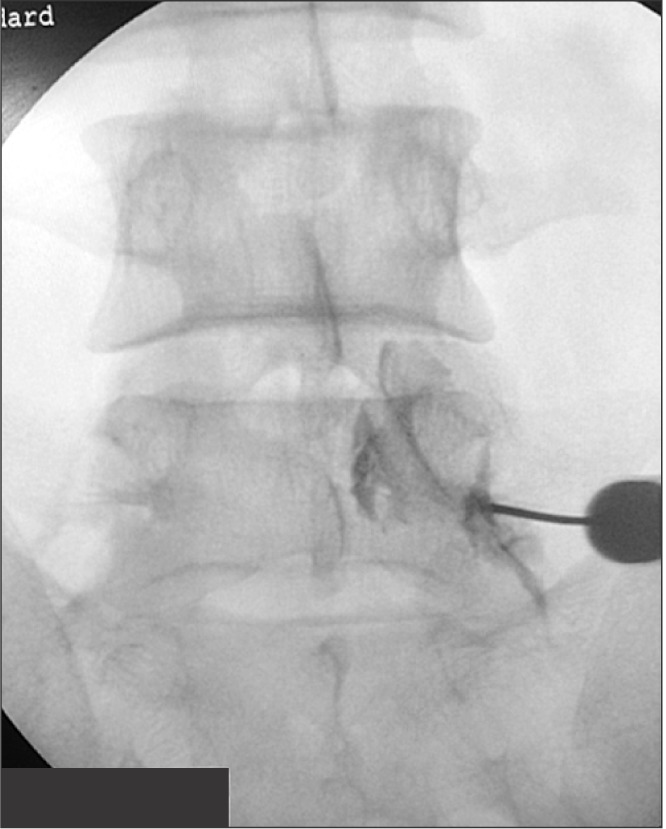
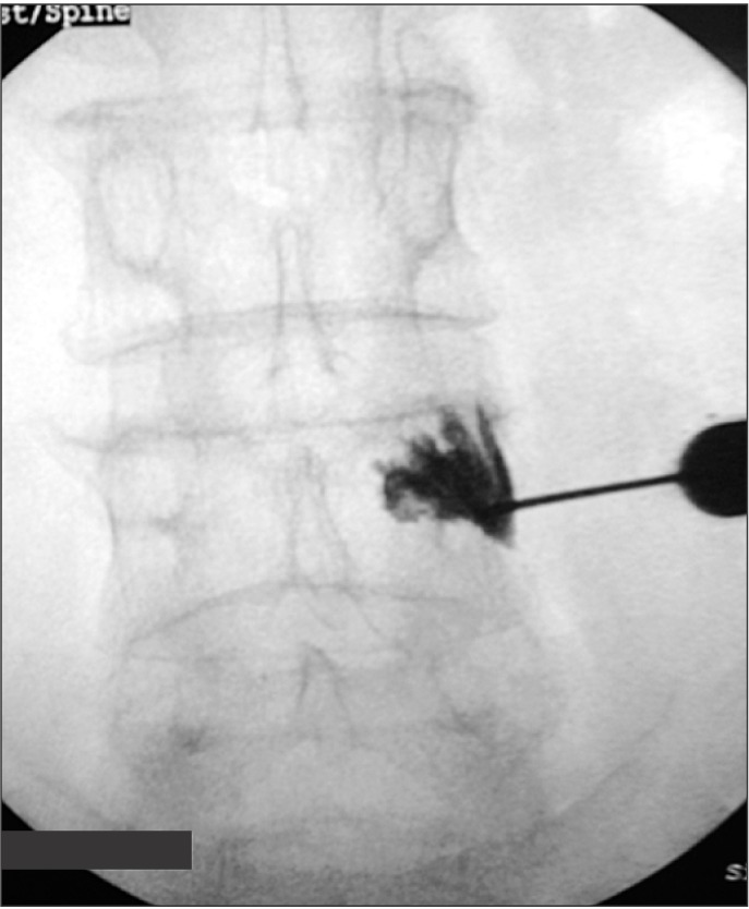
Following placement of a needle using any of these approaches, an iodine-based dye was injected to confirm the position (Fig. 3). If satisfactory spread of the dye was visualized, a mixture containing 1 mL of methyl prednisolone-based suspension and 1 mL of local anaesthetic was injected. In some patients, satisfactory spread of dye was not obtained and the attempt was considered a failure (Fig. 4). The needle had to be manipulated again until satisfactory positioning prior to injection of the medications. This was difficult and time-consuming due to the previously injected dye.
Fig. 3. Successful attempt characterized by appropriate spread of contrast along the nerve root.

Fig. 4. Failed attempt showing vague contrast spread.

All available preprocedural parameters and procedural parameters including duration of the procedure, number of C-arm exposures, and number of first attempt failures were noted and tabulated for comparison between groups. Most patients had immediate relief and their SLR improved. NRS pain score was calculated on the 3rd day. Functional outcomes were measured by the 4th week using Roland Morris Disability Questionnaire (RMDQ) score. Follow-up assessment was done every week for the first month and every month thereafter until recurrence of symptoms. Duration of pain relief among patients was calculated. Results were tabulated and statistical analysis was done.
Informed consent was obtained from all patients prior to inclusion in the study. This study was approved by the Institutional Review Board of Melmaruvathur Adhiparasakthi Institute of Medical Sciences and Research (MAPIMS&R) with EC approval dated 2016.05.02 and the study was performed in compliance with the 1964 declaration of Helsinki, its later amendments or comparable ethical standards. Statistical analysis was done using GraphPad Prism 5 (GraphPad Software Inc., San Diego, CA, USA). We used Student t-test for continuous variables and chi-square test for categorical variables. A probability p-value of less than 0.05 was considered statistically significant.
RESULTS
Based on our selection criteria, 47 patients were short-listed. They were divided into 2 groups, namely, the AP group (n = 25; mean age ± standard deviation [SD], 45 ± 5.4 years) and the oblique group (n = 22; mean age ± SD, 43.8 ± 4.7 years), depending on the approach used for SNRB. Mean duration of symptoms, preprocedural NRS pain score and RMDQ score did not show any significant difference between the groups. In addition, demographic characteristics were not significantly different between the groups (Table 1). The number of patients with various grades of nerve root compromise in each group was tabulated as per Pfirrmann's criteria, and no statistically significant difference was inferred between the groups (Table 2).
Table 1. Demographic Characteristics.
| Variable | Anteroposterior group (n = 25) | Oblique group (n = 22) | p-value* |
|---|---|---|---|
| Age (yr) | 45 ± 5.4 (33–54) | 43.8 ± 4.7 (35–52) | 0.43 |
| Sex (male:female) | 11:14 | 9:13 | |
| Duration of symptoms (mo) | 5.6 ± 1.3 (3–7) | 5.4 ± 1.3 (3–7) | 0.61 |
| NRS pain score (before procedure) | 7.8 ± 0.7 (7–9) | 7.7 ± 0.7 (7–9) | 0.44 |
| RMDQ score (before procedure) | 18.4 ± 2.4 (15–21) | 17.8 ± 1.9 (15–21) | 0.36 |
Values are presented as mean ± standard deviation (range).
NRS: numeric rating scale, RMDQ: Roland Morris Disability Questionnaire.
*A probability value p < 0.05 was considered statistically significant.
Table 2. Magnetic Resonance Imaging Based Grading of Lumbar Nerve Root Compromise.
| Grade | Anteroposterior group | Oblique group | p-value* |
|---|---|---|---|
| 1 | 14 (56) | 10 (45.45) | 0.47 |
| 2 | 9 (36) | 10 (45.45) | 0.51 |
| 3 | 2 (8) | 2 (9.09) | 0.89 |
Values are presented as number (%).
*A probability value p < 0.05 was considered statistically significant.
Procedure-related parameters such as duration, the number of C-arm exposures, and the number of first attempt failures were calculated (Table 3). These parameters showed certain differences between the groups. Duration of the procedure was significantly longer in the oblique group when compared to the AP group (p = 0.02). Similarly, the required number of C-arm exposures was greater in the oblique group than the AP group (p = 0.001). The accuracy of needle placement that was assessed by satisfactory spread of the radiopaque dye on the first attempt was 95.5% in the oblique group compared to only 72% in the AP group (p = 0.03).
Table 3. Results.
| Variable | Anteroposterior group | Oblique group | p-value* |
|---|---|---|---|
| Duration of procedure (min) | 21 ± 3.2 (17–27) | 23 ± 2.9 (19–29) | 0.02 |
| No. of C-arm exposures | 15.5 ± 6.5 (8–30) | 21.5 ± 5.1 (14–30) | 0.001 |
| No. of first attempt successes | 18 (72) | 21 (95.5) | 0.03 |
| No. of first attempt failures | 7 (28) | 1 (4.5) | - |
| NRS pain score (3rd day) | 3.3 ± 0.7 (2–4) | 3.2 ± 0.7 (2–4) | 0.61 |
| NRS pain score (4 wk) | 3.5 ± 0.9 (2–5) | 3.2 ± 0.7 (2–4) | 0.27 |
| RMDQ score (4 wk) | 6.2 ± 1.4 (4–8) | 6.5 ± 1.6 (4–8) | 0.49 |
| Duration of relief (wk) | 21.5 ± 9.6 (4–40) | 23.6 ± 9.5 (8–44) | 0.44 |
Values are presented as mean ± standard deviation (range) or number (%).
NRS: numeric rating scale, RMDQ: Roland Morris Disability Questionnaire.
*A probability value p < 0.05 was considered statistically significant.
Immediate symptomatic relief was seen in most of our patients due to the local anaesthetic effect. NRS pain score was calculated on the 3rd day following the procedure. It showed a significant decrease in pain among both groups. Furthermore, NRS pain score was calculated every week for the first month, but functional outcome assessment was delayed until the end of the first month, as patients were advised to rest and not to carry on with strenuous activities. Functional outcome assessed using RMDQ score at the 4th week showed significant improvement of the functional status in both groups, which, however, was short-lived.
Sequential follow-up showed a gradual decrease in patients with pain relief every month. Moreover, initial pain relief did not predict prognosis and duration of pain relief varied among individuals. Those patients with pain score of more than 6 in subsequent follow-ups were considered to have recurrence and went on for further management. Yet, the mean duration of pain relief in both groups showed no significant difference. On summarising, our results showed no significant difference in clinical outcomes, but the accuracy of oblique Scotty dog subpedicular approach was found to be higher; unfortunately, this demanded additional time and C-arm exposures.
DISCUSSION
The literature regarding lumbar SNRBs reveals inconsistent information on the exact needle tip position.6) However, it is understood that the nerve exits below the pedicle and hence targeting this region is ideal for a root block.2,7) Yet, variations exist for approaching this point.12) Hence, we chose to compare 2 commonly used approaches for needle placement and patients were grouped as per the approaching technique.12) Even though both these approaches have almost the same pathway of needle advancement, the procedures required to place the needle are entirely different. The foremost difference is the C-arm orientation. Our subject selection was strictly restricted to patients in whom lumbar nerve roots had to be targeted, as the two procedures were similar unlike the procedure targeting S1 that requires certain adaptations.13)
Types of disc herniation including protrusion, extrusion and sequestration in addition to the zones of location in the transverse plane are well defined in the literature.14) However, disc prolapse can also be present in asymptomatic population.15) Considering the fact that the relation between the disc and the nerve root is the main cause for symptoms, we preferred to use the MR image-based grading of lumbar nerve root compromise suggested by Pfirrmann et al.11) This classification system incorporates all anatomical varieties of disc prolapse and grades them according to the nerve root compromise. Both groups in our study consisted of patients with all three described grades of nerve root compromise as per Pfirrmann's criteria.
We used the NRS scoring for pain and RMDQ assessment of functional status as they were simple and scoring could be done just by verbal conversation.16,17,18) To confirm the placement of needles using both approaches, we injected an iodine-based contrast and checked its spread along the nerve root in an AP view. We noticed various patterns of contrast distribution as described in the literature.19) Like many authors, we used a methyl prednisolone-based suspension to obtain the desired effect.20,21)
On analysing our results, we found that the clinical outcomes following SNRBs using both these approaches were similar but operative parameters showed differences. There were certain advantages and disadvantages of each approach. The oblique Scotty dog subpedicular approach necessitated a significantly longer duration, especially for identifying the Scotty dog and maintaining the needle in an “end on” position along the direction of the C-arm throughout the procedure. This also prompted additional C-arm exposures. On the contrary, handling of the C-arm and the needle was undemanding, using the AP subpedicular approach. In some cases, however, bony resistance was felt before placing the needle in an appropriate position; in such circumstances, we had to walk over the bone towards the ideal point, looking for a giving way feeling. Once giving way feeling was noticed, a lateral view was taken to confirm the depth of the needle.
Our results showed that the oblique Scotty dog subpedicular approach was more accurate for ideal placement of the needle tip. This was statistically proven and should be considered as an important factor in selecting an approach for SNRB. The duration of pain relief offered by either of these approaches was short-lived and hence, recurrence is expected.10) An SNRB using these approaches can be used as an alternative procedure for patients not willing for surgery or presenting with an indefinite indication for surgery. It can also be used to predict surgical outcome.22)
In conclusion, we compared the operative parameters and clinical outcomes following lumbar SNRBs using the AP subpedicular approach and the oblique Scotty dog subpedicular approach. Our results showed no significant difference between these approaches in terms of clinical outcomes; however, the accuracy of the oblique Scotty dog subpedicular approach was found to be higher although this demanded additional time and C-arm exposures. We think that both these approaches can be used under appropriate circumstances depending on the individual preference and expertise, especially when one technique fails, the other can come in handy.
Footnotes
CONFLICT OF INTEREST: No potential conflict of interest relevant to this article was reported.
References
- 1.Stafford MA, Peng P, Hill DA. Sciatica: a review of history, epidemiology, pathogenesis, and the role of epidural steroid injection in management. Br J Anaesth. 2007;99(4):461–473. doi: 10.1093/bja/aem238. [DOI] [PubMed] [Google Scholar]
- 2.Patel N. Surgical disorders of the thoracic and lumbar spine: a guide for neurologists. J Neurol Neurosurg Psychiatry. 2002;73(Suppl 1):i42–i48. doi: 10.1136/jnnp.73.suppl_1.i42. [DOI] [PMC free article] [PubMed] [Google Scholar]
- 3.Shelerud RA, Paynter KS. Rarer causes of radiculopathy: spinal tumors, infections, and other unusual causes. Phys Med Rehabil Clin N Am. 2002;13(3):645–696. doi: 10.1016/s1047-9651(02)00012-8. [DOI] [PubMed] [Google Scholar]
- 4.Yeom JS, Lee JW, Park KW, et al. Value of diagnostic lumbar selective nerve root block: a prospective controlled study. AJNR Am J Neuroradiol. 2008;29(5):1017–1023. doi: 10.3174/ajnr.A0955. [DOI] [PMC free article] [PubMed] [Google Scholar]
- 5.Narozny M, Zanetti M, Boos N. Therapeutic efficacy of selective nerve root blocks in the treatment of lumbar radicular leg pain. Swiss Med Wkly. 2001;131(5-6):75–80. doi: 10.4414/smw.2001.09689. [DOI] [PubMed] [Google Scholar]
- 6.Eastley NC, Spiteri V, Newey ML. Variations in selective nerve root block technique. Ann R Coll Surg Engl. 2013;95(7):515–518. doi: 10.1308/003588413X13629960048073. [DOI] [PMC free article] [PubMed] [Google Scholar]
- 7.Pfirrmann CW, Oberholzer PA, Zanetti M, et al. Selective nerve root blocks for the treatment of sciatica: evaluation of injection site and effectiveness: a study with patients and cadavers. Radiology. 2001;221(3):704–711. doi: 10.1148/radiol.2213001635. [DOI] [PubMed] [Google Scholar]
- 8.Eckel TS, Bartynski WS. Epidural steroid injections and selective nerve root blocks. Tech Vasc Interv Radiol. 2009;12(1):11–21. doi: 10.1053/j.tvir.2009.06.004. [DOI] [PubMed] [Google Scholar]
- 9.Hui CW, Chan PH, Cheung KK. Selective nerve root block for sciatica. Hong Kong J Orthop Surg. 2005;9(1):22–27. [Google Scholar]
- 10.Arun-Kumar K, Jayaprasad S, Senthil K, Lohith H, Jayaprakash KV. The outcomes of selective nerve root block for disc induced lumbar radiculopathy. Malays Orthop J. 2015;9(3):17–22. doi: 10.5704/MOJ.1511.002. [DOI] [PMC free article] [PubMed] [Google Scholar]
- 11.Pfirrmann CW, Dora C, Schmid MR, Zanetti M, Hodler J, Boos N. MR image-based grading of lumbar nerve root compromise due to disk herniation: reliability study with surgical correlation. Radiology. 2004;230(2):583–588. doi: 10.1148/radiol.2302021289. [DOI] [PubMed] [Google Scholar]
- 12.Chiba S, Tsutsui S, Chiba T, et al. Relationship among analgesic effects, radiating pain, and radiological contrast material distribution in lumbosacral selective nerve root block. J Pain Relief. 2014;3(5):157. [Google Scholar]
- 13.Fish DE, Lee PC, Marcus DB. The S1 “Scotty dog”: report of a technique for S1 transforaminal epidural steroid injection. Arch Phys Med Rehabil. 2007;88(12):1730–1733. doi: 10.1016/j.apmr.2007.07.041. [DOI] [PubMed] [Google Scholar]
- 14.Fardon DF, Williams AL, Dohring EJ, Murtagh FR, Gabriel Rothman SL, Sze GK. Lumbar disc nomenclature: version 2.0: recommendations of the combined task forces of the North American Spine Society, the American Society of Spine Radiology and the American Society of Neuroradiology. Spine J. 2014;14(11):2525–2545. doi: 10.1016/j.spinee.2014.04.022. [DOI] [PubMed] [Google Scholar]
- 15.Boden SD, Davis DO, Dina TS, Patronas NJ, Wiesel SW. Abnormal magnetic-resonance scans of the lumbar spine in asymptomatic subjects: a prospective investigation. J Bone Joint Surg Am. 1990;72(3):403–408. [PubMed] [Google Scholar]
- 16.Hawker GA, Mian S, Kendzerska T, French M. Measures of adult pain: Visual Analog Scale for Pain (VAS Pain), Numeric Rating Scale for Pain (NRS Pain), McGill Pain Questionnaire (MPQ), Short-Form McGill Pain Questionnaire (SF-MPQ), Chronic Pain Grade Scale (CPGS), Short Form-36 Bodily Pain Scale (SF-36 BPS), and Measure of Intermittent and Constant Osteoarthritis Pain (ICOAP) Arthritis Care Res (Hoboken) 2011;63(Suppl 11):S240–S252. doi: 10.1002/acr.20543. [DOI] [PubMed] [Google Scholar]
- 17.Roland M, Morris R. A study of the natural history of back pain. part I: development of a reliable and sensitive measure of disability in low-back pain. Spine (Phila Pa 1976) 1983;8(2):141–144. doi: 10.1097/00007632-198303000-00004. [DOI] [PubMed] [Google Scholar]
- 18.Stevens ML, Lin CC, Maher CG. The Roland Morris Disability Questionnaire. J Physiother. 2016;62(2):116. doi: 10.1016/j.jphys.2015.10.003. [DOI] [PubMed] [Google Scholar]
- 19.Vassiliev D. Spread of contrast during L4 and L5 nerve root infiltration under fluoroscopic guidance. Pain Physician. 2007;10(3):461–466. [PubMed] [Google Scholar]
- 20.Tauheed N, Usmani H, Siddiqui AH. A comparison of the analgesic efficacy of transforaminal methylprednisolone alone and with low doses of clonidine in lumbo-sacral radiculopathy. Saudi J Anaesth. 2014;8(1):51–58. doi: 10.4103/1658-354X.125937. [DOI] [PMC free article] [PubMed] [Google Scholar]
- 21.Owlia MB, Salimzadeh A, Alishiri G, Haghighi A. Comparison of two doses of corticosteroid in epidural steroid injection for lumbar radicular pain. Singapore Med J. 2007;48(3):241–245. [PubMed] [Google Scholar]
- 22.Benedetti EM, Siriwetchadarak R. Selective nerve root blocks as predictors of surgical outcome: fact or fiction? Tech Reg Anesth Pain Manag. 2011;15(1):4–11. [Google Scholar]


