Abstract
Background
The purpose of this study was to report the ultrasonographic findings and clinical features of schwannoma of the hand.
Methods
We enrolled 8 patients who were initially diagnosed with ganglion by ultrasonography but finally with schwannoma by a tissue biopsy. We retrospectively analyzed the ultrasonographic findings of eight patients including echogenicity, internal homogeneity, posterior enhancement, internal vascularity, and clinical manifestations such as the occurrence site, tenderness, Tinel's sign, and paresthesia before the surgery.
Results
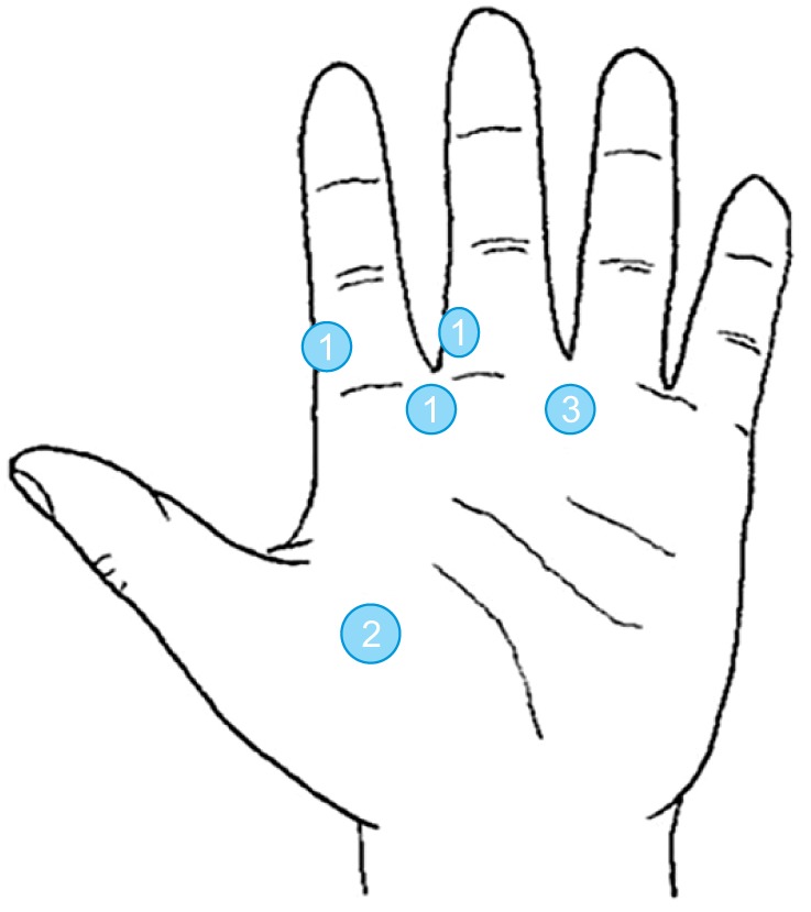
The occurrence sites were as follows: two cases on the thenar area, one case on the second web space, three cases on the third web space, one case on the radiovolar aspect of the proximal phalanx of the index finger, and one case on the radiovolar aspect of the proximal phalanx of the middle finger. Four patients suffered from tenderness and pain on presentation, and all patients had pain around the mass before presentation. Tinel's sign was present without paresthesia in one case. Ultrasonography revealed cystic lesions showing clear margins in all cases, and two of them had acoustic enhancement without internal flow.
Conclusions
It may not be easy to diagnosis schwannoma of the hand with ultrasonography alone when the lesion is small because of the similarity to the ultrasonographic findings of ganglion. Therefore, it is necessary to consider the possibility of schwannoma if a mass near the digital nerve or cutaneous nerve branch is accompanied by dull pain and tenderness.
Keywords: Hand, Schwannoma, Ultrasonography
High-resolution ultrasonography has been used as a firstline imaging tool to evaluate and diagnose a mass in the hand because this technique is noninvasive and easy to use and the hand is an anatomically superficial structure and easier to access than other joints. In particular, high-resolution ultrasonography can be used to differentiate many mass lesions of the hand. However, it is not easy to differentiate schwannoma of the hand from a cystic lesion like ganglion. The low incidence and the variable features of schwannoma of the hand can cause a misdiagnosis. We retrospectively reviewed 8 patients who were diagnosed initially with ganglion by ultrasonography but finally with schwannoma by a tissue biopsy. In this study, we report clinical and ultrasonographic features of schwannoma identified in these patients.
METHODS
All patients at Konkuk University Medical Center who were diagnosed with a ganglion within the hand by ultrasonography between March 2012 and August 2014 were reviewed. A total of 152 patients underwent excision and biopsy after being diagnosed with a ganglion within the hand by ultrasonography. Of these, 8 patients were finally diagnosed with schwannoma by a tissue biopsy after the surgery. We performed mass excision and tissue biopsy in the patients who wanted to remove the mass due to discomfort, pain, or unpleasant appearance. We retrospectively reviewed the ultrasonographic findings and clinical records of these 8 patients. The ultrasonographic findings included echogenicity, internal homogeneity, posterior enhancement, nerve continuity, and internal vascularity. The clinical records included preoperative clinical symptoms such as the occurrence site, tenderness, pain, Tinel's sign, and paresthesia. One musculoskeletal radiologist performed ultrasonography using a LOGIQ S8 (GE Healthcare, Chicago, IL, USA) with a 12-MHz transducer before the surgery. A surgeon (SJL) performed all the operations. We performed enucleation for the 8 patients under microscopic magnification to avoid damaging nerves during dissection.
RESULTS
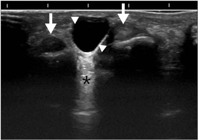
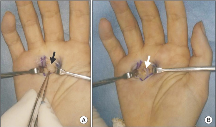
The mean age of 8 patients (2 males and 6 females) was 54 years (range, 32 to 82 years). The locations of the masses were as follows: two cases on the thenar area, one case on the second web space, three cases on the third web space, one case on the radiovolar aspect of the proximal phalanx of the index finger, and one case on the radiovolar aspect of the proximal phalanx of the middle finger (Fig. 1). Four of 8 patients complained of pain and tenderness on the mass at the time of presentation. The other 4 patients reported they had felt pain and tenderness before presentation although they did not complain of pain at presentation. All 8 patients stated that they felt dull pain around the mass and found the mass incidentally (Table 1). The duration of symptom ranged from 14 days to 2 months. One patient had a positive Tinel's sign but did not complain of any sensory changes. Ultrasonography showed a round, hypoechoic, homogenous mass with clear margins in all patients. The mean size of the mass was 3.2 mm (range, 2 to 5 mm). There was posterior acoustic enhancement in two cases, and there was no internal flow (Fig. 2). We could not definitely identify nerve continuity on ultrasonography (Table 2). Intraoperatively, we found the yellow white colored eccentric solid mass at the digital nerve. We enucleated the mass from the nerve using a microsurgical technique (Fig. 3). There was no recurrence and neurologic deficits after the surgery.
Fig. 1. The occurrence sites of schwannoma in the hand. The circled number indicates the number of occurrences.

Table 1. Clinical Findings.
| Variable | Yes | No |
|---|---|---|
| Tinel's sign | 1 | 7 |
| Sensory change | 0 | 8 |
| Tenderness | ||
| On visit | 4 | 4 |
| Before visit | 8 | 0 |
| Dull pain | ||
| On visit | 4 | 4 |
| Before visit | 8 | 0 |
Fig. 2. Ultrasonographic findings. Axial scan shows homogenous, anechoic round mass (arrow head) with posterior enhancement (asterisk) and flexor tendon of finger (arrow).

Table 2. Ultrasonography Findings.
| Variable | No. of cases |
|---|---|
| Echogenicity | |
| Anechoic-hypoechoic | 8 |
| Hyperechoic | 0 |
| Internal homogeneity | |
| Homogenous | 8 |
| Heterogeneous | 0 |
| Posterior enhancement | 2 |
| Internal vascularity | 0 |
| Nerve continuity | 0 |
Fig. 3. Intraoperative photographs. (A) The yellowish-white colored ovoid mass (black arrow) was identified in the nerve at the bifurcation between the common digital nerve of the long and the ring fingers. (B) The yellowish-white colored solitary mass was enucleated microscopically. The white arrow indicates the digital nerve.
DISCUSSION
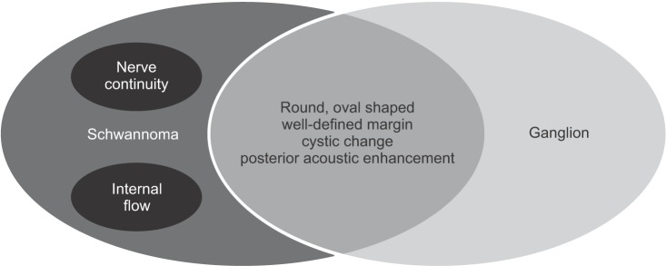
Ganglion cysts are the most common cystic lesions among masses of the hand and giant cell tumors of the tendon sheath manifest as solid tumor lesions.1,2) Schwannoma is the most common benign tumor of peripheral nerves but represents only 5%–8% of all soft tissue tumors, with the incidence of schwannoma of the hand reported as low as 1%.3,4) The method of surgical excision commonly depends on the preoperative diagnosis. Marginal excision is the general surgical treatment for ganglion cysts and benign solid tumors. Enucleation using a microsurgical technique is the common surgical treatment for schwannoma. We suspected the possibility of schwannoma in the lesions originating from the nerves during operation, and performed enucleation of the lesions under microscopic magnification to avoid damaging the nerves. Fortunately, there were no neurologic deficits after the surgery in our cases. However, White5) reported a 6% rate of nerve resection due to misdiagnosis, and Rockwell et al.4) reported that accurate differential diagnosis of schwannoma and other mass lesions is important because an incorrect preoperative diagnosis may lead to permanent neurologic deficits. Schwannoma of the hand can be difficult to distinguish from other lesions.4,5) Although magnetic resonance imaging (MRI) has proven to be the most effective imaging modality, ultrasonography has become more widely used due to its relatively lower cost.6) With the development of higher resolution techniques, the use of ultrasonography in diagnose of hand diseases has increased, and now it is the primary diagnostic tool for a mass lesion of the hand. Schwannoma has variable sonographic features. It manifests as a homogenous, hypoechoic mass and may show posterior acoustic enhancement, target appearance, pseudocystic appearance, or internal flow as unusual findings on ultrasonography.7,8,9,10) In this study, lesions of all eight cases were identified to be hypoechoic, homogenous, round masses without any internal flow. We diagnosed these masses to be ganglion cysts rather than schwannomas in consideration of the ultrasonographic findings and the low incidence of schwannomas. With an increase in the tumor size, schwannomas commonly undergo degenerative changes including cyst formation, calcification, hemorrhage, and fibrosis. During these changes, large schwannomas show variable sonographic features (homogenous or heterogeneous, hypoechoic or hyperechoic, posterior acoustic enhancement, target appearance, etc.); however, smaller lesions show pseudocystic appearance because it is difficult to detect and assess the degenerative changes within the lesions with ultrasonography.11,12,13) So, they are sometimes misdiagnosed as a lipoma, fibroma, ganglion, or xanthoma.14,15) Similar to our experience, the rate of misdiagnosis has been reported high.16,17,18) The eccentric position of the mass in the peripheral nerve and nerve continuity form the basis of the ultrasonographic diagnosis of schwannoma; however, when peripheral nerve size is < 2 mm, nerve continuity is hard to identify, which consequentially leads to a misdiagnosis.19) Because the digital and cutaneous nerves of the hand are much smaller, it can be difficult to confirm continuity of the nerves without suspecting schwannoma during ultrasonography (Fig. 4). Accurate preoperative differential diagnosis of schwannoma from solitary neurofibroma is also important. Neurofibromas involving a nerve are intimately connected and require resection of the involved nerve segment, whereas schwannomas are well encapsulated and can be removed without disruption of the nerve.20) However, there are no distinct criteria to accurately differentiate these tumors clinically. Differentiation between schwannomas and neurofibromas is known to be difficult on the basis of sonographic findings, including well-defined margins, a homogeneous hypoechoic appearance, a round to oval shape, through transmission and internal vascularity, visualization of the entering and exiting nerves, and the presence of a target sign.13,21) Several studies reported that it was possible to differentiate them only in limited cases that showed an entering or exiting nerve.7,19,22) Finding the nerve is sometimes not easy because the digital nerve of the hand or cutaneous nerve branch is small and is confused with the adjacent fat and connective tissue. We think that it is difficult to distinguish them with ultrasonography in lesions of the hand. Thus, it is recommended to diagnose schwannoma of the hand not only with ultrasonography but also using clinical signs. Generally, schwannoma is known to be a painless mass.18,23) However, Rockwell et al.4) reported that there was pain and tenderness on schwannoma of the hand unlike those of other sites. Our study showed clinical features similar to those reported by Rockwell et al.4) Four of 8 patients complained of pain and tenderness on the mass, and other 4 patients also had had pain and tenderness although the pain disappeared at the time of presentation to our hospital. All patients had pain around the mass. In the current study, there was no patients with sensory change, and Tinel's sign was positive in 1 patient, indicating that sensory change and Tinel's sign do not carry any clinical significance. Based on the findings of this study, we think that pain and tenderness may be a clue to differentiate schwannomas from other mass lesions of the hand.
Fig. 4. Ultrasonographic features of ganglion and schwannoma. Schwannoma is often hypoechoic with posterior acoustic enhancement and thus may simulate a ganglion cyst. The diagnostic clue of schwannoma is nerve continuity.
This study has some limitations. First, the number of patients was small for meaningful statistical analysis. Second, this study had a retrospective and there was no control group. We did not compare the ultrasonographic and clinical characteristics of schwannomas in the hand to those of other cystic masses with pain or tenderness. And there was also no effort to assess nerve continuity on the initial ultrasonographic examination. We think that the rarity of schwannomas in the hand and similarity to ganglion cysts led to the misdiagnosis on the ultrasonographic examination. We think the history of pain and tenderness on a mass in the hand will help differentiate schwannomas from ganglions and other mass lesions on ultrasonography. When a patient with a mass around the digital nerve complains of pain, it is advised to consider the possibility of schwannoma and identify nerve continuity using MRI if needed.
In conclusion, a schwannoma of the hand looks like a ganglion on ultrasonography. Therefore, we think that the initial diagnostic approach should be to consider the possibility of schwannoma in cases where a mass near the digital nerve or cutaneous nerve branch is accompanied by dull pain and tenderness.
Footnotes
CONFLICT OF INTEREST: No potential conflict of interest relevant to this article was reported.
References
- 1.Bianchi S, Abdelwahab IF, Zwass A, Giacomello P. Ultrasonographic evaluation of wrist ganglia. Skeletal Radiol. 1994;23(3):201–203. doi: 10.1007/BF00197461. [DOI] [PubMed] [Google Scholar]
- 2.Kransdorf MJ, Murphey MD. Imaging of soft tissue tumors. Philadelphia, PA: W.B. Saunders; 1997. pp. 275–316. [Google Scholar]
- 3.Das Gupta TK, Brasfield RD, Strong EW, Hajdu SI. Benign solitary Schwannomas (neurilemomas) Cancer. 1969;24(2):355–366. doi: 10.1002/1097-0142(196908)24:2<355::aid-cncr2820240218>3.0.co;2-2. [DOI] [PubMed] [Google Scholar]
- 4.Rockwell GM, Thoma A, Salama S. Schwannoma of the hand and wrist. Plast Reconstr Surg. 2003;111(3):1227–1232. doi: 10.1097/01.PRS.0000046039.28526.1A. [DOI] [PubMed] [Google Scholar]
- 5.White NB. Neurilemomas of the extremities. J Bone Joint Surg Am. 1967;49(8):1605–1610. [PubMed] [Google Scholar]
- 6.Simoens WA, Wuyts FL, De Beuckeleer LH, Vandevenne JE, Bloem JL, De Schepper AM. MR features of peripheral nerve sheath tumors: can a calculated index compete with radiologist's experience? Eur Radiol. 2001;11(2):250–257. doi: 10.1007/s003300000552. [DOI] [PubMed] [Google Scholar]
- 7.Reynolds DL, Jr, Jacobson JA, Inampudi P, Jamadar DA, Ebrahim FS, Hayes CW. Sonographic characteristics of peripheral nerve sheath tumors. AJR Am J Roentgenol. 2004;182(3):741–744. doi: 10.2214/ajr.182.3.1820741. [DOI] [PubMed] [Google Scholar]
- 8.Lin J, Jacobson JA, Hayes CW. Sonographic target sign in neurofibromas. J Ultrasound Med. 1999;18(7):513–517. doi: 10.7863/jum.1999.18.7.513. [DOI] [PubMed] [Google Scholar]
- 9.Chinn DH, Filly RA, Callen PW. Unusual ultrasonographic appearance of a solid schwannoma. J Clin Ultrasound. 1982;10(5):243–245. doi: 10.1002/jcu.1870100509. [DOI] [PubMed] [Google Scholar]
- 10.Hughes DG, Wilson DJ. Ultrasound appearances of peripheral nerve tumours. Br J Radiol. 1986;59(706):1041–1043. doi: 10.1259/0007-1285-59-706-1041. [DOI] [PubMed] [Google Scholar]
- 11.Murphey MD, Smith WS, Smith SE, Kransdorf MJ, Temple HT. From the archives of the AFIP: imaging of musculoskeletal neurogenic tumors: radiologic-pathologic correlation. Radiographics. 1999;19(5):1253–1280. doi: 10.1148/radiographics.19.5.g99se101253. [DOI] [PubMed] [Google Scholar]
- 12.Bacigalupo L, Bianchi S, Valle M, Martinoli C. Ultrasonography of peripheral nerves. Radiologe. 2003;43(10):841–849. doi: 10.1007/s00117-003-0963-y. [DOI] [PubMed] [Google Scholar]
- 13.Stuart RM, Koh ES, Breidahl WH. Sonography of peripheral nerve pathology. AJR Am J Roentgenol. 2004;182(1):123–129. doi: 10.2214/ajr.182.1.1820123. [DOI] [PubMed] [Google Scholar]
- 14.Kececi Y, Gurler T, Gundogan H, Bilkay U, Cagdas A. Benign giant schwannoma located in the upper arm. Ann Plast Surg. 1997;39(1):100–102. doi: 10.1097/00000637-199707000-00019. [DOI] [PubMed] [Google Scholar]
- 15.Kang HJ, Shin SJ, Kang ES. Schwannomas of the upper extremity. J Hand Surg Br. 2000;25(6):604–607. doi: 10.1054/jhsb.2000.0472. [DOI] [PubMed] [Google Scholar]
- 16.De Flaviis L, Nessi R, Del Bo P, Calori G, Balconi G. High-resolution ultrasonography of wrist ganglia. J Clin Ultrasound. 1987;15(1):17–22. doi: 10.1002/jcu.1870150105. [DOI] [PubMed] [Google Scholar]
- 17.Kehoe NJ, Reid RP, Semple JC. Solitary benign peripheral-nerve tumours: review of 32 years' experience. J Bone Joint Surg Br. 1995;77(3):497–500. [PubMed] [Google Scholar]
- 18.Holdsworth BJ. Nerve tumours in the upper limb: a clinical review. J Hand Surg Br. 1985;10(2):236–238. doi: 10.1016/0266-7681(85)90026-9. [DOI] [PubMed] [Google Scholar]
- 19.Tsai WC, Chiou HJ, Chou YH, Wang HK, Chiou SY, Chang CY. Differentiation between schwannomas and neurofibromas in the extremities and superficial body: the role of high-resolution and color Doppler ultrasonography. J Ultrasound Med. 2008;27(2):161–166. doi: 10.7863/jum.2008.27.2.161. [DOI] [PubMed] [Google Scholar]
- 20.Plate AM, Lee SJ, Steiner G, Posner MA. Tumorlike lesions and benign tumors of the hand and wrist. J Am Acad Orthop Surg. 2003;11(2):129–141. doi: 10.5435/00124635-200303000-00007. [DOI] [PubMed] [Google Scholar]
- 21.Carra BJ, Bui-Mansfield LT, O'Brien SD, Chen DC. Sonography of musculoskeletal soft-tissue masses: techniques, pearls, and pitfalls. AJR Am J Roentgenol. 2014;202(6):1281–1290. doi: 10.2214/AJR.13.11564. [DOI] [PubMed] [Google Scholar]
- 22.Peer S, Kovacs P, Harpf C, Bodner G. High-resolution sonography of lower extremity peripheral nerves: anatomic correlation and spectrum of disease. J Ultrasound Med. 2002;21(3):315–322. doi: 10.7863/jum.2002.21.3.315. [DOI] [PubMed] [Google Scholar]
- 23.Rinaldi E. Neurilemomas and neurofibromas of the upper limb. J Hand Surg Am. 1983;8(5 Pt 1):590–593. doi: 10.1016/s0363-5023(83)80133-6. [DOI] [PubMed] [Google Scholar]


