Erratum
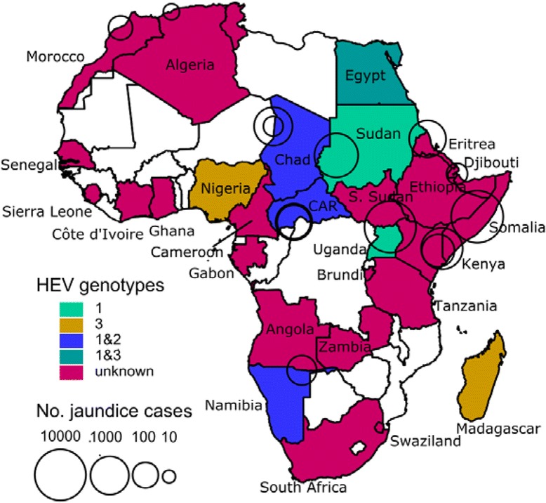
In this letter, we wish to correct errors in the previously published article [1]. Although the errors do not change the main results and conclusions described in the abstract of the original article, we believe providing the correct information is important. The major correction is about the genotype distribution of HEV in Africa. In the original article, we indicated that genotype 3 is rare and less commonly found than genotype 2 while genotype 1 is the most prevalent. The correct information is, however, that genotypes 2 and 3 were identified at a similar frequency while genotype 1 was the most prevalent. This error arose because the genotypes of HEV identified in seven Nigerian adults [89] were mistaken to be 2, when their actual genotype was 3. In what follows, we revised the relevant section named “Genotype prevalence” on page 5 of the original article and the relevant table and figure (i.e., Table 5 and Fig. 2).
Table 5.
Genotype distribution from African HEVs
| Genotype | Country | Year of sampling | Sample | RNA region tested | Source |
|---|---|---|---|---|---|
| 1 | CARa | 2002 | One fecal sample from an outbreak | NAb | [34] |
| Chad | 1984 | A patient with hepatitis E | Complete genome | [28] | |
| 2004 | Five isolates from an outbreak | ORFc2 (363 ntd) | [35] | ||
| Egypt | 1993 | Acute hepatitis patients | ORF1 (location: 55-320) | [46] | |
| 2006-8 | Acute hepatitis patients | ORF1 | [62] | ||
| 2012e | Sixteen isolates from acute hepatitis patients | ORF2 (189 nt) | [124] | ||
| Namibia | 1983 | Nine isolates from an outbreak in Kavango | ORF2 (296 nt), 3 (188 nt) | [88] | |
| Sudan | 2004 | Twenty three isolates from an outbreak | ORF2 (363 nt) | [35] | |
| Uganda | 2007 | Internally displaced persons camp | NA | [123] | |
| 2008 | Twenty four isolates from an outbreak | NA | [119] | ||
| 2 | CAR | 2002 | Three fecal samples from an outbreak | NA | [34] |
| Chad | 2004 | Four isolates from an outbreak | ORF2 (363 nt) | [35] | |
| Namibia | 1995 | Four isolates from NANB outbreak in Rundu | ORF2 (451 nt near 3'-end) | [87] | |
| 3 | Nigeria | 2000e | Ten adult acute hepatitis patients | ORF1, 2 (3'-end) | [89] |
| Egypt | 2007 | One 9 year-old acute hepatitis patient | ORF1, 2, 2/3 | [48] | |
| Mayotte | 2009 | One French acute hepatitis patient (46 yr old) | ORF2 (288 nt) | [82] | |
| Madagascar | 2009 | Slaughter house workers | ORF2,3 (1000 nt) | [81] |
aCAR; Central African Republic
bNA; not available
cORF; open reading frame
dnt; nucleotides
ePublication year
Fig. 2.

Map of Africa. Colored areas represent countries where HEV is endemic at least for some subpopulations or sporadic HEV cases or outbreaks have been detected. Circles indicate HEV outbreaks with centers and areas indicating the location and outbreak size, respectively. Different colors represent different genotypes. White areas indicate countries where no data is available
Genotype prevalence
Data on the genotypes of circulating HEV’s are available for 9 countries (16 studies). Table 5 presents a summary sorted by genotype and also provides characteristics of the sample, genomic regions tested. Genotype 1 seems to be most prevalent as it was found in Central African Republic [34], Sudan [35], Chad [28, 35], Egypt [46, 62, 124], and Namibia [88] followed by genotype 2 and 3, of which both were observed at a similar frequency. Genotype 2 was found in Central African Republic [34], Chad [35], and Namibia [87]. Genotype 3 was observed in one Egyptian child [48], one acute hepatitis patient in Mayotte (originally from France) [82], seven Nigerian adults with acute hepatitis E [89], and slaughter house workers in Madagascar [81]. Genotype prevalence can differ in neighboring countries as was demonstrated by one study in Sudan and Chad where genotype 1 was more common in Sudan and genotype 2 was more common in Chad [35]. Figure 2 shows a map of Africa where countries in which HEV infections were observed are differently colored according to HEV genotype.
We corrected additional minor errors in Tables 1 and 2 although these corrections do not cause any changes in the main text. We have made three revisions to Table 1 of the original article:
The seroprevalence of a Zambian population were 42% and 16%, which should be 40.6% and 16.0%, respectively [115]
The sample size, (n = 402), in the description of the study conducted in Ghana (the first row of Ghana) was removed to avoid duplication
The study of HEV in Sierra Leone was mistaken to be omitted in the original article with no reference included. It is now included in the revised Table 1 with the full reference [139]
Table 1.
Seroprevalence of anti-HEV antibodies in Africa. Seroprevalence varies by country and by subpopulation and studies were done under different conditions (e.g., sample size, demographics, and different diagnostic methods). Age of the sample is provided as mean (range or ± standard deviation, if available)
| Country | % sero-prevalence | Sample demographics | Sample size | Year of sampling | Diagnostic methods | Source |
|---|---|---|---|---|---|---|
| Burkina Faso | 19.1 | Blood donors | 178 | 2010-12 | IgG | [29] |
| 11.6 | Pregnant women | 189 | 2010-12 | IgG | [29] | |
| Burundi | 14.0 | Adults without chronic liver disease, 44.7 yrs old (±13.5) | 129 | 1986 | Total Ig | [30] |
| Cameroon | 14.2 | HIV-infected adults, 38.1 yrs old (±11.3)and | 289 | 2009-10 | IgG | [32] |
| 2.0 | HIV-infected children, 8.3 yrs old (±7.5) | 100 | 2009-10 | IgG | [32] | |
| CARa | 24.2 | Patients attending the center for sexually transmitted diseases | 157 | 1995b | Total Ig | [33] |
| Djibouti | 13.0 | Male peacekeepers in Haiti, 31.2 yrs old | 112 | 1998b | Total Ig | [42] |
| Egypt | 84.3 | Pregnant women, 24 yrs old (16-48) | 2,428 | 1997-2003 | Total Ig | [55] |
| 80.1 | Patients with chronic liver disease, 48 yrs old (23-62) | 518 | 2000-2 | IgG | [57] | |
| 67.6 | Residents of two rural villages, 24.5 and 26.5 yrs, respectively | 10,156 | 1997 | Total Ig | [54] | |
| 58.6 | Asymptomatic pregnant women, ~33 yrs old | 116 | 2009 | IgG | [58] | |
| 56.4 | Residents of a semi-urban village, 1-67 yrs old | 140 | 1993 | Total Ig | [51] | |
| 54.1 | Four waste water treatment plant male workers, 20-60 yrs old | 205 | 1998-9 | IgG | [116] | |
| 51.2 | Waste water treatment plant workers, 47.1 yrs old | 43 | 2011b | Total Ig | [60] | |
| 50.6 | Waste water treatment plant workers, 20-60 yrs old | 233 | 2000b | Total Ig | [61] | |
| 45.3 | Blood donors, 18-45 yrs old | 95 | 1998b | IgG | [52] | |
| 39.6 | Haemodialysis patients, 8-20 yrs old | 96 | 1998b | IgG | [52] | |
| 38.9 | Healthy females, 21.8 yrs old (16-25) | 95 | 1995 | IgG | [50] | |
| 17.2 | Residents of a hamlet, 20.9 yrs old (<1-95) | 1259 | 1992 | IgG | [49] | |
| 0.0 | Healthy controls, 20–60 yrs old | 96 | 1998-9 | IgG | [116] | |
| Gabon | 14.2 | Pregnant women, 24.6 yrs old (14-44) | 840 | 2005, 2007 | IgG | [73] |
| 0.0 | Villagers, 29 yrs old (2-80) | 35 | 1991-2 | Total Ig | [72] | |
| Ghana | 45.3 | Adult HIV patients, 40 yrs old (±9.6) | 402 | 2008-10 | IgG | [32] |
| 38.1 | Pig handlers, 36.5 yrs old (12-65) | 105 | 2009b | Total Ig | [77] | |
| 34.8 | Pig handlers, 32.9 yrs old (15-70) | 353 | 2008 | Total Ig | [75] | |
| 28.7 | Pregnant women, 28.9 yrs old (13-42) | 157 | 2008 | Total Ig | [78] | |
| 4.6 | Blood donors | 239 | 2012b | IgG | [76] | |
| 4.4 | 6-18 yr olds | 803 | 1993 | Total Ig | [74] | |
| Madagascar | 14.1 | Slaughterhouse workers | 427 | 2008-9 | Total Ig | [81] |
| Morocco | 8.5 | Blood donors | 200 | 2000-1 | IgG | [85] |
| 2.2 | men (n = 232) and women (n = 259), 27.7 yrs old (±5.9) | 491 | 1995b | IgG | [84] | |
| Nigeria | 94.0 | Control healthy adults (n = 44) | 44 | 2008-9 | Total Ig | [90] |
| 43.0 | Health care workers | 88 | 2008-9 | Total Ig | [90] | |
| 13.4 | Healthy and sick people, 29.8 yrs old (3-72) | 186 | 2007 | Total Ig | [91] | |
| Sierra Leone | 7.6 | Primary school children, 6-12 yrs old | 66 | 1998b | IgG | [139] |
| South Africa | 10.7 | Urban (n = 407) and rural (n = 360) blacks, 42 yrs old (18-85) | 767 | 1996b | Total Ig | [98,117] |
| 2.6 | Medical students | 227 | 1992 | Total Ig | [97] | |
| 1.8 | Canoeists who have been regularly exposed to waste water | 555 | 1992 | Total Ig | [97] | |
| Tanzania | 6.6 | Women, 32.1 yrs old (15-45) | 212 | 1996 | Total Ig | [114] |
| 0.2 | Healthy adults, 30.3 yrs old | 403 | 1992 | Total Ig | [112] | |
| 0.0 | Women | 180 | 1995 | Total Ig | [113] | |
| Tunisia | 46.0 | Healthy persons, > 60 yrs old | 100 | 1991 | IgG | [106] |
| 29.5 | Children with chronic haematological diseases | 34 | 1996 | IgG | [106] | |
| 28.9 | Polytransfused patients; adults (n = 59, 34.8 yrs old [20-61]) and children (n = 48, 7.3 yrs old [1-15]) | 107 | 2008-9 | IgG | [107] | |
| 22.0 | Healthy blood donors, < 40 yrs old | 100 | 1996 | IgG | [106] | |
| 12.1 | Pregnant women, 30.1 yrs old (17-52) | 404 | 2008-9 | IgG | [108] | |
| 10.0 | Healthy controls; blood donors (n = 100, 31.3 yrs old [20–58]) and children, (n = 60, 7.9 yrs old [1–15]) |
160 | 2008-9 | IgG | [107] | |
| 5.4 | Blood donors, 32.6 yrs old (± 8.6) | 687 | 2007-8 | Total Ig | [109] | |
| 4.3 | Healthy persons, 20.7 yrs old (16-25) | 1,505 | 2008b | IgG | [110] | |
| Zambia | 40.6c | Urban adults, 18–64 yrs old | 106 | 1999 | IgG | [115] |
| 16.0 | Urban children, 1–15 yrs old | 194 | 2011 | IgG | [115] |
aCAR; Central African Republic
bThe year of the publication
cThe original study reports 42%, but the actual figures indicate that 43 out of 106 specimens are positive; 43/106 = 0.4056
Table 2.
Sporadic cases caused by hepatitis E virus in Africa. Proportion of sporadic hepatitis cases attributable to HEV varies by country and by subpopulation and studies were done under different conditions (e.g., sample size, demographics, and different diagnostic methods). Age of the sample is provided as mean (range or ± standard deviation, if available)
| Country | % sero-positivity | Case demographics | No. of cases | Year of sampling | Diagnostic methods | Source |
|---|---|---|---|---|---|---|
| Chad | 48.8 | Acute or fulminant hepatitis patients, 4-64 yrs old | 41 | 1993 | IgM | [36] |
| 20.0a | Sporadic cases | 17 | 1994 | RT-PCRb | [27] | |
| Djibouti | 58.5 | Acute hepatitis patients, 21.8 yrs old (2-65) | 65 | 1992-3 | IgM | [41] |
| Egypt | 24.2 | Jaundiced patients, 1-73 yrs old | 202 | 1993 | IgM | [46] |
| 22.2 | Jaundiced children, 5 yrs old (1-11) | 261 | 1990 | IgM | [70] | |
| 21.7 | Acute hepatitis patients, 26.6 yrs old (18-60) | 143 | 1993-4 | IgM | [71] | |
| 20.2 | Acute viral hepatitis patients, 8 yrs old | 287 | 2006-8 | IgM | [62] | |
| 17.9 | Acute hepatitis patients, 15.7 (± 14.9) yrs old | 235 | 2007-8 | IgM or > = 3-fold rise in IgG | [69] | |
| 17.2 | Children with elevated level (two-fold or more) of AST and ALT | 64 | 2006d | IgM | [47] | |
| 15.7 | Acute hepatitis patients, 15.9 yrs old (1-65) | 235 | 2007-8 | IgM | [63] | |
| 15.1 | Children with acute jaundice, 6.4 yrs old (1-13) | 73 | 1987-8 | IgM | [45] | |
| 12.5 | Patients with acute hepatitis, 20.2 yrs old (4-65) | 200 | 2001-2 | IgM | [64] | |
| 6.0 | Children with minor hepatic ailments, 6 mo-10 yrs | 100 | 2004-5 | IgM | [65] | |
| 5.0 | Patients with acute on chronic liver failure, 46.4 yrs old | 100 | 2009-10 | IgM | [66] | |
| 2.1 | Acute viral hepatitis patients, 25 yrs old (2-77) | 47 | 2002-5 | IgM | [76] | |
| 2.0 | Hepatitis patients, 5.4 yrs old (1.5-15) | 50 | 2007 | RT-PCR | [48] | |
| Ethiopia | 45.6 | Acute viral hepatitis patients with NANB | 79 | 1988-91 | FABAd | [43] |
| 31.8 | Non-pregnant women with acute viral hepatitis, 30 yrs old | 22 | 1988-91 | FABA | [6] | |
| 67.9 | Pregnant women with acute viral hepatitis, 26 yrs old | 28 | 1988-91 | FABA | [6] | |
| Mayotte | 100.0 | Patients with acute jaundice, 46 yrs old | 1 | 2009 | IgM | [82] |
| Nigeria | 70.0 | Male patients with acute hepatitis, 25-33 yrs old | 10 | 1997-8 | RT-PCR | [89] |
| Senegal | 20.0 | Patients with jaundice | 30 | 1992c | IgM | [93] |
| 10.2 | Patients with viral hepatitis | 49 | 1993c | IgM | [92] | |
| Somalia | 61.1 | Native Somalis and displaced Ethiopian patients with acute hepatitis, 7-90 yrs old | 36 | 1992-3 | IgM | [96] |
| Sudan | 5.4 | Patients with fulminant hepatic failure, 38 yrs old (19-75) | 37 | 2003-4 | IgM | [103] |
| 59.0 | Children with acute clinical jaundice, ≤14 yrs old | 39 | 1987-8 | IgM | [118] |
a20% was extrapolated from the results of RT-PCR of 5 samples out of total 17 cases
bReverse transcription polymerase chain reaction
cThe year of the publication
dFABA; fluorescent antibody blocking assay, which is claimed to detect acute infection, not but past infection
The order of table cells was rearranged for Egyptian data by descending seroprevalence to make it consistent across countries. For Table 2, some of decimal points appear as middle dots in the original article, which were revised to be the same as other decimal points (i.e., periods) in the revised Table 2.
139. Hodges M, Sanders E, Aitken C. Seroprevalence of hepatitis markers; HAV, HBV, HCV and HEV amongst primary school children in Freetown, Sierra Leone. West Afr J Med. 1998; 17(1): 36-7.
Footnotes
The online version of the original article can be found under doi:10.1186/1471-2334-14-308.
Reference
- 1.Kim J-H, Nelson KE, Panzner U, Kasture Y, Labrique AB, Wierzba TF. A systematic review of the epidemiology of hepatitis E virus in Africa. BMC Infectious Diseases. 2014;14:308. doi: 10.1186/1471-2334-14-308. [DOI] [PMC free article] [PubMed] [Google Scholar]


