Abstract
Porcine circovirus type 2 (PCV2) is the primary causative agent of postweaning multisystemic wasting syndrome (PMWS) in pigs. To identify potential genetic determinants for virulence and replication, we serially passaged a PCV2 isolate 120 times in PK-15 cells. The viruses harvested at virus passages 1 (VP1) and 120 (VP120) were biologically, genetically, and experimentally characterized. The PCV2 VP120 virus replicated in PK-15 cells to a titer similar to that of the PK-15 cell line-derived nonpathogenic PCV1 but replicated more efficiently than PCV2 VP1 with a difference of about 1 log unit in the titers. The complete genomic sequences of viruses at passages 0, 30, 60, 90, and 120 were determined. After 120 passages, only two nucleotide mutations were identified in the entire genome, and both were located in the capsid gene: the mutations were located at nucleotide positions 328 (C328G) and 573 (A573C). The C328G mutation, in which a proline at position 110 of the capsid protein changed to an alanine (P110A), occurred at passage 30 and remained in the subsequent passages. The second mutation, A573C, resulting in a change from an arginine to a serine at position 191 (R191S), appeared at passage 120. To experimentally characterize the VP120 virus, 31 specific-pathogen-free pigs were randomly divided into three groups. Ten pigs in group 1 received phosphate-buffered saline as negative controls. Each pig in group 2 (11 pigs) was inoculated intramuscularly and intranasally with 104.9 50% tissue culture infective doses (TCID50) of PCV2 VP120. Each pig in group 3 (10 pigs) was similarly inoculated with 104.9 TCID50 of PCV2 VP1. Viremia was detected in 9 of 10 pigs in the PCV2 VP1 group with a mean duration of 3 weeks, but in only 4 of 11 pigs in the PCV2 VP120 group with a mean duration of 1.6 weeks. The PCV2 genomic copy numbers in serum in the PCV2 VP1 group were significantly higher than those in the PCV2 VP120 group (P < 0.0001). Gross and histopathologic lesions in pigs inoculated with PCV2 VP1 were more severe than those inoculated with PCV2 VP120 at both day 21 and 42 necropsies (P = 0.0032 and P = 0.0274, respectively). Taken together, the results from this study indicated that the P110A and R191S mutations in the capsid of PCV2 enhanced the growth ability of PCV2 in vitro and attenuated the virus in vivo. This finding has important implications for PCV2 vaccine development.
Postweaning multisystemic wasting syndrome (PMWS) was initially recognized in weaning piglets of a high-health-status Canadian herd in 1991 (17). Since then, PMWS has become an economically important disease in virtually all regions of the world that produce pigs (3, 8, 10, 13, 23, 26). PMWS primarily affects pigs between 5 and 18 weeks of age. Clinical PMWS signs include progressive weight loss, dyspnea, tachypnea, anemia, diarrhea, and jaundice (4, 11, 17). The primary causative agent of PMWS has been identified as porcine circovirus type 2 (PCV2) (2-4, 8, 10-13, 18-21). Porcine circovirus type 1 (PCV1) was discovered as a noncytopathic contaminant of the porcine kidney cell line PK-15 (30, 32) and is nonpathogenic to pigs (1, 31).
Both PCV1 and PCV2 are small, nonenveloped viruses with a single-stranded circular DNA genome of about 1.76 kb. The PCV genome contains at least two functional open reading frames (ORFs): ORF1 encodes the Rep proteins involved in viral replication (7), and ORF2 encodes the immunogenic capsid protein (7, 22, 25). PCV belongs to the Circoviridae family along with chicken anemia virus, psittacine beak and feather disease virus, and tentative members columbid circovirus, goose circovirus, and canary circovirus (5, 24, 27, 33).
The complete genomic sequences of PMWS-associated PCV2 and nonpathogenic PCV1 have been determined (13, 21). Sequence analyses revealed that PCV1 and PCV2 exhibit about 75 or 76% nucleotide sequence identity and that their genomes are organized similarly. We have previously demonstrated that the PK-15 cell-derived PCV1 replicated more efficiently in PK-15 cells than PCV2 (15) and that PCV2 caused characteristic PMWS pathological lesions in specific-pathogen-free (SPF) pigs whereas PCV1 did not (12, 14-15). The genetic determinants for PCV2 pathogenicity in pigs and for the enhanced growth ability of PCV1 in PK-15 cells are not known.
The objectives of this study are to identify the genetic determinants for PCV2 virulence and replication. A pathogenic PCV2 isolate was serially passaged in PK-15 cells 120 times, and the viruses harvested from passages 1 and 120 were biologically, genetically, and experimentally characterized. We report here that the passage 120 PCV2 virus replicated more efficiently in PK-15 cells but was attenuated in experimentally infected pigs. Two amino acid mutations identified in the capsid gene are believed to be responsible for the increased growth rate in vitro and for the attenuation in vivo.
MATERIALS AND METHODS
Virus and cell.
The PCV1 virus used in this study was originally isolated from a PK-15 cell line (ATCC CCL-33) (12). The PCV2 virus used in this study was originally isolated from a spleen tissue sample from a pig with naturally occurring PMWS (isolate 40895) (12, 13). The PK-15 cell line used in this study is free of PCV1 contamination (12).
Serial passages of PCV2 in vitro.
A homogenous PCV2 virus stock, designated virus passage 1 (VP1), was generated by transfection of PK-15 cells with the PCV2 infectious DNA clone as previously described (12). The VP1 PCV2 virus stock was then serially passaged 120 times in PK-15 cells. Briefly, the infected cells, when reaching confluency, were subcultured at a 1-to-3 ratio in minimum essential medium containing Earle's salts and l-glutamine supplemented with 2% fetal calf serum and 1× antibiotic (Invitrogen, Inc., Carlsbad, Calif.). Every 10 to 15 passages, the infected cells were harvested by freeze-thawing three times and used to inoculate a new PK-15 culture. The newly infected culture was then passaged 10 to 15 times by subculturing before repeating the freeze-thaw procedure. This procedure was repeated until virus passage 120 (VP120) was reached. The virus harvested at each passage was stored at −80°C for further analyses.
Biological characterization of PCV1, PCV2 VP1, and PCV2 VP120 viruses in PK-15 cells.
A one-step growth curve was performed to compare the growth ability of PCV1, PCV2 VP1, and PCV2 VP120 in vitro. Briefly, PK-15 cells were grown on six 12-well plates. The plates were infected, in duplicate, with PCV1, PCV2 VP1, or PCV2 VP120 at a multiplicity of infection of 0.1, respectively. After 1-h absorption, the inoculum was removed, and the cell monolayer was washed five times with phosphate-buffered saline (PBS). Maintenance minimum essential medium (2% bovine calf serum and 1× antibiotics) was subsequently added to each well, and the infected cell cultures were continuously incubated at 37°C with 5% CO2. Every 12 h, the media and infected cells from duplicate wells of each inoculated group were harvested and stored at −80°C until virus titration. The infectious titers of PCV1 and PCV2 viruses collected at different time points were determined by immunofluorescence assays specific for PCV1 or PCV2 as previously described (12, 15).
Genetic characterization of PCV2 viruses harvested at different passages.
The original PCV2 from a spleen tissue sample from a pig with PMWS (passage 0) and PCV2 viruses from passages 30, 60, 90, and 120 were genetically characterized by determining the complete genomic sequences of the viruses from each passage. Briefly, viral DNA was extracted from 100 μl of the cell culture materials collected at different passages by using DNAzol reagent according to the manufacturer's protocol (Molecular Research Center, Cincinnati, Ohio). The extracted DNA was resuspended in DNase-, RNase-, and proteinase-free water. To amplify the entire genome, three pairs of PCV2-specific primers were used to amplify three overlapping fragments: primer pair PCV2.2B (5′-TCCGAAGACGAGCGCA-3′) and PCV2.2A (5′-GAAGTAATCCTCCGATAGAGAGC-3′), primer pair PCV2.3B (5′-GTTACAAAGTTATCATCTAGAATAACAGC-3′) and PCV2.3A (5′-ATTAGCGAACCCCTGGAG-3′), and primer pair PCV2.4B (5′-AGAGACTAAAGGTGGAACTGTACC-3′) and PCV2.4A (5′-AGGGGGGACCAACAAAAT-3′). PCR was performed as follows: (i) 38 cycles, with 1 cycle consisting of denaturation (1 min at 94°C), annealing (30 s at 46°C), and extension (2 min at 72°C); and (ii) a final extension step (7 min at 72°C). The PCR products with the expected sizes were excised from 0.8% agarose gels, purified by using a Geneclean kit (Bio 101, Inc., La Jolla, Calif.), and directly sequenced for both strands using the PCR primers. The nucleotide and amino acid sequences were compiled and analyzed with the MacVector program (Oxford Molecular Ltd., Beaverton, Oreg.) using Clustal W alignment. The complete sequence of PCV2 VP120 was compared to those of PCV2 VP0 and 91 other PCV2 isolates as well as four PCV1 isolates available in the GenBank database.
Experimental characterization of PCV2 VP1 and serially passaged PCV2 VP120.
To determine the pathogenic potential of VP120 PCV2, 31 SPF pigs that were 3 to 4 weeks old were randomly assigned to three groups. Each treatment group of pigs was housed in an individual room on raised wire decks. The air in the rooms was changed 15 to 20 times per minute, and the temperature in the rooms was maintained at approximately 72°F. Before inoculation, serum samples from all piglets were tested by PCR for the presence of PCV1 or PCV2 DNA.
To maximize the efficiency of inoculation (14), each pig was inoculated with 1 ml of the inoculum intramuscularly and 3 ml intranasally. The 10 pigs in group 1 were each inoculated with PBS buffer as negative controls. Each pig in group 2 (11 pigs) received 104.9 50% tissue culture infective doses (TCID50) of PCV2 VP120, and each pig in group 3 (10 pigs) received 104.9 TCID50 of PCV2 VP1. All pigs were monitored for clinical signs of disease. Serum samples were collected from each pig at −1, 7, 14, 21, 28, 35, and 42 days postinoculation (dpi). At 21 dpi, five randomly selected pigs from each group were necropsied. The remaining pigs in each group were necropsied at 42 dpi.
Clinical evaluation.
Pigs were weighed at −1, 7, 14, 21, 28, 35, and 42 dpi. Rectal temperatures and clinical scores, ranging from 0 (normal) to 6 (severe) (12), were recorded every other day from 0 to 42 dpi. Clinical observations, including evidence of central nervous system disease, liver disease (icterus), musculoskeletal disease, and changes in body condition, were recorded daily. A team of two people performed clinical evaluations.
Gross pathology and histopathology.
Complete necropsies were performed on all pigs. The necropsy team was unaware of the inoculation status of the pigs. The percentage of lung with grossly visible pneumonia was estimated for each pig based on a previously described scoring system (12). Lesions such as the enlargement of the lymph nodes (ranging from 0 for normal to 3 for three times normal size) were scored separately. Sections for histopathologic examination were taken from the lungs (five sections) (12), heart, lymph nodes (tracheobronchial, external iliac, mesenteric, mediastinal, and superficial inguinal), tonsil, liver, thymus, spleen, small intestine, colon, and kidney. The tissues were examined by individuals unaware of the inoculation status of the pigs and given a subjective score for the severity of lung, lymph node, kidney, heart, and liver lesions (12). Lung scores ranged from 0 (normal) to 4 (severe lymphohistiocytic interstitial pneumonia). Liver scores ranged from 0 (normal) to 3 (severe lymphohistiocytic interstitial hepatitis). Lymph node scores were an estimated amount of lymphoid depletion and histiocytic replacement of follicles ranging from 0 (normal or no lymphoid depletion) to 3 (severe lymphoid depletion and histiocytic replacement of follicles) (12).
Serology.
Blood samples were collected from all pigs at −1, 7, 14, 21, 28, 35, and 42 dpi. Serum antibodies to PCV2 were detected by a modified indirect enzyme-linked immunosorbent assay based on the recombinant ORF2 capsid protein of PCV2 (25). Serum samples with a sample/positive (S/P) ratio of 0.2 or greater were considered seropositive for PCV2.
Quantitative real-time PCR.
Quantitative real-time PCR was performed to determine the PCV2 virus loads in serum samples collected at −1, 7, 14, 21, 28, 35, and 42 dpi and in tracheobronchial lymph nodes collected during necropsies at 21 and 42 dpi. Primer pair MCV1 (5′-GCTGAACTTTTGAAAGTGAGCGGG-3′) and MCV2 (5′-TCACACAGTCTCAGTAGATCATCCCA-3′) (13) was used for quantitative real-time PCR. PCR was performed in the presence of intercalating SYBR green dye (Molecular Probes, Inc., Eugene, Oreg.) as previously described (14). A standard dilution series with a known amount of pBluescript plasmid containing a single copy of the PCV2 genome (12) was run simultaneously in each reaction mixture to quantify the virus genomic copy number (14).
Immunohistochemistry (IHC).
IHC detection of PCV2-specific antigen was performed on formalin-fixed sections of lymph node, spleen, tonsil, and thymus tissues collected during necropsy at 21 and 42 dpi as previously described (12, 14, 15). The amount of PCV2 antigen distributed in the lymphoid tissues was scored by individuals unaware of the inoculation status of the pigs by assigning a score of 0 for no signal to 3 for a strong positive signal (29).
Statistical analysis.
All statistical analyses were performed using the SAS system (version 8.02) (SAS Institute Inc., Cary, N.C.). Growth characteristics of viruses were compared by linear regression analyses using the GLM procedure. Serum samples S/P ratios were compared by analysis of variance, with the MIXED procedure. The model included effects of inoculum, dpi, and their interaction. S/P ratios were dichotomized to the presence or absence of seroconversion at a S/P ratio of 0.20 and analyzed by logistic regression using the method of generalized equations in the SENROD procedure. Mean viral genomic copy numbers in sera and lymph nodes of piglets in groups 2 and 3 were compared by the Kruskal-Wallis test using the NPAR1WAY procedure and/or analysis of variance of ranked data, followed by a Bonferroni test of multiple mean ranks, using the GLM procedure. Serology and viremia data were analyzed for all pigs up to 21 dpi, and these data were analyzed separately for the pigs necropsied at 42 dpi. Clinical sign scores were dichotomized to the presence or absence of clinical signs for each examination date and per pig over the entire period of study and compared between groups by Fisher's exact test using the FREQ procedure, and by logistic regression using the LOGISTIC procedure. Gross pathological and histopathologic scores were compared by the Kruskal-Wallis test using the NPAR1WAY procedure and/or analysis of variance by using the GLM procedure, followed by a Bonferroni test of multiple means. The proportions of pigs with gross and histopathologic lesions in various tissues in the different groups of pigs were compared by Fisher's exact test using the FREQ procedure.
RESULTS
PCV2 VP120 replicated more efficiently in PK-15 cells than the wild-type PCV2 VP1 did.
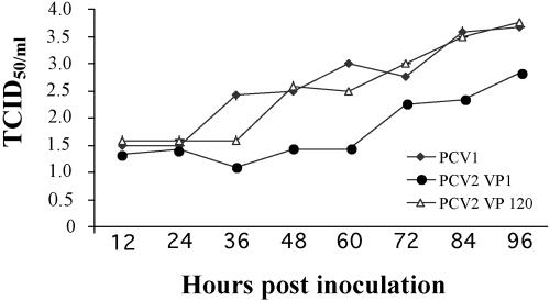
To compare the growth characteristics of nonpathogenic PCV1, wild-type PCV2 VP1, and serially passaged PCV2 VP120, one-step growth curves were performed in duplicate simultaneously for PCV1, PCV2 VP1, and PCV2 VP120. The infectious titers of viruses collected at 12-h intervals were determined by immunofluorescence assays (Fig. 1). The initial titers after infection at 12 h postinoculation were about 101.5 TCID50/ml for all three viruses. The infectious titers of PCV1 and PCV2 VP120 increased differently than wild-type PCV2 VP1 did (P = 0.0053) from 12 to 96 h. By 96 h postinfection, PCV1 and PCV2 VP120 had infectious titers of 103.66 and 103.75 TCID50/ml, whereas PCV2 VP1 had a titer of 102.83 TCID50/ml (Fig. 1).
FIG. 1.
One-step growth curves of PCV1, PCV2 VP1, and PCV2 VP120. Duplicate synchronized PK-15 cell cultures were each infected with PCV1, PCV2 VP1, or PCV2 VP120, all at an multiplicity of infection of 0.1. All three viruses had a titer of about 101.5 TCID50/ml at 12 h postinoculation. PCV1 and PCV2 VP120 replicated more efficiently in vitro than did PCV2 VP1 (P = 0.0053).
Identification of two amino acid mutations within the PCV2 capsid protein during serial passages.
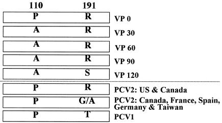
The complete genomes of the original wild-type PCV2 (VP0) and PCV2 passages 30, 60, 90, and 120 were amplified and sequenced. Sequence analyses revealed that there were a total of two nucleotide mutations, both in the capsid gene, at nucleotide positions 328 (C328G) and 573 (A573C) over the entire genome after 120 passages, and both resulted in amino acid changes. The C328G mutation appeared at passage 30 in which a proline at amino acid position 110 of the capsid was changed to an alanine (P110A) (Fig. 2). This mutation was present during the remaining passages. A second mutation, A573C, resulting in an amino acid change from arginine to serine at position 191 of the capsid (R191S), was identified only at passage 120 (Fig. 2).
FIG. 2.
Schematic diagram of amino acid mutations in the capsid protein during serial passages of PCV2 in PK-15 cells. Serial passage numbers are indicated (VP1, VP30, VP60, VP90, and VP120). Known field isolates of PCV2 and PCV1 from different geographic origins are also compared for these two mutations. US, United States.
By comparing all known PCV1 and PCV2 sequences available in the GenBank database, including 91 PCV2 and 4 PCV1 isolates, we found that the P110A mutation is unique, as all known PCV1 and PCV2 isolates have a proline at residue 110 of the capsid protein (Fig. 2). The R191S mutation, however, is variable: most PCV2 isolates from North America have an arginine, French PCV2 isolates and some Canadian PCV2 isolates have a glycine, and Spanish, Taiwanese, and German PCV2 isolates have an alanine. All nonpathogenic PCV1 isolates have a threonine residue at amino acid position 191 (Fig. 2). Overall, comparing the entire sequences of the capsid genes of PCV1 and PCV2, extensive sequence variation was observed: 33 to 35% of the nucleotides and 32 to 37% of the amino acids varied (13).
PCV2 VP120 virus exhibited reduced duration of viremia and virus loads in sera of infected pigs compared to those of the wild-type PCV2 VP1 virus.
Serum samples were collected from all control and inoculated pigs at −1, 7, 14, 21, 28, 35, and 42 dpi and assayed for PCV2 viremia by quantitative real-time PCR and for anti-PCV2 antibody by enzyme-linked immunosorbent assay. Prior to inoculation at −1 dpi, serum samples from all pigs were tested and were negative for PCV2 DNA.
Group 1 (control) pigs were negative for PCV2 viremia throughout the study (Table 1). PCV2 maternal antibodies in pigs in group 1 were detected at −1 dpi, which decreased in all pigs by 21 dpi. Seroconversion to PCV2 was not detected in control pigs (Table 2).
TABLE 1.
Detection of viremia in sera from inoculated and control pigs by real-time PCR
| Group | Inoculum | No. of pigs with viremia/no. testeda at dpi:
|
Total no. of pigs with viremia/no. tested | ||||||
|---|---|---|---|---|---|---|---|---|---|
| −1 | 7 | 14 | 21 | 28 | 35 | 4 | |||
| 1 | PBS buffer | 0/10 | 0/10 | 0/10 | 0/10 | 0/5 | 0/5 | 0/5 | 0/10 |
| 2 | PCV2 VP120 | 0/11 | 1/11 | 2/11 | 3/11 | 0/6 | 1/6 | 1/6 | 4/11 |
| 3 | PCV2 VP1 | 0/10 | 7/10 | 8/10 | 8/10 | 3/5 | 3/5 | 1/5 | 9/10 |
Five pigs from each group were necropsied at 21 dpi and the remaining pigs were necropsied at 42 dpi.
TABLE 2.
Seroconversion to PCV2 antibodies in pigs inoculated with PCV2 VP1 and VP120
| Group | Inoculum | No. of pigs with PCV2 antibodies/no. testeda at dpi:
|
||||||
|---|---|---|---|---|---|---|---|---|
| −1 | 7 | 14 | 21 | 28 | 35 | 42 | ||
| 1 | PBS buffer | 10/10b | 1/10 | 1/10 | 0/10 | 0/5 | 0/5 | 0/5 |
| 2 | PCV2 VP120 | 3/11b | 0/11 | 0/11 | 0/11 | 0/6 | 6/6 | 6/6 |
| 3 | PCV2 VP1 | 1/10b | 0/10 | 1/10 | 2/10 | 4/5 | 5/5 | 5/5 |
Five pigs from each group were necropsied at 21 dpi, and the remaining pigs were necropsied at 42 dpi.
Maternal antibodies were detected at −1 dpi but decreased in all groups between 7 and 21 dpi.
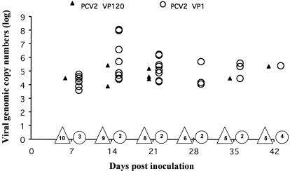
In the group 2 pigs inoculated with PCV2 VP120, viremia was first detected in 1 of 11 pigs at 7 dpi (Table 1 and Fig. 3). A total of four pigs in group 2 were viremic during the study. The average length of continuous viremia was 1.6 weeks. By 35 dpi, all group 2 pigs seroconverted to PCV2 (Table 2).
FIG. 3.
Quantitative real-time PCR results of PCV2 VP1 and PCV2 VP120 viral genomic copy numbers in 1 ml of serum sample collected at −1, 7, 14, 21, 28, 35, and 42 dpi from group 1, 2, and 3 pigs. Group 2 pigs inoculated with PCV2 VP120 that were positive for PCV2 DNA (▴) are indicated. The numbers inside the ▵ symbol on the x axis (10, 9, 8, 6, 5, and 5) are the numbers of pigs in group 2 that were negative for PCV2 viremia on the respective dpi. Group 3 pigs inoculated with PCV2 VP1 that were positive for PCV2 DNA (○) are indicated. The numbers inside the ○ symbol on the x axis (3, 2, 2, 2, 2, and 4) are the numbers of pigs in group 3 that were negative for PCV2 viremia on the respective dpi. The PCV2 genomic copy numbers are presented as the log of copy numbers per milliliter of serum (y axis).
In the group 3 pigs inoculated with PCV2 VP1, viremia was first detected in 7 of 10 pigs at 7 dpi (Table 1 and Fig. 3). Of the 10 pigs in group 3, 9 became viremic during the study, and the average length of continuous viremia was 3 weeks. All animals in group 3 seroconverted to PCV2 by 35 dpi (Table 2).
The PCV2 genomic copy numbers per milliliter of serum in positive samples ranged from 8,840 to 274,800 in group 2 pigs inoculated with PCV2 VP120 and from 26,520 to 120,000,000 in group 3 pigs inoculated with PCV2 VP1 (Fig. 3). The PCV2 genomic copy numbers per milliliter of serum were greater in group 3 pigs than in group 2 pigs at 21 dpi (P = 0.0003) and 42 dpi (P = 0.039). However, PCV2 DNA was detected in the lymph nodes of only 3 of 11 group 2 pigs and 2 of 10 group 3 pigs, and the median PCV2 genomic copy numbers per milligram of tracheobronchial lymph node did not differ in group 2 and 3 pigs (P = 0.72). The virus recovered from the sera and tracheobronchial lymph nodes of four selected pigs in groups 2 and 3 were sequenced, and sequence analyses revealed that the viruses recovered from infected pigs originated from the respective inocula.
The S/P ratios of PCV2 antibodies differed in the pigs of groups 1, 2, and 3 (P < 0.0001) and over time (P < 0.0001).
PCV2 VP120 virus is attenuated in pigs.
Mild sneezing and rough hair coat were noted in some animals from all three groups (data not shown). Two of the 10 control pigs and all of the 21 inoculated pigs had sneezing and rough hair coat (P = 0.051). Up to 21 dpi, group 2 and 3 pigs were 58 (95% confidence interval [95% CI], 13.1 to 255.0) and 41 (95% CI, 9.3 to 178.0) times more likely to show sneezing and rough hair coat at any examination date than negative-control pigs, with no difference between group 2 and 3 pigs (odds ratio for group 3 and 2 pigs [OR3vs2], 1.4 [95% CI, 0.8 to 2.6]). For the 16 pigs that were necropsied at 42 dpi, over the entire study period, again group 2 and 3 pigs were more likely to show sneezing and rough hair coat than negative-control pigs (OR2vs1, 20.4 [95% CI, 4.6 to 90.1]; OR3vs1, 71.6 [95% CI, 16.0 to 320.8]) with group 3 pigs being 3.5 (95% CI, 1.9 to 6.6) times more likely to show sneezing and rough hair coat than group 2 pigs. There were no differences in weight gain (P = 0.081) or mean rectal temperatures (P > 0.05) among any of the groups (data not shown).
At necropsies, lymph nodes of two of five pigs in group 1 were mildly enlarged; however, this was not associated with PCV2 infection, as evidenced by the lack of PCV2 DNA, PCV2 antigen, or seroconversion. At 42 dpi necropsy, all group 1 pigs had normal lymph nodes (Table 3). Group 2 pigs inoculated with PCV2 VP120 had mild to moderately enlarged lymph nodes at both 21 and 42 dpi (Table 3). The lymph nodes in group 3 pigs were moderately to severely enlarged at both 21 and 42 dpi (Table 3). Two pigs inoculated with PCV2 VP1 had visible gross pneumonia at 21 dpi. Gross pneumonia was not found in group 1 or 2 pigs at either 21 or 42 dpi.
TABLE 3.
Gross lymph node and lung lesions in control and inoculated pigs
| Group | Inoculum | No. of pigs with enlarged lymph nodesa
|
No. of pigs with gross pneumonia lesionsa
|
||
|---|---|---|---|---|---|
| 21 dpi | 42 dpi | 21 dpi | 42 dpi | ||
| 1 | PBS buffer | 2/5 (0.4) A | 0/5 (0.0) A | 0/5 (0.0) A | 0/5 (0.0) |
| 2 | PCV2 VP120 | 2/5 (0.4) A | 4/6 (1.3) B | 0/6 (0.0) A | 0/5 (0.0) |
| 3 | PCV2 VP1 | 5/5 (2.2) B | 5/5 (2.6) B | 2/5 (2.6) B | 0/5 (0.0) |
Five pigs from each group were necropsied at 21 dpi, and the remaining pigs were necropsied at 42 dpi. Values in parentheses are the mean scores of estimated lymph node enlargement (scored from 0 [normal] to 3 [severely enlarged and tan-colored lymph nodes]) and mean percentage of lungs affected by grossly visible pneumonia (0 to 100%). Different letters (A and B) indicate statistically different mean value score between groups (P < 0.05).
Microscopic lung lesions characterized by mild peribronchiolar lymphoplasmacytic and histiocytic bronchointerstitial pneumonia and liver lesions characterized by mild lymphoplasmacytic hepatitis were detected at variable levels in the pigs of all groups (Table 4). The mean histological scores for interstitial pneumonia in the lung, interstitial hepatitis in the liver, and lymphoid depletion and histiocytic replacement for lymph nodes, tonsils, and spleens are summarized in Table 4. The presence of lesions in the kidney and heart is also presented in Table 4.
TABLE 4.
Distribution of histopathologic lesions in different tissues and organs from control and inoculated pigs
| Group | Inoculum | dpi | No. of pigs with lesions/no. testeda
|
|||||||||
|---|---|---|---|---|---|---|---|---|---|---|---|---|
| Lung | Lymph nodes
|
Tonsil
|
Spleen
|
Liver | Kidney | Heart | ||||||
| LD | HR | LD | HR | LD | HR | |||||||
| 1 | PBS buffer | 21 | 0/5 (0.0) | 0/5 (0.0) | 0/5 (0.0) | 0/5 (0.0) | 0/5 (0.0) | 0/5 (0.0) | 0/5 (0.0) | 0/5 (0.0) | 1/5 | 1/5 |
| 42 | 3/5 (0.6) | 0/5 (0.0) | 0/5 (0.0) | 0/5 (0.0) | 0/5 (0.0) | 0/5 (0.0) | 0/5 (0.0) | 1/5 (0.2) | 1/5 | 0/5 | ||
| 2 | PCV2 VP120 | 21 | 2/5 (0.4) | 3/5 (0.6) | 0/5 (0.0) | 2/5 (0.4) | 0/5 (0.0) | 2/5 (0.4) | 0/5 (0.0) | 3/5 (0.6) | 2/5 | 0/5 |
| 42 | 5/6 (1.0) | 2/6 (0.3) | 2/6 (0.3) | 0/6 (0.0) | 0/6 (0.0) | 1/6 (0.2) | 1/6 (0.2) | 0/6 (0.0) | 2/6 | 0.5 | ||
| 3 | PCV2 VP1 | 21 | 2/5 (0.4) | 4/5 (1.2) | 3/5 (1.0) | 5/5 (1.2) | 1/5 (0.2) | 4/5 (1.0) | 4/5 (1.0) | 3/5 (1.2) | 3/5 | 3/5 |
| 42 | 5/5 (1.4) | 5/5 (1.4) | 3/5 (0.8) | 0/5 (0.0) | 1/5 (0.2) | 4/5 (1.0) | 3/5 (0.6) | 3/6 (0.6) | 1/5 | 2/5 | ||
Values in parentheses are mean histological scores for interstitial pneumonia, interstitial hepatitis, and lymphoid depletion (LD) and histiocytic replacement (HR) for lymph nodes, tonsils, and spleen. The severities of histopathologic lesions were significantly different (P < 0.05 by Fisher's exact test) for LD of lymph nodes at 42 dpi, LD of tonsils at 21 dpi, LD of spleen at 42 dpi, and HR of spleen at 21 dpi for groups 1, 2, and 3.
At necropsies (21 and 42 dpi), PCV2 antigen was not detected by IHC in the lymphoid tissues of the negative-control group 1 pigs. In group 2 pigs inoculated with PCV2 VP120, low levels of PCV2 antigen were detected in spleen tissues of one of five pigs at 21 dpi, in lymph node tissues of two of six pigs at 42 dpi, and in tonsil tissues of three of six pigs at 42 dpi (Table 5). In group 3 pigs inoculated with PCV2 VP1, low to high levels of PCV2 antigen were detected in lymph node tissues of five of five pigs, in tonsil tissues of four of five pigs, and in spleen tissues of four of five pigs at 21 dpi. At 42 dpi, low to moderate levels of PCV2 antigen were detected in group 3 pigs inoculated with PCV2 VP1 (Table 5).
TABLE 5.
IHC detection of PCV2 antigen in lymph nodes, tonsils, and spleens of inoculated and control pigs
| Group | Inoculum | dpi | No. of pigs with PCV2 antigen/no. testeda
|
||
|---|---|---|---|---|---|
| Lymph node | Tonsil | Spleen | |||
| 1 | PBS buffer | 21 | 0/5 (0.0) | 0/5 (0.0) | 0/5 (0.0) |
| 42 | 0/5 (0.0) | 0/5 (0.0) | 0/5 (0.0) | ||
| 2 | PCV2 VP120 | 21 | 0/5 (0.0) | 0/5 (0.0) | 1/5 (0.2) |
| 42 | 2/6 (0.3) | 3/6 (0.5) | 0/6 (0.0) | ||
| 3 | PCV2 VP1 | 21 | 5/5 (1.6) | 4/5 (1.0) | 4/5 (1.2) |
| 42 | 3/5 (0.8) | 2/5 (0.4) | 2/5 (0.4) | ||
Values in parentheses are the mean scores of the level of PCV2 antigen in lymphoid tissues (scores ranged from 0 for no antigen detected to 3 for high levels of antigen). The PCV2 antigen in lymph nodes and tonsils at 21 dpi were significantly different (P < 0.05 by Fisher's exact test) in pigs from groups 1, 2, and 3.
All gross pathological and histopathologic scores at 21 and 42 dpi were compared by analysis of variance using the GLM procedure followed by a Bonferroni test of multiple means. At 21 dpi, the mean scores of group 1 and 2 pigs are similar (P = 1.00) but differ from the mean scores of group 3 pigs (P = 0.0032). By 42 dpi, the mean scores of group 1 differ from the mean scores of group 2 (P = 0.0083) and group 3 (P = 0.0001), and the group 2 mean scores are lower than those of group 3 (P = 0.0274) (Tables 3 to 5).
DISCUSSION
PMWS associated with PCV2 infection has now become an economically important global disease. The molecular mechanisms of PCV2 replication and pathogenesis are poorly understood. We report here the identification of two amino acid mutations within the PCV2 capsid protein that are important for PCV2 attenuation in vivo and for enhanced growth ability in PK-15 cells.
PCV2 VP120 contained two amino acid mutations in the capsid protein, P110A and R191S, compared to the wild-type PCV2 parent virus (passage 0). As a result of these two mutations, PCV2 VP120 replicated more efficiently (P = 0.0053) in PK-15 cells with a difference in infectious titer of about 1 log unit compared to its parent virus. The difference in growth ability with a 1-log-unit titer increase between VP120 and VP1 virus may be insignificant for many other viruses. However, PCV is known to replicate to relatively low titers, usually below 105 TCID50 per ml, in PK-15 cells. PCV1 grows slightly better than PCV2 (14-15), likely due to cell culture adaptation, since PCV1 was originally isolated from PK-15 cells. PCV2 VP120 replicated to a similar level with the PK-15 cell-derived nonpathogenic PCV1; thus, these two mutations either alone or collectively are responsible for the enhanced growth ability of PCV2 VP120 in vitro. Allan et al. (1) attempted to infect Vero cells with PCV1. Intranuclear immune staining, characteristic of PCV1 replication, was not detected until cell culture passage 6 in Vero cells. By passage 15, PCV1 replicated in Vero cells to a level similar to that in PK-15 cells, suggesting that efficient PCV1 replication in Vero cells depended upon the number of virus passages. It would be interesting to see whether additional passages in PK-15 cells can further enhance the growth ability of PCV2.
When SPF piglets were inoculated with PCV2 VP120, fewer pigs developed viremia with shorter duration and lower PCV2 genomic copy numbers compared to pigs inoculated with PCV2 VP1. We previously showed that the nonpathogenic PK-15 cell-derived PCV1 also had a shorter mean viremia length (0.625 week) in infected pigs (15). Seroconversion in the absence of viremia has recently been demonstrated for an attenuated chimeric PCV1-2 virus (14). Viremia was not detected in pigs vaccinated with the chimeric PCV1-2 candidate vaccine virus, even though the vaccinated pigs seroconverted and were protected against challenge (14). Analyses of the gross, microscopic, and IHC mean scores revealed that the pigs inoculated with PCV2 VP120 virus had milder pathological lesions than the pigs inoculated with PCV2 VP1 virus. These results suggested that PCV2 VP 120 had adapted to grow better in PK-15 cells and is attenuated in virulence in vivo.
After 120 passages in PK-15 cells, only two amino acid mutations, both in the capsid gene, were detected in the entire PCV2 genome, suggesting that the PCV2 genome is relatively stable. This may explain why the sequences of all known PCV2 field isolates worldwide identified so far are highly conserved (13, 21). The P110A mutation occurred early (passage 30) during serial passage and involved two hydrophobic amino acids, proline and alanine. The change from proline to alanine may alter the tertiary structure of the capsid protein, as proline is often involved in the bending regions of protein structures. The uniqueness of the P110A mutation in passages 30 to 120 compared to the sequences of known PCV2 field isolates suggested a biological role for this mutation. The fact that the nonpathogenic PCV1 isolates have a proline (rather than alanine) at this position complicated the potential implication of this P110A mutation in PCV2 virulence. However, the extensive sequence variation between the capsid proteins of PCV1 and PCV2 (32 to 37% amino acids differed) suggested that it would be difficult to draw any conclusion on PCV2 virulence based on sequence comparison with the capsid protein of the nonpathogenic PCV1. The R191S mutation occurred very late during the serial passage (between passages 91 and 120) and is unique to the VP120 PCV2 virus, as all known PCV2 and PCV1 sequences so far do not have a serine residue at this position. However, glycine, alanine, and threonine substitutions at this position were found in field isolates of PCV1 and PCV2. Lekcharoensuk et al. (22) identified at least two conformational neutralizing epitopes in the PCV2 capsid protein, and the R191S mutation falls within one of these immunoreactive epitopes, suggesting that the R191S mutation may play an important role in PCV2 replication and pathogenesis. Tertiary structure changes as the results of these two amino acid mutations in the capsid protein could drastically influence its biological functions. Crystallization of the capsid protein to determine the three-dimensional structure could provide clues regarding the effects of these amino acid mutations on virus structure.
Amino acid substitutions induced by cell culture passage or chemical mutagenesis techniques have been routinely used for the attenuation of many viruses and have led to the production of numerous vaccines (6). There are many examples of a single amino acid mutation leading to the attenuation of a virus. For example, a single amino acid change in the VP3 capsid of poliovirus Sabin type 3 strain (35) and two changes in the chicken infectious bursal disease virus VP2 capsid were responsible for the acquisition of attenuated phenotypes (34). A single amino acid change in the rabies virus envelope glycoprotein (9) and three changes in Sindbis virus E1 and E2 proteins (28) also led to virus attenuation. It has also been shown that substitution of proline for leucine at residue 101 of the nonstructural 4B protein of the mosquito-borne dengue 4 virus resulted in decreased viral replication in mosquitoes but increased replication in Vero cells (16). Hence, the balancing control of efficient replication of dengue 4 virus in either mosquito or Vero cells was maintained by a single amino acid change. Within the Circoviridae family, a single amino acid mutation in the VP1 capsid protein of chicken anemia virus was found to be responsible for the pathogenicity of the virus in chickens (36). Taken together, the results from this study suggested that the P110A and R191S mutations are likely responsible for the attenuation of PCV2 VP120 in pigs and for the enhanced growth ability of PCV2 in PK-15 cells. Additional studies are under way to determine whether the two mutations act singly or collectively in modifying the PCV2 phenotype.
Acknowledgments
We thank S. M. Boyle, R. B. Duncan, L. A. Eng, J. C. Sible, and T. E. Toth of Virginia Tech and M. Gill and S. Q. Wu of Fort Dodge Animal Health, Inc., for their help and Jillian Fenaux for editorial assistance.
This study was supported in part by a grant from Fort Dodge Animal Health, Inc., Fort Dodge, Iowa, and by a grant from the U.S. Department of Agriculture National Research Initiative Competitive Grant Program (NRI 2004-35204-14213).
REFERENCES
- 1.Allan, G. M., D. P. Mackie, J. McNair, B. M. Adair, and M. S. McNulty. 1994. Production, preliminary characterization and applications of monoclonal antibodies to porcine circovirus. Vet. Immunol. Immunopathol. 43:357-371. [DOI] [PubMed] [Google Scholar]
- 2.Allan, G. M., F. McNeilly, J. P. Cassidy, G. A. Reilly, B. Adair, W. A. Ellis, and M. S. McNulty. 1995. Pathogenesis of porcine circovirus: experimental infections of colostrum deprived piglets and examination of pig fetal material. Vet. Microbiol. 44:49-64. [DOI] [PubMed] [Google Scholar]
- 3.Allan, G. M., F. McNeilly, S. Kennedy, B. Daft, E. G. Clarke, J. A. Ellis, and D. M. Haines. 1998. Isolation of porcine circovirus-like viruses from pigs with a wasting disease in the USA and Europe. J. Vet. Diagn. Investig. 10:3-10. [DOI] [PubMed] [Google Scholar]
- 4.Allan, G. M., and J. A. Ellis. 2000. Porcine circoviruses: a review. J. Vet. Diagn. Investig. 12:3-14. [DOI] [PubMed] [Google Scholar]
- 5.Bassami, M. R., D. Berryman, G. E. Wilcox, and S. R. Raidal. 1998. Psittacine beak and feather disease virus nucleotide sequence analysis and its relationship to porcine circovirus, plant circoviruses, and chicken anemia virus. Virology 249:453-459. [DOI] [PubMed] [Google Scholar]
- 6.Brooks, G. F., J. S. Butel, and S. A. Morse. 1998. Pathogenesis and control of viral diseases, p. 363-365. In Jawetz, Melnick, and Adelberg's medical microbiology, 21st ed. Appleton & Lange, Stamford, Conn.
- 7.Cheung, A. K. 2003. Transcriptional analysis of porcine circovirus. Virology 305:168-180. [DOI] [PubMed] [Google Scholar]
- 8.Choi, C., C. Chae, and E. G. Clark. 2000. Porcine postweaning multisystemic wasting syndrome in Korean pig: detection of porcine circovirus 2 infection by immunohistochemistry and polymerase chain reaction. J. Vet. Diagn. Investig. 12:151-153. [DOI] [PubMed] [Google Scholar]
- 9.Dietzschold, B., W. H. Wunner, T. J. Wiktor, A. D. Lopes, M. Lafon, C. L. Smith, and H. Koprowski. 1983. Characterization of an antigenic determinant of the glycoprotein that correlates with pathogenicity of rabies virus. Proc. Natl. Acad. Sci. USA 80:70-74. [DOI] [PMC free article] [PubMed] [Google Scholar]
- 10.Edwards, S., and J. J. Sands. 1994. Evidence of circovirus infection in British pigs. Vet. Rec. 134:680-681. [DOI] [PubMed] [Google Scholar]
- 11.Ellis, J., L. Hassard, E. Clark, J. Harding, G. Allan, P. Willson, J. Strakappe, K. Martin, F. McNeilly, B. Meehan, D. Todd, and D. Haines. 1998. Isolation of circovirus from lesions of pigs with postweaning multisystemic wasting syndrome. Can. Vet. J. 39:44-51. [PMC free article] [PubMed] [Google Scholar]
- 12.Fenaux, M., P. G. Halbur, G. Haqshenas, R. Royer, P. Thomas, P. Nawagitgul, M. Gill, T. E. Toth, and X. J. Meng. 2002. Cloned genomic DNA of type 2 porcine circovirus is infectious when injected directly into the liver and lymph nodes of pigs: characterization of clinical disease, virus distribution, and pathological lesions. J. Virol. 76:541-551. [DOI] [PMC free article] [PubMed] [Google Scholar]
- 13.Fenaux, M., P. G. Halbur, M. Gill, T. E. Toth, and X. J. Meng. 2000. Genetic characterization of type 2 porcine circovirus (PCV-2) from pigs with postweaning multisystemic wasting syndrome in different geographic regions of North America and development of a differential PCR-restriction fragment length polymorphism assay to detect and differentiate between infections with PCV-1 and PCV-2. J. Clin. Microbiol. 38:2494-2503. [DOI] [PMC free article] [PubMed] [Google Scholar]
- 14.Fenaux, M., T. Opriessnig, P. G. Halbur, F. Elvinger, and X. J. Meng. 2004. A chimeric porcine circovirus (PCV) with the immunogenic capsid gene of the pathogenic PCV2 cloned into the genomic backbone of the nonpathogenic PCV1 induces protective immunity against PCV2 infection in pigs. J. Virol. 78:6297-6303. [DOI] [PMC free article] [PubMed] [Google Scholar]
- 15.Fenaux, M., T. Opriessnig, P. G. Halbur, and X. J. Meng. 2003. Immunogenicity and pathogenicity of the chimeric infectious DNA clones between pathogenic type 2 porcine circovirus (PCV2) and nonpathogenic PCV1 in weaning pigs. J. Virol. 77:11232-11243. [DOI] [PMC free article] [PubMed] [Google Scholar]
- 16.Hanley, K. A., L. R. Manlucu, L. E. Gilmore, J. E. Blaney, C. T. Hanson, B. R. Murphy, and S. S. Whitehead. 2003. A trade-off in replication in mosquito versus mammalian systems conferred by a point mutation in the NS4B protein of dengue virus type 4. Virology 312:222-232. [DOI] [PubMed] [Google Scholar]
- 17.Harding, J. C., and E. G. Clark. 1997. Recognizing and diagnosing post-weaning multisystemic wasting syndrome (PMWS). Swine Health Prod. 5:201-203. [Google Scholar]
- 18.Harms, P. A., S. D. Sorden, P. G. Halbur, S. R. Bolin, K. M. Lager, L. Morozov, and P. S. Paul. 2001. Experimental reproduction of severe disease in CD/CD pigs concurrently infected with type 2 porcine circovirus and porcine reproductive and respiratory syndrome virus. Vet. Pathol. 38:528-539. [DOI] [PubMed] [Google Scholar]
- 19.Krakowka, S., J. A. Ellis, B. Meehan, S. Keenedy, F. McNeilly, and G. Allan. 2000. Viral wasting syndrome of swine: experimental reproduction of postweaning multisystemic wasting syndrome in gnotobiotic swine by coinfection with porcine circovirus 2 and porcine parvovirus. Vet. Pathol. 37:254-263. [DOI] [PubMed] [Google Scholar]
- 20.Ladekjaer-Mikkelsen, A. S., J. Nielsen, T. Stadejek, T. Storgaard, S. Krakowka, J. Ellis, F. McNeilly, G. Allan, and A. Botner. 2002. Reproduction of postweaning multisystemic wasting syndrome (PMWS) in immunostimulated and non-immunostimulated 3-week-old piglets experimentally infected with porcine circovirus type-2 (PCV2). Vet. Microbiol. 89:97-114. [DOI] [PMC free article] [PubMed] [Google Scholar]
- 21.Larochelle, R., R. Magar, and S. D'Allaire. 2002. Genetic characterization and phylogenetic analysis of porcine circovirus type 2 (PCV2) strains from cases presenting various clinical conditions. Virus Res. 90:101-112. [DOI] [PubMed] [Google Scholar]
- 22.Lekcharoensuk, P., I. Morozov, P. S. Paul, N. Thangthumniyom, W. Wajjawalku, and X. J. Meng. 2004. Epitope mapping of the major capsid protein of type 2 porcine circovirus (PCV2) by using chimeric PCV1 and PCV2. J. Virol. 78:8135-8145. [DOI] [PMC free article] [PubMed] [Google Scholar]
- 23.Mankertz, A., M. Domingo, J. M. Folch, P. LeCann, A. Jestin, J. Segales, B. Chmielewicz, J. Plana-Duran, and D. Soike. 2000. Characterization of PCV-2 isolates from Spain, Germany and France. Virus Res. 66:65-77. [DOI] [PubMed] [Google Scholar]
- 24.Mankertz, A., K. Hattermann, B. Ehlers, and D. Soike. 2000. Cloning and sequencing of columbid circovirus (CoCV), a new circovirus from pigeons. Arch. Virol. 145:2469-2479. [DOI] [PubMed] [Google Scholar]
- 25.Nawagitgul, P., P. A. Harms, I. Morozov, B. J. Thacker, S. D. Sorden, C. Lekcharoensuk, and P. S. Paul. 2002. Modified indirect porcine circovirus (PCV) type 2-based and recombinant capsid protein (ORF2)-based enzyme-linked immunosorbent assays for detection of antibodies to PCV. Clin. Diagn. Lab. Immunol. 9:33-40. [DOI] [PMC free article] [PubMed] [Google Scholar]
- 26.Onuki, A., K. Abe, K. Togashi, K. Kawashima, A. Taneichi, and H. Tsunemitsu. 1999. Detection of porcine circovirus from lesions of a pig with wasting disease in Japan. J. Vet. Med. Sci. 61:1119-1123. [DOI] [PubMed] [Google Scholar]
- 27.Phenix, K. V., J. H. Weston, I. Ypelaar, A. Lavazza, J. A. Smyth, D. Todd, G. E. Wilcox, and S. R. Raidal. 2001. Nucleotide sequence analysis of a novel circovirus of canaries and its relationship to other members of the genus Circovirus of the family Circoviridae. J. Gen. Virol. 82:2805-2809. [DOI] [PubMed] [Google Scholar]
- 28.Polo, J. M., and R. E. Johnston. 1990. Attenuating mutations in glycoproteins E1 and E2 of Sindbis virus produce a highly attenuated strain when combined in vitro. J. Virol. 64:4438-4444. [DOI] [PMC free article] [PubMed] [Google Scholar]
- 29.Sorden, S. D., P. A. Harms, P. Nawagitgul, D. Cavanaugh, and P. S. Paul. 1999. Development of a polyclonal-antibody-based immunohistochemical method for the detection of type 2 porcine circovirus in formalin-fixed, paraffin-embedded tissue. J. Vet. Diagn. Investig. 11:528-530. [DOI] [PubMed] [Google Scholar]
- 30.Tischer, I., H. Gelderblom, W. Vettermann, and M. A. Koch. 1982. A very small porcine virus with circular single-stranded DNA. Nature 295:64-66. [DOI] [PubMed] [Google Scholar]
- 31.Tischer, I., W. Mields, D. Wolff, M. Vagt, and W. Griem. 1986. Studies on epidemiology and pathogenicity of porcine circovirus. Arch. Virol. 91:271-276. [DOI] [PubMed] [Google Scholar]
- 32.Tischer, I., R. Rasch, and G. Tochtermann. 1974. Characterization of papovavirus and picornavirus-like particles in permanent pig kidney cell lines. Zentralbl. Bakteriol. Hyg. A 226:153-167. [PubMed] [Google Scholar]
- 33.Todd, D., J. H. Weston, D. Soike, and J. A. Smyth. 2001. Genome sequence determinations and analyses of novel circoviruses from goose and pigeon. Virology 286:354-362. [DOI] [PubMed] [Google Scholar]
- 34.van Loon, A. A., N. de Haas, I. Zeyda, and E. Mundt. 2002. Alteration of amino acids in VP2 of very virulent infectious bursal disease virus results in tissue culture adaptation and attenuation in chickens. J. Gen. Virol. 83:121-129. [DOI] [PubMed] [Google Scholar]
- 35.Westrop, G. D., K. A. Wareham, D. M. Evans, G. Dunn, P. D. Minor, D. I. Magrath, F. Taffs, S. Marsden, M. A. Skinner, G. C. Schild, and J. W. Almond. 1989. Genetic basis of attenuation of the Sabin type 3 oral poliovirus vaccine. J. Virol. 63:1338-1344. [DOI] [PMC free article] [PubMed] [Google Scholar]
- 36.Yamaguchi, S., T. Imada, N. Kaji, M. Mase, K. Tsukamoto, N. Tanimura, and N. Yuasa. 2001. Identification of a genetic determinant of pathogenicity in chicken anemia virus. J. Gen. Virol. 82:1233-1238. [DOI] [PubMed] [Google Scholar]


