Abstract
Cholangiolithiasis is a rare condition in animals wherein the choleliths are formed in the biliary tracts causing chronic inflammation. In the current study, choleliths which were firm and friable concretions of approximately 3–4 cm in diameter were found in the bile ducts of the liver of an adult cattle infected with Fasciola gigantica. Two such irregularly shaped concretions were encountered in the two biliary tracts leading to common bile duct in the liver. There were localized irregular saccular dilatation and numerous liver flukes in the bile ducts. Further, several small hard concretions of varying sizes were also present in the ducts. The histological investigation revealed chronic cholangitis with bile duct hyperplasia and cirrhosis. The co-existence of F. gigantica and choleliths indicated the physical pre-disposition for the formation of bile concretions in the bile duct which is not a common site for the occurrence of gall stones.
Keywords: Cholangiolithiasis, Choleliths, Biliary tracts, Fasciola gigantica, Cattle
Introduction
Fascioliosis is a highly pathogenic disease caused by liver fluke species, Fasciola hepatica and Fasciola gigantica and traditionally has been considered to be an important veterinary problem therefore; most information on the pathologic process of fascioliosis has been obtained from domestic herbivorous mammals. Adult flukes in the bile ducts cause inflammation and hyperplasia of the epithelium leading to cholangitis and cholecystitis resulting in mechanical obstruction of the biliary duct. Choleliths are commonly called as gall stones, which are generally located in the gall bladder. The choleliths present in the bile ducts are called as cholangiolith, which are rarely formed in bile ducts and obstruction of biliary tract due to choleliths is uncommon (Cable et al. 1997; Klopfleisch et al. 2004; Maxie 2007). Such concretions in the bile ducts are very rare and hence it is widely believed by the local people to be having a medicinal property. In local language, these choleliths are commonly called as “Gailan” in Urdu, and are believed to be useful for the treatment of partial paralysis in man. Such choleliths formed in male cattle are considered invaluable and are priced at a higher rate than those formed in the female cattle and buffaloes. It is also believed that, the Gailan is credited with enhancing aggressiveness among men due to which there is much demand of Gailan in the local market. It is stated that the bile concretions usually occur in cattle in conjunction with the fasciolosis (Gwilym et al. 1946; Sastry and Rao 2001). In the present case, cholangiolithiasis was found in the liver, along with F. gigantica during the post mortem examination of adult cattle.
Materials and methods
During routine inspection for lesions in the slaughtered cattle, the present condition was detected. The infected liver was collected from the slaughter house and transported in cold chain to the laboratory and dissection of liver was performed. The tissues of liver were collected in Neutral buffered formalin and were subjected to the routine histopathological examination by Paraffin embedding technique (Luna 1964).
The parasites were collected in normal saline, washed 3 times, processed and then stained with hematoxylene and eosine stain for identification as per the method described by Bowman (2009). The species of parasite was identified based on the morphological characters described by Soulsby (1982).
Results and discussion
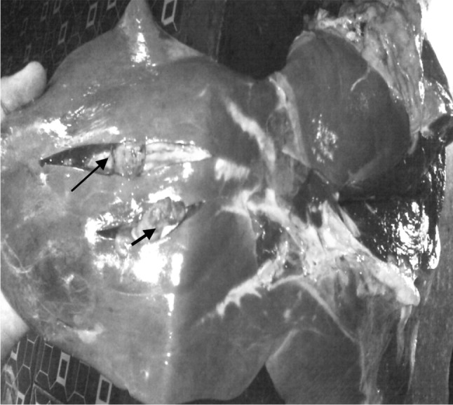
Cholangioliths approximately 3–4 cm in diameter, irregular in shape of greenish-black and greenish yellow color were found. Grossly they could be detected only by palpation of the liver. Two liths were found in the bile ducts leading to the common bile duct and were located at least 5–6 cm to the interior of the bile duct (Fig. 1). However, gallstones were not found in the gall bladder.
Fig. 1.

Yellow-green colored bile concretions in liver flukes in biliary tracts (indicated by arrows)
It has been described that, the choleliths are of mixed composition, yellow-black or green-black and friable. There may be hundreds of small stones or a few large ones in the gall bladder. The choleliths are usually faceted if they are large, but are usually light and friable (Sastry and Rao 2001; Maxie 2007). In one of the studies in cattle, several small choleliths of less than 10 mm were seen in the bile ducts and they were faceted, hard, orange to tan colored (Klopfleisch et al. 2004).
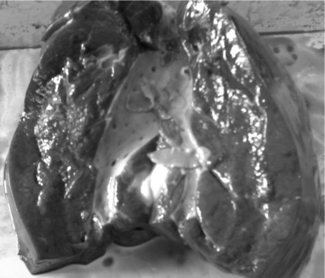
While dissecting the liver, many trematode parasites were found in the biliary tracts along with the choleliths. Based on morphological characters, the collected parasites were identified as F. gigantica (Fig. 2). The micrometry of adult fluke revealed a length of 72 mm and a width of 11 mm with a longer rounded posterior end along with a shorter cephalic cone and the ventral sucker was found to be slightly larger than the oral sucker with anteriorly located testes as described by Soulsby (1982).
Fig. 2.

Incised liver showing leaf-like the bile ducts
The bile duct showed irregular mucosal surface, epithelial hyperplasia, peri-ductular inflammation with fibrosis. Wherever there was depression in the wall, the gall stones were found either embedded or free with different size and shape. As per Klopfleisch et al. (2004) the origin of gall stones is non-specific and gall stones are asymptomatic or clinically silent. The obstruction may be caused by parasites, bacteria, bile contents, cicatrices and stenosis along with choleliths (Klopfleisch et al. 2004; Maxie 2007).
In the present case, the gall stones could have originated due to the inflammatory effect of F. gigantica on the bile ductular wall. The ductal inflammation would have caused cholangitis induced irregularity of the bile duct leading to stasis of the bile in the small spaces. The prolonged stasis causes concentration of the bile contents, especially the salts. The toxic or metabolic products of F. gigantica can form a nidus upon which, the bile contents would get precipitated to form choleliths. Inspissiated bile stained friable plugs are occasionally responsible for obstruction in liver, as reported in horses (Maxie 2007; Vegad and Katiyar 1998).
It is well documented that, in liver fluke infection, cholangitis is caused by the irritation of the spines on the tegument of the parasites as well as the toxic metabolites released by them (Sastry and Rao 2001). Several workers postulated that, their development is probably secondary to chronic and mild cholecystitis and related disturbances to resorptive activities of gall bladder. In resorptive disturbances, sometimes, the bile salts are removed at a faster rate than the stone forming components of the bile (Maxie 2007).
Further, Maxie (2007) reported that, occasionally they lodge in the bile ducts and cause jaundice and that the calcareous deposits are often seen in duct of F. gigantica infected cattle. Otherwise, the occurrence of choleliths in bile ducts is very rare (Cable et al. 1997).
In the area of lodgement of liths, the bile duct showed necrotic changes and thinning of the vessel wall, and saccular dilatation on all the sides. The reports indicates that, the large stones are known to cause pressure necrosis and ulceration of the mucosa, localized dilatation of the bile ducts and saccular diverticulum of the gall bladder (Sastry and Rao 2001; Maxie 2007; Vegad and Katiyar 1998). In an earlier case report, cholangiolithiasis with suppurative infection in a 17 year-old cow has been documented by Klopfleisch et al. (2004).
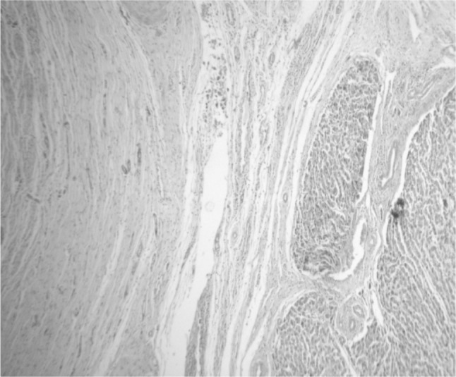
The histological investigation showed chronic cholangitis, peri-cholangitis with biliary epithelial hyperplasia (Fig. 3). This finding is in line with the observations made by Maxie (2007) who stated that, the impacted gall stones cause cholangitis or cholecystitis and obstruction. The bile ducts undergo progressive cylindric dilatation, with inflammation of the walls of the duct, portal triads partly because of the chemical irritation of the bile acids and partly due to bacterial infections (Maxie 2007). Sastry and Rao (2001) are of the opinion that gall stones are often found in the bile ducts of cattle, because of the frequency of the parasitic involvement which holds well with the present case. The present study can be concluded that, the occurrence of a cholelithiasis was a consequence of chronic fascioliasis in cattle. As a result of cholangitis and dilatation of biliary tracts caused by chronic fasciolosis, there were congregations due to retention of bile, forming choleliths of varying sizes. The histological investigation revealed chronic cholangitis with bile duct hyperplasia and cirrhosis. The co-existence of F. gigantica and choleliths indicated the physical pre-disposition for the formation of bile concretions in the bile duct which is not a common site for the occurrence of gall stones.
Fig. 3.

Histologic section of liver showing chronic fibrosis, cholangitis and pseudolobule formation
References
- Bowman DD. Georgis’ parasitology for veterinarians. 9. St. Louis: Saunders Elsevier; 2009. [Google Scholar]
- Cable CS, Rebhun WC, Fortier LA. Cholelithiasis and cholecystitis in a dairy cow. J Am Vet Med Assoc. 1997;211(7):899–900. [PubMed] [Google Scholar]
- Gwilym OD, Gaiger D. Veterinary pathology and bacteriology. 3. London: Bailliere, Tindall & Cox, University of Liverpool; 1946. [Google Scholar]
- Klopfleisch R, Polster U, Klingler K, Teifke JP. Severe but silent cholangiolithiasis in a cattle: a case report. Tierarztliche Prax Grobtiere. 2004;1:13–17. [Google Scholar]
- Luna LG. Manual of histologic staining methods of the Armed Forces Institute of Pathology. 3. London: McGraw Hill; 1964. [Google Scholar]
- Maxie GM. Jubb, Kennedy and Palmer’s pathology of domestic animals. 5. St. Louis: Saunders Elsevier; 2007. [Google Scholar]
- Sastry GA, Rao RP. Veterinary pathology. 7. New Delhi: CBS Publishers and Distributers; 2001. [Google Scholar]
- Soulsby EJL. Helminths, arthropods and protozoa of domesticated animals. 7. London: ELBS and Baillere Tindal; 1982. [Google Scholar]
- Vegad JL, Katiyar AK. A textbook of veterinary systemic pathology. New Delhi: Vikas Publishing House; 1998. [Google Scholar]


