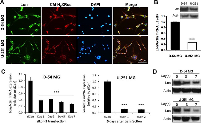
Figure 3. Successful knockdown of Lon in glioma cells.

A. Immunofluorescent staining for LONP1 (green) and CM-H2XRos (red) in D-54 and U-251 cells. Nuclei were counterstained with DAPI (blue). B. D-54 cells had a higher Lon expression than U-251 cells at both protein and mRNA levels. C. Successful knockdown of Lon mRNA was detected in D-54 cells even at Day 7 after siLon-1 transfection. SiRNAs targeting Lon (siLon-1and siLon-2) significantly decreased Lon mRNA levels in U-251 cells 5 days after transfection. D. D-54 and U-251 cells were transfected by siLon-1. Cells were collected and Western blot was used to detect LONP1 at 3 and 7 days after transfection.
