



Abstract
As one of the greatest threats facing the 21st century, antibiotic resistance is now a major public health concern. Host-defense peptides (HDPs) offer an alternative approach to combat emerging multi-drug-resistant bacteria. It is known that helical HDPs such as magainin 2 and its analogs adopt cationic amphipathic conformations upon interaction with bacterial membranes, leading to membrane disruption and subsequent bacterial cell death. We have previously shown that amphipathic sulfono-γ-AApeptides could mimic magainin 2 and exhibit bactericidal activity. In this article, we demonstrate for the first time that amphipathic helical 1:1 α/sulfono-γ-AA heterogeneous peptides, in which regular amino acids and sulfono-γ-AApeptide building blocks are alternatively present in a 1:1 pattern, display potent antibacterial activity against both Gram-positive and Gram-negative bacterial pathogens. Small angle X-ray scattering (SAXS) suggests that the lead sequences adopt defined helical structures. The subsequent studies including fluorescence microscopy and time-kill experiments indicate that these hybrid peptides exert antimicrobial activity by mimicking the mechanism of HDPs. Our findings may lead to the development of HDP-mimicking antimicrobial peptidomimetics that combat drug-resistant bacterial pathogens. In addition, our results also demonstrate the effective design of a new class of helical foldamer, which could be employed to interrogate other important biological targets such as protein–protein interactions in the future.
Graphical abstract

INTRODUCTION
One of the greatest threats facing the 21st century is antibiotic resistance.1 Multi-drug-resistant pathogens, including K. pneumoniae, P. aeruginosa, S. epidermidis, and S. aureus, have caused significant mortality and health care cost.2,3 Alternative therapeutic strategies are in an urgent need to combat drug resistance. Host-defense peptides (HDPs), small natural cationic amphipathic peptides found in most life forms, are being revisited for potential antibiotic development.4–6 It is known that the outer leaflet of bacterial membranes is mainly composed of negatively charged phospholipids such as cardiolipin and phosphatidylglycerol.7 Moreover, other negatively charged molecules are also present on their membranes, including teichoic acids identified in Gram-positive bacteria and lipopolysaccharides found in Gram-negative bacteria. As HDPs are positively charged, they could target bacterial membrane through electrostatic attraction.2 Another important feature of HDPs is that they possess amphipathic global structures containing cationic and hydrophobic domains, even though their secondary structures could be diverse.5 After docking on bacterial membranes, the hydrophobic patch of HDPs interacts with the lipid bilayer, leading to bacterial membrane disruption and cell death.2,8 It is known that some HDPs may involve intracellular targets such as ribosomes and nucleic acids; however, their initial interactions with bacterial membranes are still critical for their cellular entry.7 It should be noted that, although no therapeutic strategies are capable of escaping the mechanism of resistance development, HDPs may be preferable to conventional antibiotics, as the membrane damage caused by HDPs is the biophysical force lacking specific membrane targets, thus, it is more difficult for bacteria to develop resistance.10 In contrast, mammalian cell membranes are comprised of phosphatidylcholine, sphingomyelin, and cholesterol, most of which are zwitterionic. Negatively charged phospholipids are instead hidden in the inner leaflet of plasma membranes.2,10 As a result, the selectivity of HDPs for the bacteria over mammalian cells is considerably high.2
However, there are some drawbacks associated with the development of HDP-based antibiotics, including moderate activity, low enzymatic stability, and challenge in optimization.11 For example, Pexiganan, known as MSI-78, is a synthetic derivative of the helical HDP magainin 2.12 Although it was in phase II clinical trials for diabetic foot ulcers, it failed eventually due to its moderate antimicrobial activity.13 Nonetheless, the amphipathic feature of magainin 2, including the crystal structure of a magainin 2 analogue obtained by Gellman’s group,14 inspires the design of new antimicrobial agents. It is conceived that amphipathic helical peptidomimetics may be able to mimic the mechanism of action of magainin 2, and kill bacteria via membrane disruption.15 Compared to magainin 2, peptidomimetics are expected to be more resistant to proteolytic degradation and possess enhanced chemodiversity for optimization. To date, a few classes of helical peptidomimetics, including β-peptides,16–18 peptoids,19 and oligoureas,20 have been developed as antimicrobial agents that mimic magainin 2. We recently developed a class of helical foldamer termed “sulfono-γ-AApeptides”, as they are oligomers of N-sulfono-acylated-N-aminoethyl amino acids (Figure 1).21 Our previous studies suggest that sulfono-γ- AApeptides can form helical structures analogous to that of α-peptide in solution.21 Based on the structure, we designed a series of sulfono-γ-AApeptides that are expected to mimic magainin 2 and show broad-spectrum antimicrobial activity. As we recently found that 1:1 α/sulfono-γ-AA heterogeneous peptides (Figure 1) also adopt helical conformation in solution,22 it is also compelling to study antimicrobial activity of this class of peptidomimetics.
Figure 1.

General chemical structures of α-peptide, sulfono-γ-AApeptide, and 1:1 α/sulfono-γ-AA peptide.
EXPERIMENTAL SECTION
General Information
All Fmoc-protected α-amino acids and Rink-amide resin (0.7 mmol/g, 200–400 mesh) were purchased from Chem-Impex International, Inc. Other solvents and reagents were purchased from either Sigma-Aldrich or Fisher Scientific and used without further purification. Solid-phase synthesis of 1:1 α/sulfono-γ-AA peptides were carried out in the peptide synthesis vessel on a Burrell Wrist-Action shaker. The sequences were purified on a Waters Breeze 2 HPLC system, and desired fractions were collected and lyophilized on a Labcono lyophilizer. The molecular weight of the heterogeneous peptides was obtained on an Applied Biosystems 4700 Proteomics Analyzer.
Solid Phase Synthesis of 1
The synthesis was conducted on 100 mg Rink amide resin (0.7 mmol/g) following our reported protocol.22 The resin was swelled in DMF for 1 h before use. The Fmoc protecting group was removed by shaking the resin in 3 mL 20% Piperidine/DMF for 15 min (×2). The resin was washed with DCM (×3) and DMF (×3). A premixed solution of Fmoc-Lys (Boc) –OH (3 equiv), HOBt (6 equiv), and DIC (6 equiv) in 2 mL of DMF was added to the resin. The mixture was allowed to shake for 4 h. After wash with DCM and DMF, the Fmoc protecting group was removed following the above-mentioned protocol. Next, the N-alloc γ-AApeptide building block was coupled on the resin under the same coupling condition. The introduction of sulfonamide moieties were achieved by reacting the resin with Pd(PPh3)4 (8 mg, 0.007 mmol) and Me2NH·BH3 (25 mg, 0.42 mmol) in 3 mL of DCM for 10 min (×2), followed by the reaction with the corresponding sulfonyl chlorides (4 equiv) and DIPEA (6 equiv) in 3 mL DCM for 30 min (×2). The reaction cycles were repeated until the desired sequence was assembled on the resin. The resin was then washed with DCM and dried in vacuo. The peptide on the resin was cleaved in a 4 mL vial using the cocktail of TFA/H2O/TIS (95/2.5/2.5) for 2 h. The solvent was evaporated and the crude was analyzed and purified on an analytical (1 mL/min) and a preparative (16 mL/min) Waters HPLC systems, respectively. The protocol of 5–100% linear gradient of solvent B (0.1% TFA in acetonitrile) in A (0.1% TFA in water) over 40 min was used for purification. The HPLC trace was detected at 215 nm. The desired fraction was collected, lyophilized and confirmed on an Applied Biosystems 4700 Proteomics Analyzer.
Solid Phase Synthesis of the Sequences 2–10
The synthesis of the sequences 2–10 was carried out following the same synthetic protocol for 1.
Minimum Inhibitory Concentrations (MICs) against Bacteria
Four bacteria strains including Methicillin-resistant S. aureus (MRSA, ATCC 33591), Methicillin-resistant S. epidermidis (MRSE, RP62A), P. aeruginosa (ATCC27853), and K. pneumoniae (ATCC 13383) were used in the experiment for the test of antimicrobial activities of the 1:1 α/sulfono-γ-AA heterogeneous peptides. Briefly, single colonies of these bacteria were inoculated into 3 mL of TSB medium and allowed to grow overnight. Then the bacteria were reinoculated at 1:100 dilution and grew to mid logarithmic phase, from which 1 × 10⁶ CFU/mL suspension was made. To aliquots of 50 μL of bacteria suspension, 50 μL of 2-fold serial dilutions of peptides starting from 25 μg/mL were added. After incubation at 37 °C for 16 h, the absorption of mixtures at 600 nm wavelength was read by a Biotek Synergy HT microtiter plate reader. The MICs were determined as the lowest concentration, completely inhibiting bacteria growth. Results were repeated three times with duplicates each time.
Hemolytic Assay
The freshly drawn, K2EDTA treated human red blood cells (hRBCs) were washed with 1× PBS buffer, and centrifuged at 1000 g for 10 min. The step was repeated several times until the supernatant was clear. After the supernatant was removed, the RBCs were diluted into 5% v/v suspension, and incubated with equal volume of heterogeneous peptides at 2-fold serial-diluted concentrations at 37 °C for 1 h. After centrifugation at 3500 rpm for 10 min, 30 μL of the supernatant was transferred into 100 μL of PBS, and the absorbance at 540 nm was read. Two % Triton X-100 was used the positive control. The hemolysis percentage was calculated by the formula % hemolysis = (Abs sample − Abs PBS)/(Abs Triton − Abs PBS) × 100%. Results were repeated three times with duplicates each time.
Fluorescence Microscopy
Fluorescence microscopy was used to assess the ability of the peptidomimetic to damage bacterial membranes. Two dyes, 4′,6-diamidino-2-phenylindole dihydrochloride (DAPI) and propidium iodide (PI), were used. DAPI is capable of staining bacteria cells irrespective of cell viability, whereas PI can only stain cells with compromised membranes. After bacteria grew to mid logarithmic phase, they were incubated with peptides at 37 °C for 2 h. The mixture was centrifuged at 5000 g for 15 min, and the cell pellets were collected and washed, and were subsequently incubated with PI (5 μg/mL) then with DAPI (10 μg/mL) for 15 min on ice. After washing with buffer, 10 μL of the samples were placed on chamber slides and observed under Zeiss Axio Image Zloptical microscope using 100× oil-immersion objective.
RESULTS AND DISCUSSION
We anticipated that 1:1 α/sulfono-γ-AA peptides could be excellent candidates for antimicrobial development. The heterogeneous scaffold could further enhance the diversity of chemical groups, and therefore, potent antimicrobial agents may be identified through optimization.23 As this class of heterogeneous peptides were not investigated for their antimicrobial activity previously, our studies may shed light on the design of a new class of antibiotic agents.
In our recent report, 2D-NMR, small-angle X-ray scattering (SAXS), and CD studies suggest that the 1:1 α/sulfono-γ-AA peptides adopt helical structure in solution (Figure 2).22 According to the 2D-NMR analysis, this class of heterogeneous peptides project approximately four side chains per turn (Figure 2).22 In addition, their helical folding propensity appears to be high because the helicity is still discernible for the sequence with the length comparable to that of 9-mer peptide.22 Although their helical pitch and diameter might be different from α-peptides, we believed through manipulation of side chains, new helical sequences with cationic amphipathic structures could be properly designed. These amphipathic heterogeneous peptides in theory should mimic structure and mechanism of action of helical HDPs.
Figure 2.

Schematic representation of the helical 1:1 α/sulfono-γ-AA heterogeneous peptides. The numbers represent the position of the residue in a sequence. a and b denote the chiral side chain and the sulfonamido side chain from a sulfono-γ-AA building block, respectively.
To test our hypothesis, we designed a series of 1:1 α/sulfono-γ-AA peptides with varied distribution of cationic charge and hydrophobic groups on the helical scaffold (Figure 3), in order to identify and develop new antimicrobial agents. Sequences 1 and 2 contain same number of residues as previously reported sequence shown in Figure 2,22 while the rest of eight sequences are longer and contain even one more sulfono-γ-AA residue. As our previous results suggested that longer sequences possess better folding propensity in this class of peptidomimetics,22 all the sequences were expected to adopt good helical structures in solution. Meanwhile, the side chains of these sequences were arranged in a way in which cationic amphipathic structures could be formed. These sequences were then synthesized and tested for their antimicrobial activity against a few Gram-positive and Gram-negative bacterial pathogens, including multi-drug-resistant pathogens.24,25 Hemolytic activity was also obtained to assess their selectivity toward bacteria cells.15
Figure 3.
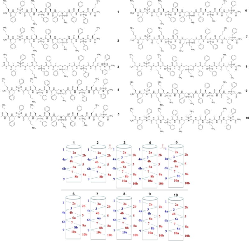
Structures of antimicrobial 1:1 α/sulfono-γ-AA heterogeneous peptides. In order to illustrate their cationic charge (colored in blue) and hydrophobic group (colored in red) distribution, the sequences are schematically shown on the helical scaffold.
As shown above, the rational design of amphipathic sequences led to the discovery of 1:1 α/sulfono-γ-AA heterogeneous peptides with potent antimicrobial activity against a few clinically relevant multidrug-resistant bacterial strains. It is also noted that none of these sequences are toxic, as they all exhibit low hemolytic activity. This is somewhat different from our previously reported homogeneous antimicrobial sulfono-γ-AApeptides,15 which show certain hemolytic activity at high concentrations. In the meantime, it appears that the activity of these peptide-mimetics correlates with their sequences well. As shown in Table 1, although sequences 1 and 2 were designed to be amphipathic, and they do show antimicrobial activity against both Gram-positive and Gram-negative bacteria, the activity is quite weak. Two reasons may account for this. First, since the sequences of 1 and 2 are shorter than other sequences, the interaction of sequences with bacterial membrane are not strong.27,30 The antimicrobial activity of membrane-acting peptides/peptidomimetics is highly dependent on their charge and hydrophobicity. Insufficient number of cationic and hydrophobic groups in a sequence could not acquire strong association of antimicrobial sequences with bacterial membranes. This is consistent to our previous findings.15 Second, sequences of 1 and 2 may not adopt a well-defined helical confirmation due to their shorter lengths, and therefore, their structures may not be sufficiently amphipathic. Compared with 1, sequences 3 and 4, with one more hydrophobic sulfono-γ-AA building block (containing two hydrophobic side chains) at the C-terminus, display enhanced antibacterial activity, particularly toward Gram-positive bacteria MRSA and MRSE. It may suggest that Gram-positive bacteria, which possess one layer of plasma membranes, are more sensitive to hydrophobic interaction of antimicrobial agents compared to Gram-negative bacteria containing both inner and outer membranes. Acetylation at the N-terminal end lead to augmented hemolytic activity, similar to previously reported antimicrobial sulfono-γ-AApeptides.15 The same trend is observed for sequences 5 and 6. Compared with previous sequences, replacement of the hydrophobic side chain at the position 8b with a cationic side chain lead to both 5 and 6 which exhibit remarkable activity against both Gram-positive and Gram-negative bacteria. As a matter of fact, the most potent sequence 6 show MIC < 5 μg/mL against all tested strains. It should be noted that 6 is even much more potent than Pexiganan,16–20,31 yet no hemolytic activity was observed under the experimental condition. It is interesting that sulfono-γ-AApeptide building blocks containing two cationic side chains are not favorable for antimicrobial activity of this class of sequences, as seen for sequences 7–10. For instance, in the sequence 7, the sulfono-γ-AA residue 6 contains two cationic side chains (6a and 6b), whereas the sequence 7 only possesses weak antimicrobial activity, even though it has the same cationic charge/hydrophobicity ratio as the sequence 6. Although the sequence 10 largely regains activity with the cationic side chain introduced at position 8b, it is still not as active as the lead sequence 6. One plausible explanation is that the sulfono-γ-AA residue bearing two adjacent cationic charges may lead to electrostatic repulsion, which destabilizes the helical folding conformation, thus, preventing the formation of defined amphipathic structure.
Table 1.
Antimicrobial and Hemolytic Activities of 1:1 α/Sulfono-γ-AA Peptidesa
| MIC (μg/mL)
|
||||||
|---|---|---|---|---|---|---|
| sequences | MRSA (Gram+) |
MRSE (Gram+) |
K. pneumonia (Gram−) |
P. aeruginosa (Gram−) |
hemolysis (HC50, μg/mL) |
selectivity index HC50/ MIC of MRSA |
| 1 | 12.5–25 | 6.3–12.5 | 12.5–25 | >25 | >250 | >10 |
| 2 | 6.3 | 6.3–12.5 | 12.5–25 | >25 | >250 | >39 |
| 3 | 6 | 5 | 6.3–12.5 | >25 | 200 | 33 |
| 4 | 6.3 | 5 | 12.5–25 | >25 | >250 | >39 |
| 5 | 5 | 3 | 12.5–25 | 12.5–25 | 230 | 46 |
| 6 | 3 | 3 | 4 | 4 | >250 | >83 |
| 7 | 12.5–25 | 5.0 | >25 | >25 | >250 | >10 |
| 8 | >25 | 6–12 | >25 | >25 | >250 | 10 |
| 9 | >25 | >25 | >25 | >25 | >250 | 10 |
| 10 | 8 | 6 | 10 | 10 | >250 | 31 |
| pexiganan | 16 | 8 | 32 | 16 | ||
The helicity of lead sequences was also evaluated by SAXS (small-angle X-ray scattering) studies. This is because these sequences are a new class of heterogeneous peptidomimetics, CD (circular dichroism) may provide ambiguous results, since the backbone is different from regular α-peptides.32 On the other hand, SAXS emerges to be an excellent technique to quickly assess the solution structures of peptides or peptidomimetics.6,21,22,33,34 Herein we used a Kratky plot to analyze the helicity of the lead 1:1 α/sulfono-γ-AA peptide sequences 1, 3, 4, 5, and 6. The peak around 0.1 Å−1 and the minimum in the q range of 0.2–0.4 Å−1 indicate the presence of the helix-rich globular protein, whereas a slope lacking maximum and minimum in the middle q range suggests the existence of a random coil (Figure S2).33,34 As shown in Figure 4, although the sequence 1 displays certain helicity, the lack of defined bell shape suggests the sequence is at least partially unfolded in solution, probably due to its short length. All other sequences adopt well-defined helical structures in solution, since they all show characteristic maximum around 0.1 Å−1 and minimum in the q range of 0.2–0.4 Å−1. The data also indicate that the helicity of 3 and 5 is stronger than that of 4 and 6, respectively. This is consistent with our previous observation that N-terminal acetylation of the sequence could enhance helical folding propensity.15 It also suggests that the acetylation could deteriorate antimicrobial activity,27,35 as 6 is much more potent than 5.
Figure 4.
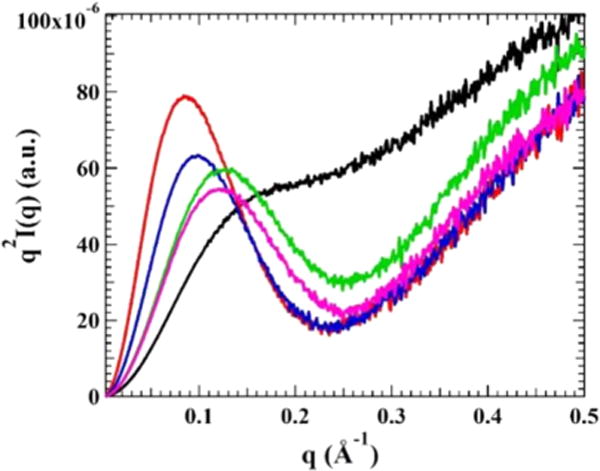
Krakty plot of 1, 3, 4, 5, and 6 measured in pH 7.4 PBS buffer. The peak around 0.1 Å−1 and the minimum in the q range of 0.2–0.4 Å−1 suggest 3 (red), 4 (blue), 5 (green), and 6 (magenta) form well-defined helical structures.
We hypothesized that 1:1 α/sulfono-γ-AA heterogeneous peptides exert their activity by mimicking HDPs such as magainin, because they adopt amphipathic structures that lead to disruption of bacterial membranes. Therefore, fluorescence microscopy was employed to assess the ability of the peptide 6 to compromise membranes of S. aureus.36 The membrane permeable dye 4′,6-diamidino-2-phenylindole (DAPI) and the DNA intercalator propidium iodide (PI; Figure 5) were used in the study. DAPI stain membranes of both dead and live cells with blue fluorescence, but PI could only fluoresce in red color when cell membranes are damaged. As shown in Figure 5, bacterial cells in the control group are only visible under DAPI channel. In contrast, incubation of bacteria with 6 led to the visibility of bacteria under both PI and DAPI channel, suggesting the membranes of S. aureus were compromised.
Figure 5.
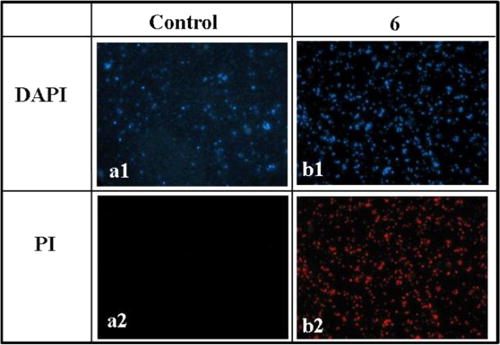
Fluorescence micrographs of S. aureus that are treated or not treated with 10 μg/mL 1:1 α/sulfono-γ-AA peptide 6 for 2 h: (a1) control, no treatment, DAPI stained; (a2) control, no treatment, PI stained; (b1) treatment with 6, DAPI stained; (b2) treatment with 6, PI stained.
It is believed that HDPs exert bactericidal action rapidly through the disruption of bacterial membranes.29 Since 1:1 α/sulfono-γ-AA heterogeneous peptides were designed to mimic HDPs, their action on bacterial killing were also expected to be fast. Thus, the time kill study was carried out to test the efficiency of 6 to kill MRSA. Compound 6 was studied at 4-, 8-, and 16-fold of the MIC. Cell viability was determined by colony count in agar plates in a time-dependent fashion at above-mentioned concentrations (Figure 6). In all cases, 6 could eradicate bacteria completely in 2 h, suggesting the mechanism of action is analogous to that of magainin.
Figure 6.

Time-kill curves of 6 for MRSA. The killing activity was monitored for the first 2 h. The concentrations were 4× MIC, 8× MIC, and 16× MIC, respectively.
CONCLUSIONS
To summarize, we identified the first example of 1:1 α/sulfono-γ-AA heterogeneous peptide foldamers with potent antimicrobial activity toward multidrug-resistant Gram-positive and Gram-negative bacteria. These sequences adopt defined amphipathic helical structures, and are likely to kill bacteria via disruption of bacterial membranes based time-killing studies and fluorescence microscopy. As their mechanism of action is similar to that of HDPs, their further development may lead to a new class of helical foldamer combating emerging antibioticresistant pathogens. In addition, the effective design of this class of antimicrobial agents suggest the potential of 1:1 α/sulfono-γ-AA heterogeneous peptides as a new class of foldamer for the interrogation of other important biological targets such as protein–protein interactions.
Supplementary Material
Acknowledgments
This work is supported by NSF CAREER 1351265 and NIH 1R01GM112652-01A1.
Footnotes
Supporting Information
The Supporting Information is available free of charge on the ACS Publications website at DOI: 10.1021/acs.biomac.6b00286.
HPLC traces, characterizations, and experimental details (PDF).
Notes
The authors declare no competing financial interest.
References
- 1.Niu Y, Wang RE, Wu H, Cai J. Future Med Chem. 2012;4:1853–1862. doi: 10.4155/fmc.12.111. [DOI] [PubMed] [Google Scholar]
- 2.Marr AK, Gooderham WJ, Hancock REW. Curr Opin Pharmacol. 2006;6:468–472. doi: 10.1016/j.coph.2006.04.006. [DOI] [PubMed] [Google Scholar]
- 3.Antimicrobial resistance: global report on surveillance. World Health Organization; Geneva, Switzerland: 2014. [Google Scholar]
- 4.Brown KL, Hancock REW. Curr Opin Immunol. 2006;18:24–30. doi: 10.1016/j.coi.2005.11.004. [DOI] [PubMed] [Google Scholar]
- 5.Hancock REW, Sahl HG. Nat Biotechnol. 2006;24:1551–1557. doi: 10.1038/nbt1267. [DOI] [PubMed] [Google Scholar]
- 6.Mercer DK, O’Neil DA. Future Med Chem. 2013;5:315–337. doi: 10.4155/fmc.12.213. [DOI] [PubMed] [Google Scholar]
- 7.Matsuzaki K. Biochim Biophys Acta, Biomembr. 2009;1788:1687–1692. doi: 10.1016/j.bbamem.2008.09.013. [DOI] [PubMed] [Google Scholar]
- 8.Kuroda K, Caputo GA. Wiley Interdiscip Rev Nanomed Nanobiotechnol. 2013;5:49–66. doi: 10.1002/wnan.1199. [DOI] [PubMed] [Google Scholar]
- 9.Tew GN, Scott RW, Klein ML, DeGrado WF. Acc Chem Res. 2010;43:30–39. doi: 10.1021/ar900036b. [DOI] [PMC free article] [PubMed] [Google Scholar]
- 10.Hancock REW, Brown KL, Mookherjee N. Immunobiology. 2006;211:315–322. doi: 10.1016/j.imbio.2005.10.017. [DOI] [PubMed] [Google Scholar]
- 11.Chongsiriwatana NP, Patch JA, Czyzewski AM, Dohm MT, Ivankin A, Gidalevitz D, Zuckermann RN, Barron AE. Proc Natl Acad Sci U S A. 2008;105:2794–2799. doi: 10.1073/pnas.0708254105. [DOI] [PMC free article] [PubMed] [Google Scholar]
- 12.Matsuzaki K. Biochim Biophys Acta, Rev Biomembr. 1998;1376:391–400. doi: 10.1016/s0304-4157(98)00014-8. [DOI] [PubMed] [Google Scholar]
- 13.Gottler LM, Ramamoorthy A. Biochim Biophys Acta, Biomembr. 2009;1788:1680–1686. doi: 10.1016/j.bbamem.2008.10.009. [DOI] [PMC free article] [PubMed] [Google Scholar]
- 14.Hayouka Z, Mortenson DE, Kreitler DF, Weisblum B, Forest KT, Gellman SH. J Am Chem Soc. 2013;135:15738–15741. doi: 10.1021/ja409082w. [DOI] [PMC free article] [PubMed] [Google Scholar]
- 15.Li Y, Wu H, Teng P, Bai G, Lin X, Zuo X, Cao C, Cai J. J Med Chem. 2015;58:4802–4811. doi: 10.1021/acs.jmedchem.5b00537. [DOI] [PubMed] [Google Scholar]
- 16.Karlsson AJ, Pomerantz WC, Weisblum B, Gellman SH, Palecek SP. J Am Chem Soc. 2006;128:12630–12631. doi: 10.1021/ja064630y. [DOI] [PubMed] [Google Scholar]
- 17.Porter EA, Weisblum B, Gellman SH. J Am Chem Soc. 2002;124:7324–7330. doi: 10.1021/ja0260871. [DOI] [PubMed] [Google Scholar]
- 18.Porter EA, Wang X, Lee HS, Weisblum B, Gellman SH. Nature. 2000;404:565–565. doi: 10.1038/35007145. [DOI] [PubMed] [Google Scholar]
- 19.Patch JA, Barron AE. J Am Chem Soc. 2003;125:12092–12093. doi: 10.1021/ja037320d. [DOI] [PubMed] [Google Scholar]
- 20.Violette A, Fournel S, Lamour K, Chaloin O, Frisch B, Briand JP, Monteil H, Guichard G. Chem Biol. 2006;13:531–538. doi: 10.1016/j.chembiol.2006.03.009. [DOI] [PubMed] [Google Scholar]
- 21.Wu H, Qiao Q, Hu Y, Teng P, Gao W, Zuo X, Wojtas L, Larsen RW, Ma S, Cai J. Chem – Eur J. 2015;21:2501–2507. doi: 10.1002/chem.201406112. [DOI] [PubMed] [Google Scholar]
- 22.Wu H, Qiao Q, Teng P, Hu Y, Antoniadis D, Zuo X, Cai J. Org Lett. 2015;17:3524–3527. doi: 10.1021/acs.orglett.5b01608. [DOI] [PubMed] [Google Scholar]
- 23.Horne WS, Gellman SH. Acc Chem Res. 2008;41:1399–1408. doi: 10.1021/ar800009n. [DOI] [PMC free article] [PubMed] [Google Scholar]
- 24.Li Y, Smith C, Wu H, Teng P, Shi Y, Padhee S, Jones T, Nguyen AM, Cao C, Yin H, Cai J. ChemBioChem. 2014;15:2275–2280. doi: 10.1002/cbic.201402264. [DOI] [PMC free article] [PubMed] [Google Scholar]
- 25.Li Y, Smith C, Wu H, Padhee S, Manoj N, Cardiello J, Qiao Q, Cao C, Yin H, Cai J. ACS Chem Biol. 2014;9:211–217. doi: 10.1021/cb4006613. [DOI] [PMC free article] [PubMed] [Google Scholar]
- 26.Wu H, Niu Y, Padhee S, Wang RE, Li Y, Qiao Q, Bai G, Cao C, Cai J. Chem Sci. 2012;3:2570–2575. [Google Scholar]
- 27.Niu Y, Padhee S, Wu H, Bai G, Harrington L, Burda WN, Shaw LN, Cao C, Cai J. Chem Commun. 2011;47:12197–12199. doi: 10.1039/c1cc14476f. [DOI] [PMC free article] [PubMed] [Google Scholar]
- 28.Thaker HD, Som A, Ayaz F, Lui D, Pan W, Scott RW, Anguita J, Tew GN. J Am Chem Soc. 2012;134:11088–11091. doi: 10.1021/ja303304j. [DOI] [PMC free article] [PubMed] [Google Scholar]
- 29.Ge Y, MacDonald DL, Holroyd KJ, Thornsberry C, Wexler H, Zasloff M. Antimicrob Agents Chemother. 1999;43:782–788. doi: 10.1128/aac.43.4.782. [DOI] [PMC free article] [PubMed] [Google Scholar]
- 30.Padhee S, Hu Y, Niu Y, Bai G, Wu H, Costanza F, West L, Harrington L, Shaw LN, Cao C, Cai J. Chem Commun. 2011;47:9729–9731. doi: 10.1039/c1cc13684d. [DOI] [PubMed] [Google Scholar]
- 31.Claudon P, Violette A, Lamour K, Decossas M, Fournel S, Heurtault B, Godet J, Mély Y, Jamart-Grégoire B, Averlant-Petit M-C, Briand J-P, Duportail G, Monteil H, Guichard G. Angew Chem, Int Ed. 2010;49:333–336. doi: 10.1002/anie.200905591. [DOI] [PubMed] [Google Scholar]
- 32.Kritzer JA, Hodsdon ME, Schepartz A. J Am Chem Soc. 2005;127:4118–4119. doi: 10.1021/ja042933r. [DOI] [PMC free article] [PubMed] [Google Scholar]
- 33.Putnam CD, Hammel M, Hura GL, Tainer JA. Q Rev Biophys. 2007;40:191–285. doi: 10.1017/S0033583507004635. [DOI] [PubMed] [Google Scholar]
- 34.Sagar A, Peddada N, Solanki Ak, Choudhary V, Garg R. Biochem Biophys Res Commun. 2013;435:740–744. doi: 10.1016/j.bbrc.2013.05.055. [DOI] [PubMed] [Google Scholar]
- 35.Schmitt MA, Weisblum B, Gellman SH. J Am Chem Soc. 2004;126:6848–6849. doi: 10.1021/ja048546z. [DOI] [PubMed] [Google Scholar]
- 36.Niu Y, Padhee S, Wu H, Bai G, Qiao Q, Hu Y, Harrington L, Burda WN, Shaw LN, Cao C, Cai J. J Med Chem. 2012;55:4003–9. doi: 10.1021/jm300274p. [DOI] [PubMed] [Google Scholar]
Associated Data
This section collects any data citations, data availability statements, or supplementary materials included in this article.