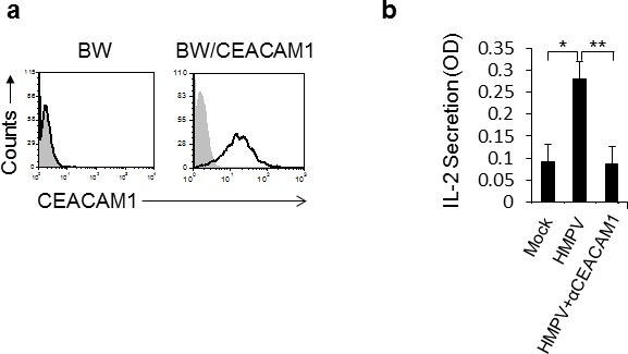
Figure 2. The expression of CEACAM1 on HMPV-infected A549 cells is functional.

a. FACS analysis of parental BW cells and BW cells transfected to expressed a chimeric protein composed of the extracellular portion of CEACAM1 fused to mouse zeta chain (BW/CEACAM1). The filled gray histogram is the background staining and the empty black histogram is the staining of CEACAM1. b. IL-2 secretion from the BW/CEACAM1 cells following 48h incubation with the indicated A549 cells that were either mock infected (Mock), infected with HMPV (HMPV) or infected with HMPV and blocked with anti CEACAM1 mAb (HMPV+αCEACAM1). Values are shown as means ± SEM. The figure shows data from three experiments combined. *p <0.05, **p < 0.02.
