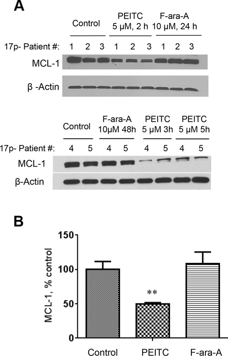
Figure 6. Decrease of MCL-1 protein induced by PEITC in CLL cells with 17p-deletion.
(A) MCL-1 protein levels were measured by Western Blotting in 17p- CLL cells before and after treatment with 5 μM PEITC for 2–5 h, or with 10 μM F-ara-A for 24–48 h. (B) Quantitative analysis of MCL-1 protein levels by Image J, **p < 0.01, between groups.

