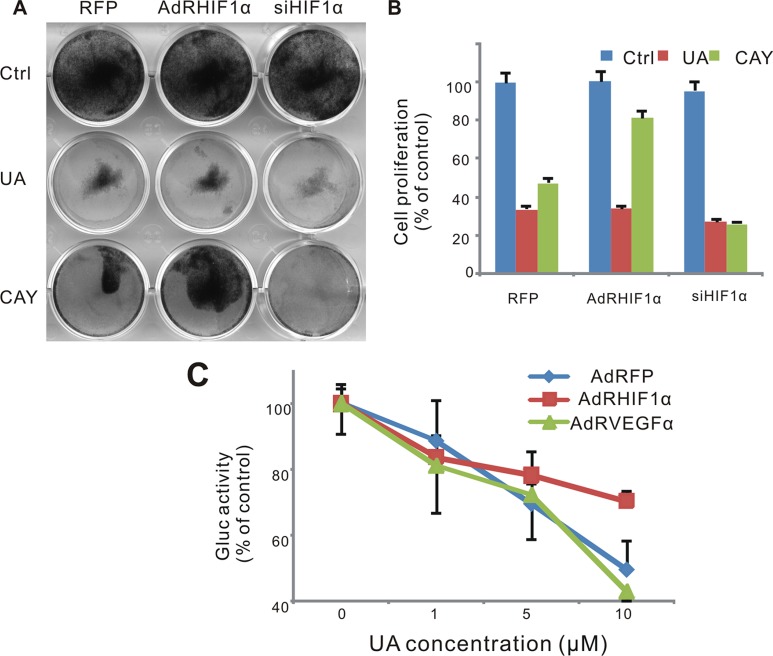
Figure 6. Effect of UA in hypoxia pathway in MDA-MB-231 cells.
(A) Cell was infected with AdRFP, AdRHIF-1α or AdRsiHIF-1α virus for 16 h, followed by UA treatment in 1% FBS DMEM medium for 48 h, cell survival was tested by crystal violet assay. (B) Cell stained with crystal violet (show in Figure 6A) were dissolved with 20% acetic acid and measured at 570 nm. (C) MDA-MB-231 -HIF-1α-GLuc cells were infected with AdRFP, AdRHIF-1α or AdRVEGF and treated with UA in serum free DMEM medium for 2 hour. The Gaussia luciferase (GLuc) activity of the cell medium was measured by the Gaussia Luciferase Assay kit. The inhibition was presented as the percentage of the control group.

