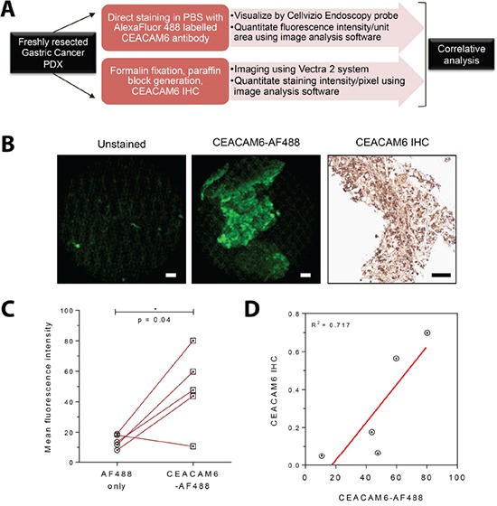
Figure 5. Ex vivo staining of a fluorescently conjugated antibody to CEACAM6 and visualization with a confocal laser endomicroscopy probe.

A. Schematic diagram of the experimental plan. Freshly resected patient derived xenografts (PDXs) of gastric cancer were divided into two pieces - one section was used for direct fluorescent staining and the remainder for immunohistochemistry. B. Representative images from freshly resected gastric cancer xenograft tumors without fixation showing clearly visible fluorescence signal from the CEACAM6 antibody conjugated to Alexa Fluor 488 (imaged with a Cellvizio confocal laser endomicroscopy probe, compatible with commercial endoscopy equipment). CEACAM6 IHC staining of the same xenograft is shown for reference. Scale bar=50μm. C. Analysis of signal intensity of CEACAM6-AF 488 stained tumors in comparison to unstained controls, as shown in B, n=5. Paired t test. D. Correlative analysis of Cellvizio CEACAM6-AF 488 fluorescence intensity values with CEACAM6 IHC staining intensity, as shown in B, n=5.
